
- Laboratory of Integrative Brain Sciences, Department of Biology, Waseda University, and Center for Medical Life Science of Waseda University, Shinjuku-ku, Tokyo, Japan
It is now established that the brain and other nervous systems have the capability of forming steroids de novo, the so-called “neurosteroids.” The pioneering discovery of Baulieu and his colleagues, using rodents, has opened the door to a new research field of “neurosteroids.” In contrast to mammalian vertebrates, little has been known regarding de novo neurosteroidogenesis in the brain of birds. We therefore investigated neurosteroid formation and metabolism in the brain of quail, a domestic bird. Our studies over the past two decades demonstrated that the quail brain possesses cytochrome P450 side-chain cleavage enzyme (P450scc), 3β-hydroxysteroid dehydrogenase/Δ5-Δ4-isomerase (3β-HSD), 5β-reductase, cytochrome P450 17α-hydroxylase/c17,20-lyase (P45017α,lyase), 17β-HSD, etc., and produces pregnenolone, progesterone, 5β-dihydroprogesterone (5β-DHP), 3β, 5β-tetrahydroprogesterone (3β, 5β-THP), androstenedione, testosterone, and estradiol from cholesterol. Independently, Schlinger’s laboratory demonstrated that the brain of zebra finch, a songbird, also produces various neurosteroids. Thus, the formation and metabolism of neurosteroids from cholesterol is now known to occur in the brain of birds. In addition, we recently found that the quail brain expresses cytochrome P4507α and produces 7α- and 7β-hydroxypregnenolone, previously undescribed avian neurosteroids, from pregnenolone. This paper summarizes the advances made in our understanding of neurosteroid formation and metabolism in the brain of domestic birds. This paper also describes what are currently known about physiological changes in neurosteroid formation and biological functions of neurosteroids in the brain of domestic and other birds.
Introduction
Peripheral steroid hormones cross the blood–brain barriers and act on brain cells through intracellular receptor-mediated mechanisms that regulate several important brain neuronal functions. Therefore, the brain traditionally has been considered to be a target site of peripheral steroid hormones. In contrast to this classical concept, new findings over the past two decades have shown that the brain itself also synthesizes steroids de novo from cholesterol through mechanisms at least partly independent of peripheral steroidogenic glands. Such steroids synthesized de novo in the brain and other nervous systems are called “neurosteroids.” Baulieu and colleagues have opened the door to a new research field of “neurosteroids” from their studies using rodents. In contrast to mammalian vertebrates, little has been known regarding de novo neurosteroidogenesis in the brain of birds.
Birds have always served as excellent animal models for understanding the actions of peripheral steroids on brain and behavior. Thus, the investigation of neurosteroid synthesis and action in birds may be also useful. We therefore characterized neurosteroids formed from cholesterol in the avian brain using the Japanese quail Coturnix japonica. A series of our studies over the past two decades demonstrated that the quail brain possesses cytochrome P450 side-chain cleavage enzyme (P450scc), 3β-hydroxysteroid dehydrogenase/Δ5-Δ4-isomerase (3β-HSD), 5β-reductase, cytochrome P450 17α-hydroxylase/c17,20-lyase (P45017α,lyase), 17β-HSD, etc., and produces pregnenolone, progesterone, 5β-dihydroprogesterone (5β-DHP), 3β, 5β-tetrahydroprogesterone (3β, 5β-THP), androstenedione, testosterone, and estradiol from cholesterol (Tsutsui and Yamazaki, 1995; Usui et al., 1995; Tsutsui et al., 1997a,b, 1999, 2003a,b; Ukena et al., 1999a,b, 2001; Matsunaga et al., 2001, 2002; Tsutsui and Schlinger, 2001). Schlinger and colleagues also undertook similar studies in the zebra finch Taeniopygia guttata (Vanson et al., 1996; Schlinger et al., 1999; Freking et al., 2000; Soma et al., 2004). Thus, the formation and metabolism of several neurosteroids from cholesterol appear to occur in the brain of birds.
In addition to mammals and birds, the formation and metabolism of neurosteroids from cholesterol is now well documented in amphibians (Mensah-Nyagan et al., 1994, 1996a,b, 1999; Beaujean et al., 1999; Takase et al., 1999, 2002; Inai et al., 2003; Matsunaga et al., 2004; Do-Rego et al., 2007; Bruzzone et al., 2010) and fish (Sakamoto et al., 2001). Accordingly, de novo neurosteroidogenesis in the brain from cholesterol is considered to be a conserved property across vertebrates (for reviews, see Baulieu, 1997; Tsutsui et al., 1999, 2000, 2003a,b, 2006; Compagnone and Mellon, 2000; Mellon and Vaudry, 2001; Tsutsui and Mellon, 2006; Do-Rego et al., 2009). However, biosynthetic pathways of neurosteroid in birds, as well as other vertebrates may still not be fully mapped (for a review, see Tsutsui et al., 2006). This conclusion is supported by the discovery that the quail brain synthesizes two previously undescribed avian neurosteroids, 7α- and 7β-hydroxypregnenolone, from pregnenolone (Tsutsui et al., 2008). Furthermore, cytochrome P4507α catalyzing the conversion of pregnenolone to 7α-hydroxypregnenolone was identified in the quail brain (Tsutsui et al., 2008).
This review summarizes neurosteroid formation and metabolism in the brain of domestic birds, in particular the quail, obtained by a series of our studies and the related studies with the ring dove (Clark et al., 1999; Tsutsui et al., 1999; Lea et al., 2001), by the collaboration with the laboratory of Lea at the University of Central Lancashire, Preston, UK. Based on recent findings, this review also highlights the current knowledge regarding physiological changes in neurosteroid formation and biological functions of neurosteroids in the avian brain.
Background of the Discovery of Neurosteroids in the Avian Brain
A great deal was known about the brain as a target site of peripheral steroid hormones more than 20 years ago. Steroid hormones supplied by the peripheral steroidogenic glands regulate several important brain functions during development which persist into adulthood in vertebrates. Peripheral steroid hormones cross the blood–brain barriers, due to their chemically lipid solubility, and act on brain tissues through intracellular receptor-mediated mechanisms that regulate the transcription of specific genes (Fuxe et al., 1977; McEwen, 1991). By diverse actions on the brain, peripheral steroids, in particular sex steroids, have profound effects on behavior of vertebrate animals. Birds have contributed significantly to our understanding of the mechanisms of steroid actions on several kinds of reproductive behaviors, such as courtship, copulatory, aggressive, and parental behaviors. Extensive studies using a variety of wild and captive, intact and castrated, reproductive and non-reproductive birds have established a relationship between the blood level of androgens, estrogens, or progestins and the expression of typical reproductive behaviors (Ottinger and Brinkley, 1978, 1979; Tsutsui and Ishii, 1981; Balthazart, 1983; Wingfield and Marler, 1988; Schlinger and Callard, 1991; Wingfield and Farner, 1993).
Gonadal androgens, for instance, act on the brain to influence several male reproductive behaviors in birds. Castration of adult male birds leads to decreases or losses of aggressive, courtship, and copulatory behaviors and replacement therapy with androgens restores these behaviors (Adkins and Adler, 1972; Arnold, 1975; Pröve, 1978; Tsutsui and Ishii, 1981; Ishii and Tsutsui, 1982; Balthazart, 1983). Many of the brain regions that control a variety of reproductive behaviors contain a high proportion of cells that concentrate androgenic hormones in male birds (Arnold et al., 1976; Korsia and Bottjer, 1989; Watson and Adkins-Regan, 1989). Therefore, the brain traditionally has been considered to be a target site of peripheral steroids. In addition to direct steroid actions, the metabolism of peripheral steroids in brain tissues can result in biotransformation and the production of biologically active metabolites. Androgenic action in the brain is often mediated by the enzymatic activity of cytochrome P450 aromatase (P450arom) which catalyzes the conversion of androgens to estrogens. Both cytochrome P450arom and estrogen receptors are expressed in several brain regions including the hypothalamus and preoptic area which are involved in the control of reproductive behaviors in birds (Schlinger and Callard, 1987, 1989a,b,c, 1991; Balthazart et al., 1990a,b, 1991).
On the other hand, new findings from several laboratories have established unequivocally that the nervous system itself forms neurosteroids de novo from cholesterol. This new concept originated from the observations made by Baulieu and colleagues. They found that several neurosteroids, such as pregnenolone, dehydroepiandrosterone, and their esters, accumulate in high quantities in the brain of several mammalian species (Corpéchot et al., 1981, 1983; Robel and Baulieu, 1985; Lanthier and Patwardhan, 1986; Robel et al., 1986, 1987; Jo et al., 1989; Mathur et al., 1993). The brain content of these neurosteroids remain constant even after the removal of peripheral steroids by procedures, such as adrenalectomy, castration, and hypophysectomy. Extensive studies on mammals have established that the brain and other nervous systems have the capability of forming neurosteroids de novo from cholesterol (Corpéchot et al., 1981, 1983; Robel and Baulieu, 1985; Robel et al., 1986, 1987; Jo et al., 1989).
In birds, the new concept of de novo neurosteroidogenesis in the brain originated from our studies in the 1990s. We demonstrated that various neurosteroids are formed from cholesterol in the brain of quail (Tsutsui and Yamazaki, 1995; Usui et al., 1995; Tsutsui et al., 1997; Tsutsui et al., 1999, 2003a; Ukena et al., 1999; Ukena et al., 2001; Matsunaga et al., 2001, 2002; Tsutsui and Schlinger, 2001). Independently, Schlinger’s laboratory at the University of California, Los Angeles, USA also contributed to this area by the studies using zebra finches (Vanson et al., 1996; Schlinger et al., 1999; Freking et al., 2000; Soma et al., 2004). The formation and metabolism of neurosteroids is now known to occur in the brain of birds in general.
Expression of Cytochrome P450scc and Pregnenolone Formation in the Avian Brain
Pregnenolone, a 3β-hydroxy-Δ5-steroid, is known to be the common precursor of all steroid hormones in peripheral steroidogenic glands including the gonads and adrenals (Figure 1). The formation of pregnenolone is initiated by the cleavage of the cholesterol side-chain by cytochrome P450scc, a rate-limiting mitochondrial enzyme originally found in peripheral steroidogenic glandular cells (Figure 1). Therefore, the demonstration of pregnenolone formation from cholesterol is essential to establish de novo neurosteroidogenesis in the brain of birds.
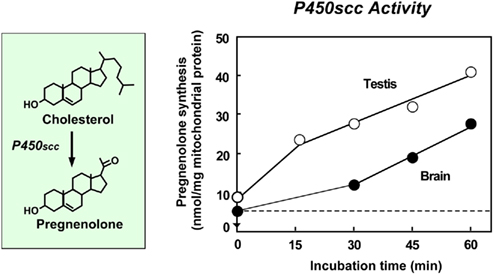
Figure 1. Pregnenolone formation from cholesterol in the quail brain. Biochemical analysis of the formation of pregnenolone from cholesterol using intact mitochondria derived from the quail brain. Incubations were performed with 200 μM cholesterol, 0.25 mg of mitochondrial protein, 10 mM isocitrate and 10 mM succinate at 41°C for different times. See Tsutsui and Yamazaki (1995) for details.
Expression and Enzymatic Activity of Cytochrome P450scc in the Avian Brain
Tsutsui and Yamazaki (1995) measured the concentration of pregnenolone in the brain of adult quail using a specific radioimmunoassay. The pregnenolone concentration in adult birds was much higher in the brain than in plasma (Tsutsui and Yamazaki, 1995). The accumulation of pregnenolone in the quail brain may be largely independent of peripheral steroidogenic glands because a high level of pregnenolone persisted in the hypophysectomized birds (Tsutsui and Yamazaki, 1995). Subsequently, the formation of pregnenolone from cholesterol was found in intact mitochondria derived from the quail brain (Tsutsui and Yamazaki, 1995; Figure 1). To investigate the presence of cytochrome P450scc in the quail brain, Tsutsui and Yamazaki (1995) carried out Western immunoblot analysis with an antibody against purified bovine P450scc after SDS-gel electrophoresis of brain homogenates. In the brain, the antibody against P450scc predominantly recognized a protein band of electrophoretic mobility in the proximity of bovine P450scc. A similar result was obtained in the brain of the ring dove, another domestic bird (Clark et al., 1999; Tsutsui et al., 1999; Lea et al., 2001), and the zebra finch, a songbird (Freking et al., 2000). Taken together, these biochemical and immunochemical studies indicate that avian brains possess cytochrome P450scc and produce pregnenolone from cholesterol (for reviews, see Tsutsui et al., 1997a,b, 1999; Tsutsui and Schlinger, 2001; Tsutsui et al., 2003a).
The presence of cytochrome P450scc and pregnenolone formation in the brain are considered as a conserved property of vertebrates, because amphibians also possess cytochrome P450scc in the brain (Takase et al., 1999; Inai et al., 2003), like birds (Tsutsui and Yamazaki, 1995; Tsutsui et al., 1997a,b, 1999; Clark et al., 1999; Freking et al., 2000; Lea et al., 2001) and mammals (Corpéchot et al., 1981, 1983; Robel and Baulieu, 1985; Lanthier and Patwardhan, 1986; Robel et al., 1986, 1987; Hu et al., 1987; Le Goascogne et al., 1987; Jung-Testas et al., 1989; Jo et al., 1989; Baulieu and Robel, 1990; Iwahashi et al., 1990; Baulieu, 1991; Papadopoulos et al., 1992; Mathur et al., 1993; Mellon and Deschepper, 1993; Compagnone et al., 1995; Kohchi et al., 1998; Ukena et al., 1998).
Localization of Cytochrome P450scc in the Avian Brain
To understand the formation of pregnenolone in the avian brain, we need to clarify the localization of steroidogenic cells expressing cytochrome P450scc in the brain. Immunohistochemical studies of the quail brain using antiserum against cytochrome P450scc (Usui et al., 1995; Tsutsui et al., 1997a) indicated that clusters of immunoreactive cells are detected in the hyperstriatum accessorium, the ventral portions of the archistriatum and the corticoid area, the preoptic area, the anterior hypothalamus, and the dorsolateral thalamus. Western immunoblot analysis confirmed the presence of cytochrome P450scc in the immunohistochemically identified brain regions (Usui et al., 1995; Tsutsui et al., 1997a). In mammals, glial cells were first accepted to play a role in neurosteroid formation and metabolism in the brain. Both oligodendrocytes and astrocytes are the primary site for pregnenolone synthesis (Hu et al., 1987; Jung-Testas et al., 1989; Baulieu and Robel, 1990; Akwa et al., 1991; Baulieu, 1991; Papadopoulos et al., 1992). This may be also true for the presence of cytochrome P450scc in glial cells located in the telencephalic and diencephalic regions of the quail (Usui et al., 1995; Tsutsui et al., 1997a) and the ring dove (Lea et al., 2001).
The concept of de novo neurosteroidogenesis in neurons in the brain had been uncertain in all vertebrates. In the middle 1990s, however we found that the Purkinje cell, a cerebellar neuron, possesses cytochrome P450scc, and produces pregnenolone from cholesterol (Usui et al., 1995; Tsutsui et al., 1997a). In immunohistochemical studies, the striking observation in the quail brain was the distribution of immunoreactive cells in the cerebellar cortex. The distribution of immunoreactive cell bodies and fibers in the cerebellar cortex coincided with the location of somata and dendrites of Purkinje cells (Usui et al., 1995; Tsutsui et al., 1997a). Western immunoblot analysis also confirmed the presence of cytochrome P450scc in this neuron (Usui et al., 1995; Tsutsui et al., 1997a). These findings in birds have provided the first evidence for the location of cytochrome P450scc in the neuron in the brain. Our biochemical and molecular studies on mammals and amphibians further identified the presence of cytochrome P450scc in the Purkinje cell (Ukena et al., 1998, 1999b; Takase et al., 1999; Tsutsui and Ukena, 1999; Tsutsui et al., 2000, 2003b; Inai et al., 2003).
Expression of Cytochrome P4507α and Formation of 7α- and 7β-Hydroxypregnenolone in the Avian Brain
As mentioned above, pregnenolone formation from cholesterol in the brain has been documented in birds (Figure 1). In the avian brain, we recently identified 7α- and 7β-hydroxypregnenolone as novel pregnenolone metabolites that are not known to be precursors of progestins, androgens, or estrogens (Tsutsui et al., 2008; Figure 2). Subsequently, we demonstrated that 7α-hydroxypregnenolone is converted from pregnenolone through the enzymatic activity of cytochrome P4507α (Tsutsui et al., 2008; Figure 2).
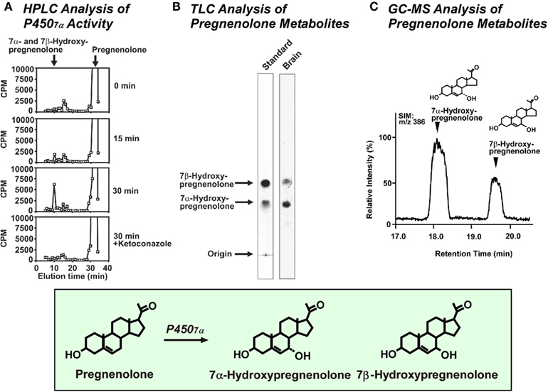
Figure 2. Conversion of pregnenolone to 7α- and 7β-hydroxypregnenolone in the quail brain. (A) HPLC profile of unknown metabolites of pregnenolone by using a reversed-phase column. Quail brain homogenates were incubated with 3H-pregnenolone, and the extracts were subjected to HPLC. The ordinate indicates the radioactivity measured in each HPLC fraction, and the arrows indicate elution positions of standard steroids, pregnenolone and 7α- and 7β-hydroxypregnenolone. (B) Autoradiography of the unknown pregnenolone metabolites (right column) and standard steroids 7α- and 7β-hydroxypregnenolone (left column) on TLC under the same condition as in (A). (C) GC–selected ion monitoring (SIM) mass trace of m/z 386 in the extract from quail brain homogenates. The arrowheads indicate the retention times of 7α-hydroxypregnenolone and 7β-hydroxypregnenolone. See Tsutsui et al. (2008) for details.
Identification, Expression, and Enzymatic Activity of Cytochrome P4507α in the Avian Brain
We initially found that the quail brain actively produces unknown neurosteroids from pregnenolone. We therefore sought to identify these avian neurosteroids from the brain of adult quail by using biochemical techniques combined with high-performance liquid chromatography (HPLC), thin-layer chromatography (TLC), and gas chromatography–mass spectrometry (GC–MS) analyses (Tsutsui et al., 2008). Quail brain homogenates were incubated with tritiated pregnenolone as a precursor, and radioactive metabolites were analyzed by reversed-phase HPLC. Several non-radioactive steroids were used as reference standards for HPLC analysis, and 7α-hydroxypregnenolone and its stereoisomer 7β-hydroxypregnenolone exhibited the same retention time of the radioactive peak under a similar chromatographic condition (Tsutsui et al., 2008; Figure 2A). The detection of 3H-7α- and 7β-hydroxypregnenolone was feasible by HPLC because the radioactive pregnenolone was labeled with 3H at multiple positions, including the 7α- or 7β-position. The HPLC peak was collected and subjected to TLC. Quail brain homogenates produced two metabolites from 3H-pregnenolone corresponding to the positions of the 7α- and 7β-hydroxypregnenolone standards (Tsutsui et al., 2008; Figure 2B). The metabolites of pregnenolone were further analyzed by GC–MS. Based on GC–selected ion monitoring (SIM) analysis (m/z 386), the metabolites had retention times that were identical to 7α-hydroxypregnenolone and 7β-hydroxypregnenolone (Tsutsui et al., 2008; Figure 2C).
7α-Hydroxypregnenolone is synthesized from pregnenolone through the enzymatic activity of cytochrome P4507α (Figure 2). In order to prove that 7α-hydroxypregnenolone is synthesized in the brain, it is therefore necessary to show that brain expresses P4507α. A full length, 2341 bp cDNA prepared from the quail brain tissue was identified to encode a putative cytochrome P4507α (Tsutsui et al., 2008). The putative quail P4507α open reading frame was initiated with a methionine at nucleotide 72 and terminates with a TGA codon at nucleotide 1581, encoding a protein of 503 amino acids. The enzymatic activity of this putative quail P4507α was demonstrated using homogenates of COS-7 cells transfected with the putative quail P4507α cDNA (Tsutsui et al., 2008). HPLC analyses demonstrated that the homogenate converts pregnenolone to 7α- and/or 7β-hydroxypregnenolone (Tsutsui et al., 2008). Subsequently, 7α-hydroxypregnenolone but not 7β-hydroxypregnenolone synthesis was confirmed by GC–MS (Tsutsui et al., 2008). Although it is still unclear whether cytochrome P4507α can also convert pregnenolone to 7β-hydroxypregnenolone, the presence of 7β-hydroxypregnenolone as well as 7α-hydroxypregnenolone is evident in the quail brain (Tsutsui et al., 2008; Figure 2).
The production of 7α-hydroxypregnenolone in the brain may be a conserved property of vertebrates because this neurosteroid has also been identified in the brain of newts (Matsunaga et al., 2004) and mammals (Akwa et al., 1992; Doostzadeh and Morfin, 1997; Weill-Engerer et al., 2003; Yau et al., 2003). Recently, a cDNA encoding cytochrome P4507α was identified in the newt brain tissue (Haraguchi et al., 2010). The homogenate of COS-7 cells transfected with the newt P4507α cDNA also converted pregnenolone into 7α-hydroxypregnenolone (Haraguchi et al., 2010).
Localization of Cytochrome P4507α in the Avian Brain
To understand the localization of cytochrome P4507α in the avian brain, the biosynthesis and concentrations of 7α- and 7β-hydroxypregnenolone were compared in different regions of the quail brain in both sexes by HPLC and GC–MS analyses, respectively (Tsutsui et al., 2008). The two neurosteroids were found predominantly in the diencephalon and were very low in other brain regions (Tsutsui et al., 2008). The biosynthesis and concentrations of 7α- and 7β-hydroxypregnenolone in the diencephalon were found to be sexually differentiated being much higher in males than in females (Tsutsui et al., 2008). Such a sexual dimorphism of cytochrome P4507α only occurs in the diencephalon (Tsutsui et al., 2008). There are similar sex differences in 3β-HSD and cytochrome P450arom in the avian brain (Schlinger and Callard, 1987; Soma et al., 2004; Tam and Schlinger, 2007).
Tsutsui et al. (2008) further investigated the expression of cytochrome P4507α by in situ hybridization to identify the cells producing 7α-hydroxypregnenolone in the quail brain. In the male diencephalon, the expression of cytochrome P4507α mRNA was localized in the nucleus preopticus medialis, the nucleus paraventricularis magnocellularis, the nucleus ventromedialis hypothalami, the nucleus dorsolateralis anterior thalami and the nucleus lateralis anterior thalami (Tsutsui et al., 2008).
Expression of 3β-HSD and Progesterone Formation in the Avian Brain
The biosynthesis of progesterone is performed by 3β-HSD that catalyzes oxidation and isomerization of Δ5-3β-hydroxysteroids (pregnenolone and dehydroepiandrosterone) into Δ4-ketosteroids (progesterone and androstenedione, respectively). 3β-HSD is highly expressed in the peripheral steroidogenic glands (Mason, 1993; Figure 3). Thus, the demonstration of 3β-HSD expression and progesterone production is essential in order to understand the biosynthetic pathway of neurosteroids in the avian brain.
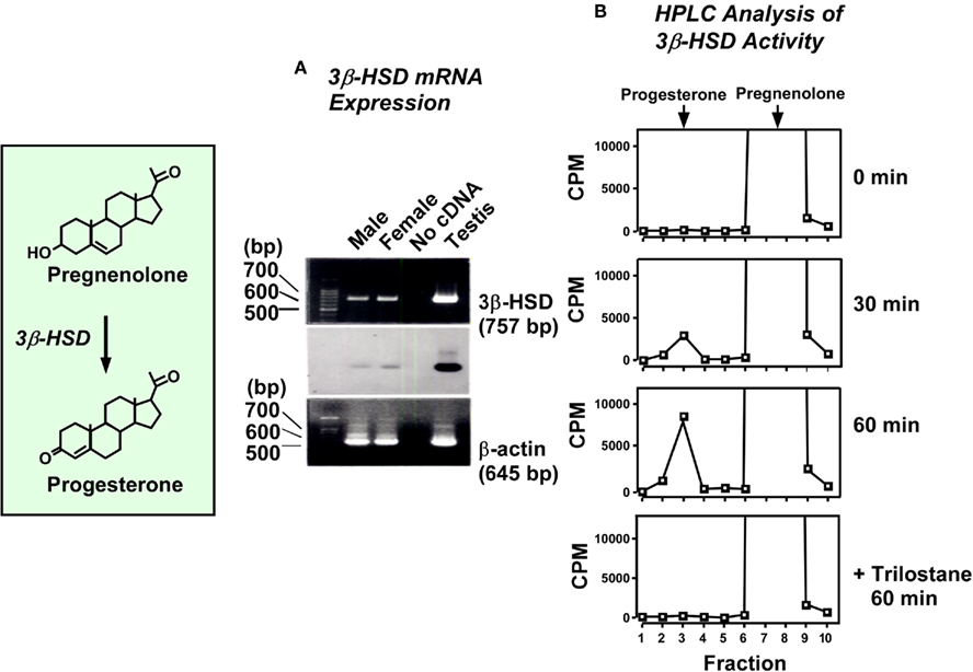
Figure 3. Progesterone formation from pregnenolone in the quail brain. (A) RT-PCR analysis of 3β-HSD mRNA in the quail brain. Upper panel shows a result of the gel electrophoresis of PT-PCR products for chicken 3β-HSD, and middle panel shows an identification of the band by Southern hybridization using digoxigenin-labeled oligonucleotide probe for chicken 3β-HSD. The lane labeled “No cDNA” was performed without template as the negative control. Lower panel shows a result of the RT-PCR for chicken β-actin as the internal control. (B) HPLC analysis of neurosteroids extracted from quail brain slices after different incubation times with 3H-pregnenolone using a reversed-phase column. The column was eluted with a linear gradient of 40–70% acetonitrile. The ordinate indicates the radioactivity measured in each HPLC fraction. The arrows indicate the elution positions of pregnenolone and progesterone. The brain slices were also incubated with trilostane, a specific inhibitor of 3β-HSD, for 60 min and subjected to HPLC analysis. See Ukena et al. (1999a) for details.
Expression and Enzymatic Activity of 3β-HSD in the Avian Brain
Our biochemical and molecular studies have demonstrated the expression of 3β-HSD and the formation of progesterone in the brain of adult quail. RT-PCR analysis together with Southern hybridization showed the expression of 3β-HSD mRNA in the quail brain in both sexes (Ukena et al., 1999a; Figure 3A). By using biochemical techniques combined with HPLC analysis, Ukena et al. (1999a) demonstrated that, in the quail brain, pregnenolone is converted to progesterone (Figure 3B). The biosynthesis of progesterone increased with time and was completely abolished by trilostane, an inhibitor of 3β-HSD (Ukena et al., 1999a; Figure 3B). These studies indicate that the quail brain expresses 3β-HSD and converts pregnenolone to progesterone (Figure 3).
The expression of 3β-HSD and its enzymatic activity were also found in the brain of zebra finches (Vanson et al., 1996) and ring doves (Lea et al., 2001). Thus, the avian brain possesses not only cytochrome P450scc but also 3β-HSD and produces progesterone de novo from cholesterol. The expression of both 3β-HSD protein and its mRNA has also been reported in mammalian brains (Dupont et al., 1994; Guennoun et al., 1995; Sanne and Krueger, 1995; Kohchi et al., 1998; Ukena et al., 1999b). In addition, 3β-HSD activity has been demonstrated biochemically in the brain of mammals (Weidenfeld et al., 1980; Akwa et al., 1993; Kabbadj et al., 1993; Ukena et al., 1999b), amphibians (Mensah-Nyagan et al., 1994), and fish (Sakamoto et al., 2001).
Localization of 3β-HSD in the Avian Brain
Based on the biochemical analysis, we further analyzed the activity of 3β-HSD in all brain regions of the adult quail. The enzymatic activity in the telencephalon and diencephalon was higher than that in the mesencephalon (Ukena et al., 1999a). The progesterone level was also high in the telencephalon and diencephalon and low in the mesencephalon (Ukena et al., 1999a). It has been reported that rat 3β-HSD mRNA is expressed in several brain regions in particular, olfactory bulb, striatum, cortex, thalamus, hypothalamus, habenula, septum, hippocampus, and cerebellum (Guennoun et al., 1995). Although the exact site showing 3β-HSD expression is still obscure in the quail brain, the regional distribution of 3β-HSD reported in the rat brain correlates with that of cytochrome P450scc in the quail brain. Therefore, sites having both steroidogenic enzymes can synthesize progesterone de novo from cholesterol.
Expression of 5β-Reductase and Progesterone Metabolism in the Avian Brain
To understand the pathway of progesterone metabolism, we investigated progesterone metabolites in the brain of adult quail. Biochemical analysis together with HPLC and TLC revealed that the quail brain produces 5β-DHP from progesterone (Ukena et al., 2001; Figure 4). We further demonstrated that the quail brain produces not only 5β-DHP but also 3β, 5β-THP from progesterone (Tsutsui et al., 2003a; Figure 4). In birds, 5β-reduction also represents a route of androgen metabolism in the brain (Massa and Sharp, 1981; Schlinger and Callard, 1987).
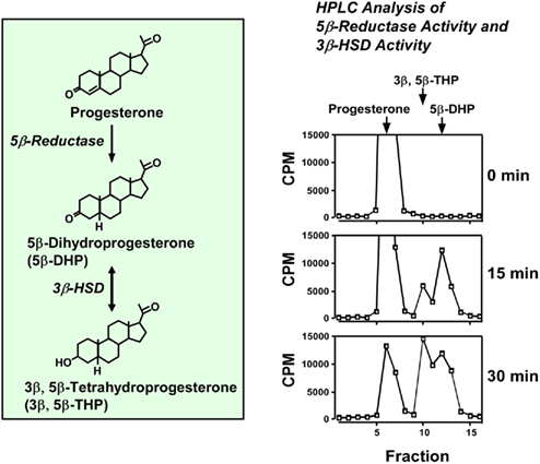
Figure 4. Conversion of progesterone to 5β-DHP and 3β, 5β-THP in the quail brain. HPLC analysis of neurosteroids extracted from quail brain homogenates after different incubation times with 3H-progesterone using a reversed-phase column. The column was eluted with an isocratic elution of 70% acetonitrile. The ordinate indicates the radioactivity measured in each HPLC fraction. The arrows indicate the elution positions of progesterone, 5β-DHP and 3β, 5β-THP. See Ukena et al. (2001) for details.
Thus, the progesterone metabolites, 5β-DHP and 3β, 5β-THP, are produced by the enzymatic activities of 5β-reductase and 3β-HSD, and accumulate in the avian brain as neurosteroids (Figure 4). In contrast to birds, progesterone is converted to 5α-DHP and 3α, 5α-THP due to 5α-reductase and 3α-HSD in mammals (for reviews, see Baulieu, 1997; Compagnone and Mellon, 2000).
Expressions of Cytochrome P45017α,Lyase and 17β-HSD and Androgen Formation in the Avian Brain
Another pathway of progesterone metabolism is mediated by cytochrome P45017α,lyase which, in addition to converting pregnenolone to dehydroepiandrosterone via 17α-hydroxypregnenolone, also converts progesterone to androstenedione via 17α-hydroxyprogesterone (Figures 5 and 8). Both of these metabolic pathways was demonstrated in the quail brain using biochemical techniques combined with HPLC analysis, and by RT-PCR analysis of cytochrome P45017α,lyase mRNA (Matsunaga et al., 2001, 2002; Figure 5). We further demonstrated that the avian brain expresses 17β-HSD that is needed to convert androstenedione to testosterone (Matsunaga et al., 2002; Figure 6).
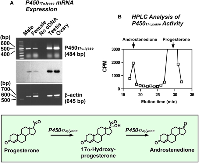
Figure 5. Conversion of progesterone to androstenedione in the quail brain. (A) RT-PCR analysis of cytochrome P45017α,lyase mRNA in the quail brain. Upper panel shows a result of the gel electrophoresis of RT-PCR products for chicken P45017α,lyase, and middle panel shows an identification of the band by Southern hybridization using digoxigenin-labeled oligonucleotide probe for chicken P45017α,lyase. The lane labeled “No cDNA” was performed without template as the negative control. Lower panel shows a result of the RT-PCR for chicken β-actin as the internal control. (B) HPLC analysis of neurosteroids extracted from quail diencephalic slices after 60 min incubation with 3H-progesterone using a reversed-phase column. The column was eluted with a 30-min linear gradient of 40–70% acetonitrile, followed by an isocratic elution of 70% acetonitrile. The ordinate indicates the radioactivity measured in each HPLC fraction. The arrows indicate the elution positions of progesterone and androstenedione. See Matsunaga et al. (2001, 2002) for details.
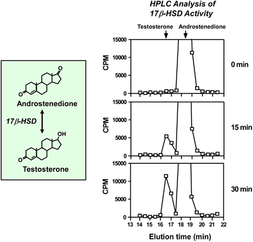
Figure 6. Androgen formation from androstenedione in the quail brain. HPLC analysis of neurosteroids extracted from quail diencephalic slices after different incubation times with 3H-androstenedione using a reversed-phase column. The column was eluted with a 30-min linear gradient of 40–70% acetonitrile, followed by an isocratic elution of 70% acetonitrile. The ordinate indicates the radioactivity measured in each HPLC fraction. The arrows indicate the elution positions of androstenedione and testosterone. See Matsunaga et al. (2002) for details.
Expression and Enzymatic Activity of Cytochrome P45017α,Lyase in the Avian Brain
In contrast to the presence of cytochrome P450scc, 3β-HSD, and 5β-reductase, limited information has been available for cytochrome P45017α,lyase in the brain of birds as well as other vertebrates. We therefore investigated the expression of cytochrome P45017α,lyase in the brain of adult quail (Matsunaga et al., 2001). RT-PCR analysis followed by Southern hybridization indicated the expression of cytochrome P45017α,lyase mRNA in the quail brain in both sexes (Matsunaga et al., 2001; Figure 5A). Employing biochemical techniques combined with HPLC analysis, the conversion of progesterone to 17α-hydroxyprogesterone (Matsunaga et al., 2001) and subsequently to androstenedione (Matsunaga et al., 2002; Figure 5B) was also found in quail brain slices. Thus, it appears that the avian brain produces androstenedione from progesterone (Figure 5). The expression of cytochrome P45017α,lyase in the brain was also detected in mammals (Compagnone et al., 1995; Strömstedt and Waterman, 1995; Kohchi et al., 1998). Therefore, the expression of cytochrome P45017α,lyase in the brain is considered to be a conserved property of vertebrates.
Localization of Cytochrome P45017α,Lyase in the Avian Brain
Birds showed clearly a region-dependent expression of cytochrome P45017α,lyase in the brain (Matsunaga et al., 2001). Based on RT-PCR analysis together with Southern hybridization, cytochrome P45017α,lyase mRNA was highly expressed in the quail diencephalon and mesencephalon (Matsunaga et al., 2001). This result is in agreement with the findings reported in mammals (Compagnone et al., 1995; Strömstedt and Waterman, 1995; Kohchi et al., 1998). According to Kohchi et al. (1998), the cytochrome P45017α,lyase mRNA expression was high in the mesencephalon, but it was very weak in the cerebrum and cerebellum. Strömstedt and Waterman (1995) also found, using RT-PCR analysis followed by Southern blots, a higher expression of the cytochrome P45017α,lyase mRNA in the brain stem of rats and mice. In addition, Compagnone et al. (1995) reported that the rat embryonic cells expressing cytochrome P45017α,lyase are located in the mesencephalic region as well as the medulla and spinal cord.
We further characterized the site showing cytochrome P45017α,lyase expression by in situ hybridization. In the quail brain, the expression of cytochrome P45017α,lyase mRNA was localized in the preoptic area, the anterior hypothalamus, the dorsolateral thalamus, the optic tectum and the ventral midbrain (Matsunaga et al., 2001). In addition to these diencephalic and mesencephalic regions, the expression was also localized in the septum, the hyperstriatum accessorium, the ventral portions of the archistriatum, and the cerebellar Purkinje cells (Matsunaga et al., 2001). In the cerebellum, only Purkinje cells expressed cytochrome P45017α,lyase mRNA. Such a regional distribution of cytochrome P45017α,lyase (Matsunaga et al., 2001) precisely correlates with that of cytochrome P450scc in the same avian species (Usui et al., 1995; Tsutsui et al., 1997a). Accordingly, both steroidogenic P450 enzymes may be co-localized in the several restricted brain regions. Indeed, cerebellar Purkinje cells possess both cytochrome P450scc (Usui et al., 1995; Tsutsui et al., 1997a,b) and cytochrome P45017α,lyase (Matsunaga et al., 2001) in quail. A similar co-localization of cytochrome P450scc and 3β-HSD has been obtained in the rat Purkinje cell (Ukena et al., 1998, 1999b; Tsutsui and Ukena, 1999; Tsutsui et al., 1999, 2000, 2003b; Tsutsui, 2008a,b,c).
Expression, enzymatic activity, and localization of 17β-HSD in the avian brain
Androstenedione is a precursor of androgens (Figure 6). To determine whether androgens are synthesized in the brain independently of other steroidogenic sites, such as the gonads or adrenals, the demonstration of the presence of 17β-HSD, a key enzyme for androgen biosynthesis, is also required in the avian brain (Figure 6). To clarify the production of androgens in the avian brain, therefore, we examined the activity of 17β-HSD, which converts androstenedione to testosterone, using the brain of adult quail. Employing biochemical techniques combined with HPLC analysis, the conversion of androstenedione to testosterone was found in quail brain slices (Matsunaga et al., 2002; Figure 6). Based on the biochemical analysis, we further analyzed the activity of 17β-HSD in all brain regions of the quail (Matsunaga et al., 2002). A clear difference in 17β-HSD activity among different brain regions was evident. The enzymatic activity in the diencephalon was higher than those in other brain regions (Matsunaga et al., 2002), indicating that de novo testosterone synthesis in the diencephalon is relatively high in birds.
Expression of Cytochrome P450arom and Estrogen Formation in the Avian Brain
As mentioned above, the expression of cytochrome P45017α,lyase and 17β-HSD and the formation of androgens have been demonstrated in the quail brain (Matsunaga et al., 2001, 2002). On the other hand, it has been well established that the brain of quail and other birds possesses cytochrome P450arom, which converts testosterone to estradiol (Schlinger and Callard, 1987, 1989a,b,c, 1991; Balthazart et al., 1990a,b, 1991; Figure 7). Therefore, not only androgens but also estrogens may be synthesized directly de novo in the avian brain. Recent progresses in the studies of physiological changes in the expression of cytochrome P450arom and their regulatory mechanisms are summarized in Section “Physiological Changes in Neurosteroid Formation and Biological Functions of Neurosteroids in the Avian Brain.”
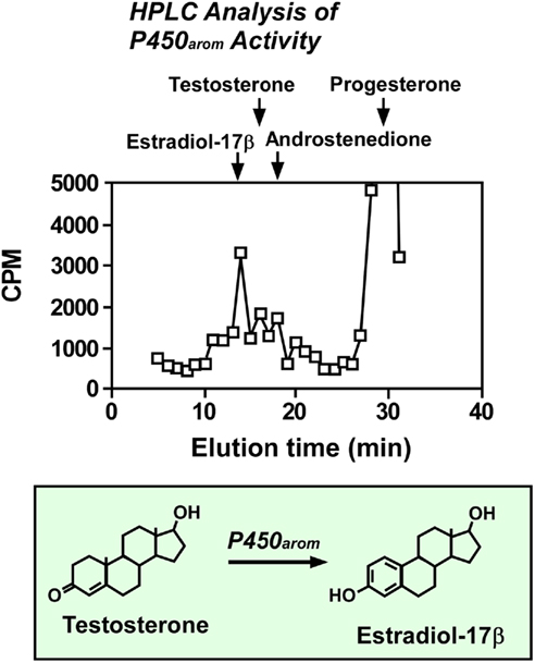
Figure 7. Estrogen formation from androgen in the quail brain. HPLC analysis of neurosteroids extracted from quail diencephalic slices after 60 min incubation with 3H-progesterone using a reversed-phase column. The column was eluted with a 30-min linear gradient of 40–70% acetonitrile, followed by an isocratic elution of 70% acetonitrile. The ordinate indicates the radioactivity measured in each HPLC fraction. The arrows indicate the elution positions of progesterone, androstenedione, testosterone, and estradiol-17β. See Matsunaga et al. (2002) for details.
In birds, both cytochrome P450arom and estrogen receptors are expressed in several brain regions including the hypothalamus and preoptic area which are involved in the control of reproductive behaviors in birds (Schlinger and Callard, 1987, 1989a,b,c, 1991; Balthazart et al., 1990a,b, 1991). Therefore, testosterone produced in these brain regions may be converted to estradiol. In fact, we detected, biochemically, the formation of estradiol from progesterone in the quail diencephalon including the hypothalamus and preoptic area (Matsunaga et al., 2002; Figure 7). Taken together, testosterone is synthesized from progesterone by cytochrome P45017α,lyase and 17β-HSD in the diencephalon, and subsequently testosterone is converted to estradiol by P450arom (Figure 8).
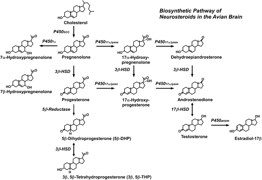
Figure 8. Biosynthetic pathway for neurosteroids in the quail brain. The arrows indicate biosynthetic pathways of neurosteroids identified in the quail brain. See the text for details.
As shown in Figure 8, the production of sex steroids requires the coordinate action of several steroidogenic enzymes that start with cholesterol, and then lead to a cascade of reactions that ultimately produce several kinds of neurosteroids. I discuss recent studies in birds whether sex steroids are synthesized in the brain independently of other steroidogenic sites, such as the gonads and/or adrenals, or sex steroid synthesis in the brain is dependent on gonadal and/or adrenal steroids in the following Section “Physiological Changes in Neurosteroid Formation and Biological Functions of Neurosteroids in the Avian Brain.”
Physiological Changes in Neurosteroid Formation and Biological Functions of Neurosteroids in the Avian Brain
Physiological changes in neurosteroid formation and biological functions of neurosteroids are becoming clear in domestic and other birds. It is well known in birds (Tsutsui et al., 2008) as well as other vertebrates (Tsutsui, 1931; Iwata et al., 2000) that the locomotor activity of males is higher than that of females. As described in Section “Expression of Cytochrome P4507α and Formation of 7α- and 7β-Hydroxypregnenolone in the Avian Brain,” the quail brain synthesizes two previously undescribed avian neurosteroids, 7α- and 7β-hydroxypregnenolone, from pregnenolone (Tsutsui et al., 2008). In view of the sex difference in concentrations of diencephalic 7α- and 7β-hydroxypregnenolone, it seemed possible that these neurosteroids play a role in the control of locomotor activity of males. Because the male quail displays a robust locomotor activity rhythm when held under typical light/dark lighting schemes (Wilson, 1972; Wada, 1979), this bird may serve as an excellent animal model to demonstrate the biological function of 7α- and 7β-hydroxypregnenolone. Both neurosteroids were therefore administered intra-cerebroventricularly to male quail during night, when their activity is low, to test whether they affect locomotor activity (Tsutsui et al., 2008). A stimulatory dose-dependent effect of 7α-hydroxypregnenolone was observed (Tsutsui et al., 2008). In contrast, 7β-hydroxypregnenolone did not influence locomotor activity (Figure 2; Tsutsui et al., 2008). In contrast to males, the locomotor activity and diencephalic concentration of 7α-hydroxypregnenolone in females were constantly low during the same observation period, i.e., from lights on to noon (Tsutsui et al., 2008). Thus, increased diencephalic 7α-hydroxypregnenolone may contribute to the higher locomotor activity in males. The low level of 7α-hydroxypregnenolone synthesis and concentration in the female diencephalon suggests that this neurosteroid may not play a role in female locomotor activity. Since other vertebrates also exhibit a clear sex difference in locomotor activity (Tsutsui, 1931; Iwata et al., 2000), it is suggested that sex differences in diencephalic 7α-hydroxypregnenolone may be a key factor controlling sex differences in vertebrate locomotor activity.
A ubiquitous property of vertebrates is fluctuation of locomotor activity over the 24-h circadian cycle (Saper et al., 2005). The endogenous diurnal rhythm of melatonin is known to control the diurnal locomotor rhythm in vertebrates including birds (Binkley et al., 1971; John et al., 1978; Cassone and Menaker, 1984; Chabot and Menaker, 1992; Hau and Gwinner, 1994; Warren and Cassone, 1995), which suggested that melatonin may regulate diencephalic 7α-hydroxypregnenolone synthesis, and thereby influence locomotor activity. This hypothesis was tested in experiments involving melatonin manipulation in male quail (Tsutsui et al., 2008). A combination of pinealectomy (Px) plus orbital enucleation (Ex) increased the production and concentration of 7α-hydroxypregnenolone and the expression of cytochrome P4507α in the diencephalon. Conversely, melatonin administration to Px/Ex quail decreased the production and concentration of 7α-hydroxypregnenolone and the expression of cytochrome P4507α in the diencephalon. Further, the inhibitory effect of melatonin on 7α-hydroxypregnenolone synthesis was abolished by luzindole, a melatonin receptor antagonist (Tsutsui et al., 2008). It is therefore considered that melatonin acts to reduce cytochrome P4507α expression through melatonin receptor-mediated mechanisms. Melatonin derived from the pineal gland and eyes therefore appears to act as a potent inhibitory factor of 7α-hydroxypregnenolone synthesis in the quail. This hypothesis is supported by earlier studies showing that melatonin treatment decreases locomotor activity in quail (Murakami et al., 2001; Nakahara et al., 2003) and other birds (Murakami et al., 2001).
On the other hand, there is evidence for neurosteroidal activation of territorial behavior in the song sparrow Melospiza melodia (Soma et al., 1999, 2000). It is known that territorial behavior of this species is expressed in the non-breeding season, Although circulating testosterone levels are low in non-breeding male song sparrows, the androgen precursor dehydroepiandrosterone is present in blood (Soma and Wingfield, 2001). Because the brain of zebra finch expresses 3β-HSD and cytochrome P450arom and produces estrogens from dehydroepiandrosterone (Vanson et al., 1996; Soma et al., 2004; Tam and Schlinger, 2007), it is considered that these steroidogenic enzymes may function in the brain of song sparrow during the non-breeding season to produce estrogens from dehydroepiandrosterone originated from the peripheral gland that activate territorial aggression. Because the brain of zebra finch also expresses cytochrome P450scc and cytochrome P45017α,lyase (London et al., 2003, 2006, 2010; London and Schlinger, 2007), dehydroepiandrosterone may also be produced de novo from cholesterol in the brain of these birds as in quail. More research is needed to evaluate the function of neurosteroids produced in the brain from cholesterol de novo and the role of central metabolism of steroids originally coming from the periphery.
As was detected in zebra finches, song sparrows expressed 3β-HSD and cytochrome P450arom in the brain (Soma et al., 2003; Pradhan et al., 2008, 2010). Importantly, both steroidogenic enzymes are subject to seasonal regulation. Cytochrome P450arom is elevated in the non-breeding season in the brain in keeping with a role for estrogen activation of aggressive behavior during the non-breeding season (Soma et al., 2003). 3β-HSD is expressed and active in the song sparrow brain (Pradhan et al., 2008, 2010). 3β-HSD is also elevated during the non-breeding season. It is important to clarify the mechanisms that regulate the expression of these steroidogenic enzymes in the avian brain.
Interestingly, there are several reports showing changes in neurosteroid formation in relation to social interactions. A recent study showed that within the caudomedial nidopallium marked changes in estradiol occurred when males were exposed to females or to conspecific zebra finch song (Remage-Healey et al., 2008). Estrogens produced in the local brain region are thought to rapidly strengthen auditory encoding and guide song preference in a songbird (Remage-Healey et al., 2010). Moreover, changes in estradiol formation were reduced by exposure to fadrozole, an inhibitor of cytochrome P450arom, or to glutamate as in quail hypothalamus (Balthazart et al., 2006). These findings suggest rapid control of cytochrome P450arom activity by glutamatergic inputs (Balthazart et al., 2006). Thus, cytochrome P450arom is subject to rapid regulation. In quail hypothalamic explants, cytochrome P450arom undergoes Ca2+-dependent phosphorylation that reduces cytochrome P450arom activity within minutes (Balthazart et al., 2001a,b, 2003). Treatments of these explants with K+ or with glutamate receptor agonists produce a similar rapid inhibition of cytochrome P450arom activity (Balthazart et al., 2001b). These results suggest that the flow of Ca2+ through voltage-gated Ca2+ channels serves as a key regulatory signal for rapid estrogen production.
Synaptic estrogen formation in the brain is also becoming clear in songbirds and other birds (Naftolin et al., 1996; Peterson et al., 2005) as in mammals (Hojo et al., 2008). Compartmentalization of cytochrome P450arom within presynaptic boutons is considered to be crucial for providing sex- and song-specific estrogenic signals in the songbird brain (Peterson et al., 2005; Remage-Healey et al., 2009).
Mode of Action of Neurosteroids in the Avian Brain
To understand the mode of action of 7α-hydroxypregnenolone on locomotion, Matsunaga et al. (2004) first found that 7α-Hydroxypregnenolone acts as a neuronal activator to stimulate locomotor activity of breeding newts by means of the dopaminergic system (Matsunaga et al., 2004). As described in Section “Expression of Cytochrome P4507α and Formation of 7α- and 7β-Hydroxypregnenolone in the Avian Brain,” the expression of cytochrome P4507α mRNA was localized in several diencephalic regions, such as the nucleus preopticus medialis, the nucleus paraventricularis magnocellularis, the nucleus ventromedialis hypothalami, the nucleus dorsolateralis anterior thalami, and the nucleus lateralis anterior thalami (Tsutsui et al., 2008) in the male quail brain. In quail (Tsutsui et al., 2008) as in newts (Matsunaga et al., 2004), 7α-hydroxypregnenolone increased the concentration of dopamine in the telencephalic region that encompasses the striatum (Sanberg, 1983; Sharp et al., 1987; Bardo et al., 1990). In birds, dopaminergic neurons that are located in the mesencephalic region, including the area ventralis and the substantia nigra, project to the telencephalon notably the striatum (Mezey and Csillag, 2002; Hara et al., 2007). Interestingly, the telencephalic region is enriched with dopamine D1 and D2 receptors in birds (Ball et al., 1995; Levens et al., 2000). Accordingly, 7α-hydroxypregnenolone synthesized actively in the diencephalon, by acting on dopamine neurons localized in the area ventralis and the substantia nigra, may induce dopamine release from their termini in the striatum, and consequently increase locomotor activity in male quail as in male newts.
The fact that 7α-hydroxypregnenolone acutely increases locomotor activity in quail suggests that this neurosteroid may act through a non-genomic rather than a genomic mechanism. It has been reported that in rats, the progesterone metabolite 3α,5α-tetrahydroprogesterone (3α,5α-THP; allopregnanolone) exerts its effects on locomotion (Wieland et al., 1995) and dopamine release (Bullock et al., 1997; Rougé-Pont et al., 2002) via a non-genomic pathway. Allopregnanolone may act through modulation of GABAA receptors, since allopregnanolone is a potent allosteric modulator of GABAA receptors (Paul and Purdy, 1992; Lambert et al., 1995) and dopaminergic neurons are regulated by GABAergic transmission (Laviolette and van der Kooy, 2001). Whether the acute actions of 7α-hydroxypregnenolone on dopamine release and locomotor activity in quail are mediated through GABAA receptors remain to be determined.
On the other hand, there are several reports showing the mode of acute actions of estrogen (Tremere et al., 2009; Remage-Healey et al., 2010; Tremere and Pinaud, 2011). The neuromodulatory role of estradiol on burst firing of neurons in the caudomedial nidopallium was demonstrated in the songbird (Tremere and Pinaud, 2011). Acute estrogen actions in the zebra finch may be dependent on a membrane-specific receptor. Further study is needed to demonstrate the molecular mechanisms underlying acute actions of estrogen in the brain of birds.
Conclusion and Future Directions
In conclusion, the quail brain possesses several kinds of steroidogenic enzymes, such as cytochrome P450scc, cytochrome P4507α, 3β-HSD, 5β-reductase, cytochrome P45017α,lyase, 17β-HSD and cytochrome P450arom, and produces pregnenolone, 7α-hydroxypregnenolone, progesterone, 5β-DHP, 3β, 5β-THP, androstenedione, testosterone, and estradiol from cholesterol (Figure 8). The brain of other birds, such as ring dove, zebra finch etc., also produces several neurosteroids. However, the biosynthetic pathway of neurosteroids in the avian brain from cholesterol may be still incomplete, because we recently found that the quail brain expresses cytochrome P4507α and actively produces 7α- and 7β-hydroxypregnenolone, previously undescribed avian neurosteroids (Figure 8). Thus, to complete our knowledge of the biosynthetic pathway for neurosteroids in the avian brain, further biochemical studies are needed.
Diurnal and seasonal changes in neurosteroid formation are becoming clear in birds. Social interactions also change neurosteroid formation in birds. Further, several important biological functions of neurosteroids are also becoming clear in birds. However, more research is needed to evaluate the function of neurosteroids produced in the brain from cholesterol de novo and the role of central metabolism of steroids originally coming from the periphery. In addition, it is important to clarify the molecular mechanisms of the regulation of neurosteroid formation and the mode of action of neurosteroids in the avian brain.
Conflict of Interest Statement
The author declares that the research was conducted in the absence of any commercial or financial relationships that could be construed as a potential conflict of interest.
Acknowledgments
The author thanks Mariko Usui, Takeshi Yamazaki, Shiro Kominami, Kazuyoshi Ukena, Yoko Honda, Masahiro Matsunaga, Hirotaka Sakamoto, Yuto Inai, Minoru Takase, Chie Kohchi, Kazuhiko Inoue, Hitomi Miyabara, Takayoshi Ubuka, and Shogo Haraguchi for their work cited in this manuscript. This work was supported by Grants-in-Aid for Scientific Research from the Ministry of Education, Science and Culture, Japan (18107002, 22132004, and 22227002 to Kazuyoshi Tsutsui).
References
Adkins, E., and Adler, N. (1972). Hormonal control of behavior in the Japanese quail. J. Comp Physiol. Psychol. 81, 27–36.
Akwa, Y., Morfin, R. F., Robel, P., and Baulieu, E.-E. (1992). Neurosteroid metabolism: 7α-Hydroxylation of dehydroepiandrosterone and pregnenolone by rat brain microsomes. Biochem. J. 288, 959–964.
Akwa, Y., Sananés, N., Gouézou, M., Robel, P., Baulieu, E.-E., and Le Goascogne, C. (1993). Astrocytes and neurosteroids: metabolism of pregnenolone and dehydroepiandrosterone. Regulation by cell density. J. Cell Biol. 121, 135–143.
Akwa, Y., Young, J., Kabbadj, K., Sancho, M. J., Zucman, D., Vourc’h, C., Jung-Testas, I., Hu, Z. Y., Le Goascogne, C., Jo, D. H., Corpéhot, C., Simon, P., Baulieu, E.-E., and Robel, P. (1991). Neurosteroids: biosynthesis, metabolism and function of pregnenolone and dehydroepiandrosterone in the brain. J. Steroid Biochem. Mol. Biol. 40, 71–81.
Arnold, A. (1975). The effects of castration and androgen replacement on song, courtship, and aggression in zebra finches (Poephila guttata). J. Exp. Zool. 191, 309–326.
Arnold, A., Nottebohm, F., and Pfaff, D. W. (1976). Hormone-concentrating cells in vocal control and other areas of the brain of the zebra finch. J. Comp. Neurol. 165, 487–512.
Ball, G. F., Casto, J. M., and Balthazart, J. (1995). Autoradiographic localization of D1-like dopamine receptors in the forebrain of male and female Japanese quail and their relationship with immunoreactive tyrosine hydroxylase. J. Chem. Neuroanat. 9, 121–133.
Balthazart, J. (1983). “Hormonal correlates of behavior,” in Avian Biology, eds D. S. Farner, and K. C. Parker (New York: Academic Press), 221–365.
Balthazart, J., Baillien, M., and Ball, G. F. (2001a). Rapid and reversible inhibition of brain aromatase activity. J. Neuroendocrinol. 13, 63–73.
Balthazart, J., Baillien, M., and Ball, G. F. (2001b). Phosphorylation processes mediate rapid changes of brain aromatase activity. J. Steroid Biochem. Mol. Biol. 79, 261–277.
Balthazart, J., Baillien, M., and Ball, G. F. (2006). Rapid control of brain aromatase activity by glutamatergic inputs. Endocrinology 147, 359–366.
Balthazart, J., Baillien, M., Charlier, T., and Ball, G. (2003). Calcium-dependent phosphorylation processes control brain aromatase in quail. Eur. J. Neurosci. 171, 591–606.
Balthazart, J., Foidart, A., and Harada, N. (1990a). Immunocytochemical localization of aromatase in the brain. Brain Res. 514, 327–333.
Balthazart, J., Foidart, A., Surlemont, C., Vockel, A., and Harada, N. (1990b). Distribution of aromatase in the brain of the Japanese quail, ring dove, and zebra finch: an immunocytochemical study. J. Comp. Neurol. 301, 276–288.
Balthazart, J., Foidart, A., Surlemont, C., and Harada, N. (1991). Neuroanatomical specificity in the co-localization of aromatase and estrogen receptors. J. Neurobiol. 22, 143–157.
Bardo, M. T., Bowling, S. L., and Pierce, R. C. (1990). Changes in locomotion and dopamine neurotransmission following amphetamine, haloperidol, and exposure to novel environmental stimuli. Psychopharmacology (Berl.) 101, 338–343.
Baulieu, E.-E. (1997). Neurosteroids: of the nervous system, by the nervous system, for the nervous system (review). Recent Prog. Horm. Res. 52, 1–32.
Baulieu, E.-E., and Robel, P. (1990). Neurosteroids: a new brain function? J. Steroid Biochem. Mol. Biol. 37, 395–403.
Beaujean, D., Mensah-Nyagan, A. G., Do-Rego, J. L., Luu-The, V., Pelletier, G., and Vaudry, H. (1999). Immunocytochemical localization and biological activity of hydroxysteroid sulfotransferase in the frog brain. J. Neurochem. 72, 848–857.
Binkley, S., Kluth, E., and Menaker, M. (1971). Pineal function in sparrows: circadian rhythms and body temperature. Science 174, 311–314.
Bruzzone, F., Do-Rego, J. L., Luu-The, V., Pelletier, G., Vallarino, M., and Vaudry, H. (2010). Immunohistochemical localization and biological activity of 3β-hydroxysteroid dehydrogenase and 5α-reductase in the brain of the frog, Rana esculenta, during development. J. Chem. Neuroanat. 39, 35–50.
Bullock, A. E., Clark, A. L., Grady, S. R., Robinson, S. F., Slobe, B. S., Marks, M. J., and Collins, A. C. (1997). Neurosteroids modulate nicotinic receptor function in mouse striatal and thalamic synaptosomes. J. Neurochem. 68, 2412–2423.
Cassone, V. M., and Menaker, M. (1984). Is the avian circadian system a neuroendocrine loop? J. Exp. Zool. 232, 539–549.
Chabot, C. C., and Menaker, M. (1992). Circadian feeding and locomotor rhythms in pigeons and house sparrows. J. Biol. Rhythms 7, 287–299.
Clark, J. A., Tsutsui, K., Ukena, K., and Lea, R. W. (1999). “Changes in central progesterone during the reproductive cycle of the ring dove (Streptopelia risoria),” in 15th National Meeting of the British Neuroscience Association, Harrogate, 15.
Compagnone, N. A., Bulfone, A., Rubenstein, J. L., and Mellon, S. H. (1995). Steroidogenic enzyme P450c17 is expressed in the embryonic central nervous system. Endocrinology 136, 5212–5223.
Compagnone, N. A., and Mellon, S. H. (2000). Neurosteroids: biosynthesis and function of these novel neuromodulators (review). Front. Neuroendocrinol. 21, 1–56.
Corpéchot, C., Robel, P., Axelson, M., Sjövall, J., and Baulieu, E.-E. (1981). Characterization and measurement of dehydroepiandrosterone sulfate in rat brain. Proc. Natl. Acad. Sci. U.S.A. 78, 4704–4707.
Corpéchot, C., Synguelakis, M., Talha, S., Axelson, M., Sjövall, J., Vihko, R., Baulieu, E.-E., and Robel, P. (1983). Pregnenolone and its sulfate ester in rat brain. Brain Res. 270, 119–125.
Doostzadeh, J., and Morfin, R. (1997). Effects of cytochrome P450 inhibitors and of steroid hormones on the formation of 7-hydroxylated metabolites of pregnenolone in mouse brain microsomes. J. Endocrinol. 155, 343–350.
Do-Rego, J. L., Seong, J. Y., Burel, D., Leprince, J., Luu-The, V., Tsutsui, K., Tonon, M.-C., Pelletier, G., and Vaudry, H. (2009). Neurosteroid biosynthesis: enzymatic pathways and neuroendocrine regulation by neurotransmitters and neuropeptides (review). Front. Neuroendocrinol. 30, 259–301.
Do-Rego, J. L., Tremblay, Y., Luu-The, V., Repello, E., Vallarino, M., Belanger, A., Pelletier, G., and Vaudry, H. (2007). Immunocytochemical localization and biological activity of the steroidogenic enzyme cytochrome P450 17α-hydroxylase/C17, 20-lyase (P450C17) in the frog brain and pituitary. J. Neurochem. 100, 251–268.
Dupont, E., Simard, J., Luu-The, V., Labrie, F., and Pelletier, G. (1994). Localization of 3β-hydroxysteroid dehydrogenase in rat brain as studied by in situ hybridization. Mol. Cell. Neurosci. 5, 119–123.
Freking, F., Nazairians, T., and Schlinger, B. A. (2000). The expression of the sex steroid-synthesizing enzymes CYP11A1, 3β-HSD, CYP17, and CYP 19 in gonads and adrenals of adult and developing zebra finches. Gen. Comp. Endocrinol. 119, 140–151.
Fuxe, K., Hokfelt, T., Eneroth, P., Gustafsson, J. A., and Skett, P. (1977). Prolactin-like immunoreactivity: localization in nerve terminals of rat hypothalamus. Science 196, 899–900.
Guennoun, R., Fiddes, R. J., Gouézou, M., Lombès, M., and Baulieu, E.-E. (1995). A key enzyme in the biosynthesis of neurosteroids, 3β-hydroxysteroid dehydrogenase/Δ5-Δ4-isomerase (3β-HSD), is expressed in rat brain. Brain Res. Mol. Brain Res. 30, 287–300.
Hara, E., Kubikova, L., Hessler, N. A., and Jarvis, E. D. (2007). Role of the midbrain dopaminergic system in modulation of vocal brain activation by social context. Eur. J. Neurosci. 25, 3406–3416.
Haraguchi, S., Koyama, T., Hasunuma, I., Vaudry, H., and Tsutsui, K. (2010). Prolactin increases the synthesis of 7α-hydroxypregnenolone, a key factor for induction of locomotor activity, in breeding male newts. Endocrinology 151, 2211–2222.
Hau, M., and Gwinner, E. (1994). Melatonin facilitates synchronization of sparrow circadian rhythms to light. J. Comp. Physiol. A 175, 343–347.
Hojo, Y., Murakami, G., Mukai, H., Higo, S., Hatanake, Y., Ogiue-Ikeda, M., Ishii, H., Kimotot, T., and Kawato, S. (2008). Estrogen synthesis in the brain-Role in synaptic plasticity and memory. Mol. Cell. Endocrinol. 290, 31–43.
Hu, Z. Y., Bourreau, E., Jung-Testas, I., Robel, P., and Baulieu, E.-E. (1987). Neurosteroids: oligodendrocyte mitochondria convert cholesterol to pregnenolone. Proc. Natl. Acad. Sci. U.S.A. 84, 8215–8219.
Inai, Y., Nagai, K., Ukena, K., Oishi, T., and Tsutsui, K. (2003). Seasonal changes in neurosteroids in the urodele brain and environmental factors inducing their changes. Brain Res. 959, 214–225.
Ishii, S., and Tsutsui, K. (1982). “Hormonal control of aggressive behavior in male Japanese quail.” in Aspects of Avian Endocrinology: Practical and Theoretical Implications, eds C. G. Scanes, M. A. Ottinger, A. D. Kenney, J. Balthazart, J. Cronshaw, and J. C. Jones (Texas: Texas Tech Press), 125–131.
Iwahashi, K., Ozaki, H. S., Tsubaki, M., Ohnishi, J., Takeuchi, Y., and Ichikawa, Y. (1990). Studies of the immunohistochemical and biochemical localization of the cytochrome P-450scc-linked monooxygenase system in the adult rat brain. Biochim. Biophys. Acta 1035, 182–189.
Iwata, T., Toyoda, F., Yamamoto, K., and Kikuyama, S. (2000). Hormonal control of urodele reproductive behavior. Comp. Biochem. Physiol. B Biochem. Mol. Biol. 126, 221–229.
Jo, D. H., Abdallah, M. A., Young, J., Baulieu, E.-E., and Robel, P. (1989). Pregnenolone, dehydroepiandrosterone, and their sulfate and fatty acid esters in the rat brain. Steroids 54, 287–297.
John, T. M., Itoh, S., and George, J. C. (1978). On the role of the pineal in thermoregulation in the pigeon. Horm. Res. 9, 41–56.
Jung-Testas, I., Hu, Z. Y., Baulieu, E.-E., and Robel, P. (1989). Neurosteroids: biosynthesis of pregnenolone and progesterone in primary cultures of rat glial cells. Endocrinology 125, 2083–2091.
Kabbadj, K., El-Etr, M., Baulieu, E.-E., and Robel, P. (1993). Pregnenolone metabolism in rodent embryonic neurons and astrocytes. Glia 7, 170–175.
Kohchi, C., Ukena, K., and Tsutsui, K. (1998). Age- and region-specific expressions of the messenger RNAs encoding for steroidogenic enzymes P450scc, P450c17 and 3β-HSD in the postnatal rat brain. Brain Res. 801, 233–238.
Korsia, S., and Bottjer, S. W. (1989). Developmental changes in the cellular composition of a brain nuvleus involved with song learning in zebra fiches. Neuron 3, 451–460.
Lambert, J. J., Belelli, D., Hill-Venning, C., and Peters, J. A. (1995). Neurosteroids and GABAA receptor function. Trends Pharmacol. Sci. 16, 295–303.
Lanthier, A., and Patwardhan, V. V. (1986). Sex steroids and 5-en-3β-hydroxysteroids in specific regions of the human brain and cranial nerves. J. Steroid Biochem. 25, 445–449.
Laviolette, S. R., and van der Kooy, D. (2001). GABAA receptors in the ventral tegmental area control bidirectional reward signalling between dopaminergic and non-dopaminergic neural motivational systems. Eur. J. Neurosci. 13, 1009–1015.
Le Goascogne, C., Robel, P., Gouézou, M., Sananès, N., Baulieu, E.-E., and Waterman, M. (1987). Neurosteroids: cytochrome P-450scc in rat brain. Science 237, 1212–1215.
Lea, R. W., Clark, J. A., and Tsutsui, K. (2001). Changes in central steroid receptor expression, steroid synthesis and dopaminergic activity related to the reproductive cycle of the ring dove (review). Microsc. Res. Tech. 55, 12–26.
Levens, N., Green, T. A., Akins, C. K., and Bardo, M. T. (2000). Dopamine D2-like receptor binding in the brain of male Japanese quail (Coturnix japonica). Neurosci. Lett. 22, 77–80.
London, S., Monks, D. A., Wade, J., and Schlinger, B. A. (2006). Widespread capacity for steroid synthesis in the avian brain and song system. Endocrinology 147, 5975–5987.
London, S., and Schlinger, B. A. (2007). Steroidogenic enzymes along the ventricular proliferative zone in the developing songbird brain. J. Comp. Neurol. 502, 507–521.
London, S. E., Boulter, J., and Schlinger, B. A. (2003). Cloning of the zebra finch androgen synthetic enzyme CYP17: a study of its neural expression throughout posthatch development. J. Comp. Neurol. 467, 496–508.
London, S. E., Itoh, Y., Lance, V. A., Wise, P. M., Ekanayake, P. S., Oyama, R. K., Arnold, A. P., and Schlinger, B. A. (2010). Neural expression and post-transcriptional dosage compensation of the steroid metabolic enzyme 17β-HSD type 4. BMC Neurosci. 1147.
Mason, J. I. (1993). The 3β-hydroxysteroid dehydrogenase gene family of enzymes. Trends Endocrinol. Metab. 4, 199–203.
Massa, R., and Sharp, P. J. (1981). Conversion of testosterone to 5β-reduced metabolites in the neuroendocrine tissues of the maturing cockerel. J. Endocrinol. 88, 263–269.
Mathur, C., Prasad, V. V., Raju, V. S., Welch, M., and Lieberman, S. (1993). Steroids and their conjugates in the mammalian brain. Proc. Natl. Acad. Sci. U.S.A. 90, 85–88.
Matsunaga, M., Ukena, K., Baulieu, E.-E., and Tsutsui, K. (2004). 7α-Hydroxypregnenolone acts as a neuronal activator to stimulate locomotor activity of breeding newts by means of the dopaminergic system. Proc. Natl. Acad. Sci. U.S.A. 101, 17282–17287.
Matsunaga, M., Ukena, K., and Tsutsui, K. (2001). Expression and localization of the cytochrome P450 17α-hydroxylase/c17,20-lyase in the avian brain. Brain Res. 899, 112–122.
Matsunaga, M., Ukena, K., and Tsutsui, K. (2002). Androgen biosynthesis in the quail brain. Brain Res. 948, 180–185.
McEwen, B. S. (1991). Steroid hormones are multifunctional messengers in the brain. Trends Endocrinol. Metab. 2, 62–67.
Mellon, S. H., and Deschepper, C. F. (1993). Neurosteroid biosynthesis: genes for adrenal steroidogenic enzymes are expressed in the brain. Brain Res. 629, 283–292.
Mellon, S. H., and Vaudry, H. (2001). Biosynthesis of neurosteroids and regulation of their synthesis (review). Int. Rev. Neurobiol. 46, 33–78.
Mensah-Nyagan, A. G., Do-Rego, J. L., Beaujean, D., Luu-The, V., Pelletier, G., and Vaudry, H. (1999). Neurosteroids: expression of steroidogenic enzymes and regulation of steroid biosynthesis in the central nervous system (review). Pharmacol. Rev. 51, 63–81.
Mensah-Nyagan, A. G., Do-Rego, J. L., Feuilloley, M., Marcual, A., Lange, C., Pelletier, G., and Vaudry, H. (1996a). In vivo and in vitro evidence for the biosynthesis of testosterone in the telencephalon of the female frog. J. Neurochem. 67, 413–422.
Mensah-Nyagan, A. G., Feuilloley, M., Do-Rego, J. L., Marcual, A., Lange, C., Tonon, M. C., Pelletier, G., and Vaudry, H. (1996b). Localization of 17β-hydroxysteroid dehydrogenase and characterization of testosterone in the brain of the male frog. Proc. Natl. Acad. Sci. U.S.A. 93, 1423–1428.
Mensah-Nyagan, A. G., Feuilloley, M., Dupont, E., Do-Rego, J. L., Leboulenger, F., Pelletier, G., and Vaudry, H. (1994). Immunocytochemical localization and biological activity of 3β-hydroxysteroid dehydrogenase in the central nervous system of the frog. J. Neurosci. 14, 7306–7318.
Mezey, S., and Csillag, A. (2002). Selective striatal connections of midbrain dopaminergic nuclei in the chick (Gallus domesticus). Cell Tissue Res. 308, 35–46.
Murakami, N., Kawano, T., Nakahara, K., Nasu, T., and Shiota, K. (2001). Effect of melatonin on circadian rhythm, locomotor activity and body temperature in the intact house sparrow, Japanese quail and owl. Brain Res. 889, 220–224.
Naftolin, F., Horvath, T. L., Jakab, R. L., Leranth, C., Harada, N., and Balthazart, J. (1996). Aromatase immunoreactivity in axon terminals of the vertebrate brain. An immunocytochemical study on quail, rat, monkey and human tissues. Neuroendocrinology 63, 149–155.
Nakahara, K., Kawano, T., Shiota, K., and Murakami, N. (2003). Effects of microinjection of melatonin into various brain regions of Japanese quail on locomotor activity and body temperature. Neurosci. Lett. 345, 117–120.
Ottinger, M. A., and Brinkley, H. J. (1978). Testosterone and sex-related behavior and morphology: relationship during maturation and in the adult Japanese quail. Horm. Behav. 11, 175–182.
Ottinger, M. A., and Brinkley, H. J. (1979). Testosterone and sex related physical characteristics during the maturation of the male Japanese quail (Coturnix coturnix japonica). Biol. Reprod. 20, 905–909.
Papadopoulos, V., Guarneri, P., Krueger, K. E., Guidotti, A., and Costa, E. (1992). Pregnenolone biosynthesis in C6-2B glioma cell mitochondria: regulation by a mitochondrial diazepam binding inhibitor receptor. Proc. Natl. Acad. Sci. U.S.A. 89, 5113–5117.
Peterson, R. S., Yarram, L., Schlinger, B. A., and Saldanha, C. J. (2005). Aromatase is pre-synaptic and sexually dimorphic in the adult zebra finch brain. Proc. Biol. Sci. 272, 2089–2096.
Pradhan, D. S., Newman, A. E., Wacker, D. W., Wingfield, J. C., Schlinger, B. A., and Soma, K. K. (2010). Aggressive interactions rapidly increase androgen synthesis in the brain during the non-breeding season. Horm. Behav. 57, 381–389.
Pradhan, D. S., Yu, Y., and Soma, K. K. (2008). Rapid estrogen regulation of DHEA metabolism in the male and female songbird brain. J. Neurochem. 104, 244–253.
Pröve, V. E. (1978). Quantitative untersuchungen zu wechselbeziehungen zwischen balzaktivitat und testosteronitern bei mannlichen Zebrafinken (Taeniopygia guttata). Ethology 48, 47–67.
Remage-Healey, L., Coleman, M. J., Oyama, R. K., and Schlinger, B. A. (2010). Brain estrogens rapidly strengthen auditory encoding and guide song preference in a songbird. Proc. Natl. Acad. Sci. U.S.A. 107, 3852–3857.
Remage-Healey, L., Maidment, N. T., and Schlinger, B. A. (2008). Forebrain steroid levels fluctuate rapidly during social interactions. Nat. Neurosci. 11, 1327–1334.
Remage-Healey, L., Oyama, R. K., and Schlinger, B. A. (2009). Elevated aromatase activity in forebrain synaptic terminals during song. J. Neuroendocrinol. 21, 191–199.
Robel, P., and Baulieu, E.-E. (1985). Neuro-steroids, 3β-hydroxy-Δ5-derivatives in the rodent brain. Neurochem. Int. 7, 953–958.
Robel, P., Bourreau, E., Corpéchot, C., Dang, D. C., Halberg, F., Clarke, C., Haug, M., Schlegel, M. L., Synguelakis, M., Vourch, C., and Baulieu, E. E. (1987). Neuro-steroids: 3β-hydroxy-Δ5-derivatives in rat and monkey brain. J. Steroid Biochem. 27, 649–655.
Robel, P., Corpéchot, C., Clarke, C., Groyer, A., Synguelakis, M., Vourc’h, C., and Baulieu, E.-E. (1986). “Neuro-steroids: 3β-hydroxy-Δ5-derivatives in the rat brain,” in Neuroendocrine Molecular Biology, eds G. Fink, A. J. Harmar, and K. W. McKerns (New York: Plenum Press), 367–377.
Rougé-Pont, F., Mayo, W., Marinelli, M., Gingras, M., Moal, M. L., and Piazza, P. V. (2002). The neurosteroid allopregnanolone increases dopamine release and dopaminergic response to morphine in the rat nucleus accumbens. Eur. J. Neurosci. 16, 169–173.
Sakamoto, H., Ukena, K., and Tsutsui, K. (2001). Activity and localization of 3β-hydroxysteroid dehydrogenase/Δ5-Δ4-isomerase in the zebrafish central nervous system. J. Comp. Neurol. 439, 291–305.
Sanberg, P. R. (1983). Dopaminergic and cholinergic influences on motor behavior in chickens. J. Comp. Psychol. 97, 59–68.
Sanne, J. L., and Krueger, K. E. (1995). Expression of cytochrome P450 side-chain cleavage enzyme and 3β-hydroxysteroid dehydrogenase in the rat central nervous system: a study by polymerase chain reaction and in situ hybridization. J. Neurochem. 65, 528–536.
Saper, C. B., Lu, J., Chou, T. C., and Gooley, J. (2005). The hypothalamic integrator for circadian rhythms. Trends Neurosci. 28, 152–157.
Schlinger, B. A., and Callard, G. V. (1987). A comparison of aromatase, 5α, and 5β-reductase activities in the brain and pituitary of male and female quail (Coturnix coturnix japonica). J. Exp. Zool. 242, 171–180.
Schlinger, B. A., and Callard, G. V. (1989a). Aromatase activity in quail brain: correlation with aggressiveness. Endocrinology 124, 437–443.
Schlinger, B. A., and Callard, G. V. (1989b). Estrogen receptors in quail brain: a functional relationship to aromatase and aggressiveness. Biol. Reprod. 40, 268–275.
Schlinger, B. A., and Callard, G. V. (1989c). Localization of aromatase in synaptosomal and microsomal subfractions of quail (Coturnix coturnix japonica) brain. Neuroendocrinology 49, 434–441.
Schlinger, B. A., and Callard, G. V. (1991). “Brain-steroid interactions and the control of aggressive behavior in birds,” in Neuroendocrine Perspectives, eds R. M. MacLeod, and E. Muller (New York: Springer-Verlag), 1–43.
Schlinger, B. A., Lane, N. I., Grisham, W., and Thompson, L. (1999). Androgen synthesis in a songbird: a study of cyp17 (17α-hydroxylase/c17,20-lyase) activity in the zebra finch. Gen. Comp. Endocrinol. 113, 46–58.
Sharp, T., Zetterström, T., Ljungberg, T., and Ungerstedt, U. (1987). A direct comparison of amphetamine-induced behaviours and regional brain dopamine release in the rat using intracerebral dialysis. Brain Res. 401, 322–330.
Soma, K. K., Alday, N. A., Hau, M., and Schlinger, B. A. (2004). Dehydroepiandrosterone metabolism by 3β-hydroxysteroid dehydrogenase/Δ5-Δ4-isomerase in adult zebra finch brain: sex difference and rapid effect of stress. Endocrinology 145, 1668–1677.
Soma, K. K., Schlinger, B. A., Wingfield, J. C., and Saldanha, C. J. (2003). Brain aromatase, 5α-reductase, and 5β-reductase change seasonally in wild male song sparrows: relationship to aggressive and sexual behavior. J. Neurobiol. 56, 209–221.
Soma, K. K., Sullivan, K., and Wingfield, J. C. (1999). Combined aromatase inhibitor and antiandrogen treatment decreases territorial aggression in a wild songbird during the nonbreeding season. Gen. Comp. Endocrinol. 115, 442–453.
Soma, K. K., Sullivan, K. A., Tramontin, A. D., Saldanha, C. J., Schlinger, B. A., and Wingfield, J. C. (2000). Acute and chronic effects of an aromatase inhibitor on territorial aggression in breeding and nonbreeding male song sparrows. J. Comp. Physiol. A 186, 759–769.
Soma, K. K., and Wingfield, J. C. (2001). Dehydroepiandrosterone in songbird plasma: seasonal regulation and relationship to territorial aggression. Gen. Comp. Endocrinol. 123, 144–155.
Strömstedt, M., and Waterman, M. R. (1995). Messenger RNAs encoding steroidogenic enzymes are expressed in rodent brain. Brain Res. Mol. Brain Res. 34, 75–88.
Takase, M., Ukena, K., and Tsutsui, K. (2002). Expression and localization of cytochrome P45011β,aldo mRNA in the frog brain. Brain Res. 950, 288–296.
Takase, M., Ukena, K., Yamazaki, T., Kominami, S., and Tsutsui, K. (1999). Pregnenolone, pregnenolone sulfate and cytochrome P450 side-chain cleavage enzyme in the amphibian brain and their seasonal changes. Endocrinology 140, 1936–1944.
Tam, H., and Schlinger, B. A. (2007). Activities of 3β-HSD and aromatase in slices of developing and adult zebra finch brain. Gen. Comp. Endocrinol. 150, 26–33.
Tremere, L. A., Jeong, J. K., and Pinaud, R. (2009). Estradiol shapes auditory processing in the adult brain by regulating inhibitory transmission and plasticity-associated gene expression. J. Neurosci. 29, 5949–5963.
Tremere, L. A., and Pinaud, R. (2011). Brain-generated estradiol drives long-term optimization of auditory coding to enhance the discrimination of communication signals. J. Neurosci. 31, 3271–3289.
Tsutsui, K. (2008a). Neurosteroid synthesis and action in the cerebellum during development (review). Cerebellum 7, 502–504.
Tsutsui, K. (2008b). Neurosteroids in the Purkinje cell: biosynthesis, mode of action and functional significance (review). Mol. Neurobiol. 37, 116–125.
Tsutsui, K. (2008c). Progesterone biosynthesis and action in the developing neuron (review). Endocrinology 149, 2757–2761.
Tsutsui, K., Inoue, K., Miyabara, H., Suzuki, S., Ogura, Y., and Haraguchi, S. (2008). 7α-Hydroxypregnenolone mediates melatonin action underlying diurnal locomotor rhythms. J. Neurosci. 28, 2158–2167.
Tsutsui, K., and Ishii, S. (1981). Effects of sex steroids on aggressive behavior of adult male Japanese quail. Gen. Comp. Endocrinol. 44, 480–486.
Tsutsui, K., Matsunaga, M., Miyabara, H., and Ukena, K. (2006). Neurosteroid biosynthesis in the quail brain (review). J. Exp. Zool. 305A, 733–742.
Tsutsui, K., Matsunaga, M., and Ukena, K. (2003a). Biosynthesis and biological actions of neurosteroids in the avian brain (review). Avian Poul. Biol Rev. 14, 63–78.
Tsutsui, K., Ukena, K., and Sakamoto, H. (2003b). A novel aspect of the cerebellum: biosynthesis of neurosteroids in the Purkinje cell (review). Cerebellum 2, 215–222.
Tsutsui, K., and Mellon, S. H. (2006). Neurosteroids in the brain neuron: biosynthesis, action and medicinal impact on neurodegenerative disease (review). Central Nerv. Syst. Agents Med. Chem. 6, 73–82.
Tsutsui, K., and Schlinger, B. A. (2001). “Steroidogenesis in the avian brain,” in Avian Endocrinology, eds A. Dawson, and C. M. Chaturvedi (New Delhi: Narosa Publishing House), 59–77.
Tsutsui, K., and Ukena, K. (1999). Neurosteroids in the cerebellar Purkinje neuron and their actions (review). Int. J. Mol. Med. 4, 49–56.
Tsutsui, K., Ukena, K., Takase, M., Kohchi, C., and Lea, R. W. (1999). Neurosteroid biosynthesis in vertebrate brains (review). Comp. Biochem. Physiol. C Pharmacol. Toxicol. Endocrinol. 124, 121–129.
Tsutsui, K., Ukena, K., Usui, M., Sakamoto, H., and Takase, M. (2000). Novel brain function: biosynthesis and actions of neurosteroids in neurons (review). Neurosci. Res. 36, 261–273.
Tsutsui, K., and Yamazaki, T. (1995). Avian neurosteroids. I. Pregnenolone biosynthesis in the quail brain. Brain Res. 678, 1–9.
Tsutsui, K., Yamazaki, T., Usui, M., Furukawa, Y., Ukena, K., Kohchi, C., and Kominami, S. (1997a). ”P450scc activity in the brain,” in Perspectives in Avian Endocrinology, eds S. Harvey, and R. J. Etches (Bristol: Journal of Endocrinol Ltd.), 427–436.
Tsutsui, K., Usui, M., Yamazaki, T., Ukena, K., and Kominami, S. (1997b). “Neurosteroids in the avian brain,” in Frontiers in Environmental and Metabolic Endocrinology, ed. S. K. Maitra (Burdwan: Burdwan press), 151–159.
Tsutsui, Y. (1931). Notes on the behavior of the common Japanese newt, Diemyctylus pyrrhogaster BOIE. I. Breeding habit. Mem. Col. Sci. Kyoto Imp. Univ. Ser. B 7, 159–179.
Ukena, K., Honda, Y., Inai, Y., Kohchi, C., Lea, R. W., and Tsutsui, K. (1999a). Expression and activity of 3β-hydroxysteroid dehydrogenase/Δ5-Δ4-isomerase in different regions of the avian brain. Brain Res. 818, 536–542.
Ukena, K., Kohchi, C., and Tsutsui, K. (1999b). Expression and activity of 3β-hydroxysteroid dehydrogenase/Δ5-Δ4-isomerase in the rat Purkinje neuron during neonatal life. Endocrinology 140, 805–813.
Ukena, K., Honda, Y., Lea, R. W., and Tsutsui, K. (2001). Developmental changes in progesterone biosynthesis and metabolism in the quail brain. Brain Res. 898, 190–194.
Ukena, K., Usui, M., Kohchi, C., and Tsutsui, K. (1998). Cytochrome P450 side-chain cleavage enzyme in the cerebellar Purkinje neuron and its neonatal change in rats. Endocrinology 139, 137–147.
Usui, M., Yamazaki, T., Kominami, S., and Tsutsui, K. (1995). Avian neurosteroids. II. Localization of a cytochrome P450scc-like substance in the quail brain. Brain Res. 678, 10–20.
Vanson, A., Arnold, A. P., and Schlinger, B. A. (1996). 3β-Hydroxysteroid dehydrogenase/isomerase and aromatase activity in primary cultures of developing zebra finch telencephalon: dehydroepiandrosterone as substrate for synthesis of androstenedione and estrogens. Gen. Comp. Endocrinol. 102, 342–350.
Wada, M. (1979). Photoperiodic control of LH secretion in Japanese quail with special reference to the photoinducible phase. Gen. Comp. Endocrinol. 39, 141–149.
Warren, W. S., and Cassone, V. M. (1995). The pineal gland: photoreception and coupling of behavioral, metabolic, and cardiovascular circadian outputs. J. Biol. Rhythms 10, 64–79.
Watson, J. T., and Adkins-Regan, E. (1989). Neuroanatomical localization of sex steroid-concentrating cells in the Japanese quail (Coturnix japonica): autoradiography with [3H] testosterone, [3H] estradiol, and [3H] dihydrotestosterone. Neuroendocrinology 49, 51–64.
Weidenfeld, J., Siegel, R. A., and Chowers, I. (1980). In vitro conversion of pregnenolone to progesterone by discrete brain areas of the male rat. J. Steroid Biochem. 13, 961–963.
Weill-Engerer, S., David, J. P., Sazdovitch, V., Liere, P., Schumacher, M., Delacourte, A., Baulieu, E.-E., and Akwa, Y. (2003). In vitro metabolism of dehydroepiandrosterone (DHEA) to 7α-hydroxy-DHEA and Δ5-androstene-3β,17β-diol in specific regions of the aging brain from Alzheimer’s and non-demented patients. Brain Res. 969, 117–125.
Wieland, S., Belluzzi, J. D., Stein, L., and Lan, N. C. (1995). Comparative behavioral characterization of the neuroactive steroids 3 alpha-OH,5 alpha-pregnan-20-one and 3 alpha-OH,5 beta-pregnan-20-one in rodents. Psychopharmacology (Berl.) 118, 65–71.
Wilson, W. O. (1972). A review of the physiology of Coturnix (Japanese quail). Worlds Poult. Sci. J. 28, 413–429.
Wingfield, J. C., and Farner, D. S. (1993). “Endocrinology of reproduction in wild species,” in Avian Biology, eds D. S. Farner, J. R. King, and K. C. Parkes (New York: Academic Press), 163–327.
Wingfield, J. C., and Marler, P. (1988). “Endocrine basis of communication in reproduction and aggression,” in The Physiology of Reproduction, eds E. Knobil, and J. Neill (New York: Raven Press), 1647–1677.
Keywords: neurosteroids, neurosteroidogenesis, steroidogenic enzymes, neurosteroid function, brain, quail, domestic birds
Citation: Tsutsui K (2011) Neurosteroid biosynthesis and function in the brain of domestic birds. Front. Endocrin. 2:37. doi: 10.3389/fendo.2011.00037
Received: 14 May 2011; Paper pending published: 13 June 2011;
Accepted: 05 September 2011; Published online: 21 September 2011.
Edited by:
Hubert Vaudry, University of Rouen, FranceReviewed by:
Jacques Balthazart, University of Liege, BelgiumJohn C. Wingfield, University of California, USA
Copyright: © 2011 Tsutsui. This is an open-access article subject to a non-exclusive license between the authors and Frontiers Media SA, which permits use, distribution and reproduction in other forums, provided the original authors and source are credited and other Frontiers conditions are complied with.
*Correspondence: Kazuyoshi Tsutsui, Laboratory of Integrative Brain Sciences, Department of Biology, Waseda University, and Center for Medical Life Science of Waseda University, 2-2 Wakamatsu-cho, Shinjuku-ku, Tokyo 162-8480, Japan. e-mail: k-tsutsui@waseda.jp