 Efstathios Giaouris1*Even Heir2Mickaël Desvaux3Michel Hébraud3Trond Møretrø2Solveig Langsrud2Agapi Doulgeraki4George-John Nychas4Miroslava Kačániová5Katarzyna Czaczyk6Hülya Ölmez7Manuel Simões8
- 1Department of Food Science and Nutrition, Faculty of the Environment, University of the Aegean, Myrina, Lemnos Island, Greece
- 2Nofima, Norwegian Institute of Food, Fisheries and Aquaculture Research, Ås, Norway
- 3INRA, UR454 Microbiologie, Centre Auvergne-Rhône-Alpes, Saint-Genès-Champanelle, France
- 4Laboratory of Microbiology and Biotechnology of Foods, Department of Food Science and Human Nutrition, Faculty of Foods, Biotechnology and Development, Agricultural University of Athens, Athens, Greece
- 5Department of Microbiology, Faculty of Biotechnology and Food Sciences, Slovak University of Agriculture in Nitra, Nitra, Slovakia
- 6Department of Biotechnology and Food Microbiology, Poznan University of Life Sciences, Poznań, Poland
- 7TÜBİTAK Marmara Research Center, Food Institute, Gebze, Kocaeli, Turkey
- 8Laboratory for Process Engineering, Environment, Biotechnology and Energy, Department of Chemical Engineering, Faculty of Engineering, University of Porto, Porto, Portugal
A community-based sessile life style is the normal mode of growth and survival for many bacterial species. Under such conditions, cell-to-cell interactions are inevitable and ultimately lead to the establishment of dense, complex and highly structured biofilm populations encapsulated in a self-produced extracellular matrix and capable of coordinated and collective behavior. Remarkably, in food processing environments, a variety of different bacteria may attach to surfaces, survive, grow, and form biofilms. Salmonella enterica, Listeria monocytogenes, Escherichia coli, and Staphylococcus aureus are important bacterial pathogens commonly implicated in outbreaks of foodborne diseases, while all are known to be able to create biofilms on both abiotic and biotic surfaces. Particularly challenging is the attempt to understand the complexity of inter-bacterial interactions that can be encountered in such unwanted consortia, such as competitive and cooperative ones, together with their impact on the final outcome of these communities (e.g., maturation, physiology, antimicrobial resistance, virulence, dispersal). In this review, up-to-date data on both the intra- and inter-species interactions encountered in biofilms of these pathogens are presented. A better understanding of these interactions, both at molecular and biophysical levels, could lead to novel intervention strategies for controlling pathogenic biofilm formation in food processing environments and thus improve food safety.
Introduction
For many years it was believed that microorganisms inhabit the planet mainly in a planktonic form, as free-living cells, but it is now widely accepted that most of them reside primarily in biofilms. These are assemblages of microorganisms adherent to each other and/or to a surface and embedded in a scaffold of self-produced extracellular polymeric substances (EPS; Stoodley et al., 2002; Hall-Stoodley et al., 2004). The last decades, biofilm formation by bacterial pathogens has attracted much attention, mainly in the medical and food processing fields, due to its potential risks, including antimicrobial resistance, persistence, and virulence factor production (Stewart and Costerton, 2001; Chmielewski and Frank, 2003; Jahid and Ha, 2014; Bridier et al., 2015). For simplicity, most of the research on biofilms has taken a reductionist approach, where single species biofilms have been extensively investigated. However, biofilms in nature mostly comprise multiple species, where inter-species interactions can shape the development, structure and function of these communities (Yang et al., 2011b; Elias and Banin, 2012; Rendueles and Ghigo, 2012; Burmølle et al., 2014). Therefore, in recent years there has gradually been a shift in focus toward examining the complexity and interactions in multi-species biofilms. The use of high throughput and high resolution methods has facilitated this development in revealing complex microbial interactions including genetic, metabolite exchange and signaling to occur between microorganisms in biofilm communities (Møller et al., 1998; Madsen et al., 2012; McLean and Kakirde, 2013). Such interactions may influence growth and survival of biofilm community members and also their potential virulence properties which could on their turn influence the overall pathogenicity of such structures (Peters et al., 2012a).
Living in biofilms allows bacteria to interact with each other and function as a group for coordinated activities (Nadell et al., 2009). These cell-to-cell interactions influence both the temporal and spatial formation of a highly organized community architecture and are roughly categorized as either cooperative or competitive (James et al., 1995; Shirtliff et al., 2002; Wuertz et al., 2004; Moons et al., 2009). Their significance was first realized and thoroughly described for bacteria residing in the oral cavity (Kolenbrander et al., 2006), while equivalent patterns were later revealed in biofilm bacteria isolated from various non-host environments, including artificial habitats, such as food processing. As an example, dental plaque is a well-recognized biofilm community, which is characterized by its broad biodiversity (>700 species) and high cell density (1011 cells/g wet wt), with the intra- and inter-species interactions encountered inside this to be well described (Kuramitsu et al., 2007; Hojo et al., 2009; Huang et al., 2011; Diaz, 2012; Guo et al., 2014). For instance, bacterial coaggregation is a main type of cooperative interactions encountered among oral bacteria that facilitate coadhesion of bacterial pairs to the tooth surface (Rickard et al., 2003). In terms of coaggregation, the succession of bacterial biofilms is tightly controlled by specific cell surface-associated receptor-ligand interactions and this often results in enhanced levels of multi-species biofilm formation. Thus, some attached pioneering bacteria will be recognized and serve as anchor for secondary colonizers, while the close contact resulting from coaggregation events facilitates cooperation between the different species.
In a true cooperative partnership, all species profit in some way from the presence of others, leading to an enhanced overall fitness of the biofilm consortium. This can be achieved through the provision of biofilm formation capacity to the community, by producing substances that can serve as nutrients for the cohabiting species, by removing metabolites that would otherwise slow down growth, or by any combination of all these. Bacteria can also collaborate to degrade compounds, which often results in a three dimensional organization whereby structures are formed that facilitate the transfer of the primary degradation product toward secondary degraders that are usually clustered around the primary degrader. Cooperative metabolic interactions are also suggested by the observation that certain bacterial species can modify the local microenvironment, making it more suitable for the growth of other organisms, for instance by changing the pH or concentration of oxygen. Thus, organisms which are able to metabolize oxygen could favor the growth of nearby anaerobic organisms (Stewart and Franklin, 2008).
Common competitive interactions are antagonism for limiting nutrient sources, oxygen and available space to colonize. One important factor in determining the bacterial composition of a biofilm is clearly the availability of nutrients with the competition for nutrients and other growth parameters to certainly be an important driving force for the development of biofilm structure. Thus, numerous experimental data obtained in the laboratory show how different microorganisms may effectively outcompete others as a result of better utilization of a given energy source (Wuertz et al., 2004). Competition may also be done through the production of compounds (e.g., bacteriocins, organic acids, biosurfactants, enzymes) that may inactivate, inhibit the growth of, or prevent attachment of other species or even provoke detachment of their cells from the biofilm structures (Rendueles and Ghigo, 2012).
It is well accepted that bacteria in biofilms are more protected against various stresses than planktonic exponentially growing cells (Anderson and O’Toole, 2008; Coenye, 2010). Several studies have further suggested that interactions between biofilm bacteria could influence their relative resistance (Burmølle et al., 2006; Simões et al., 2009; Uhlich et al., 2010; van der Veen and Abee, 2011; Giaouris et al., 2013; Lee et al., 2014b). In the presence of toxic compounds, the relative abundance of the different species in a population shifts toward the species best equipped to deal with the compound. However, sensitive strains may profit from protection by more resistant populations. The most straightforward case is when the resistant species completely detoxifies the compound. In other cases, the sensitive strain is protected through physical shielding by the resistant strain. Interestingly, advanced imaging techniques have identified that microorganisms are clustered within biofilms and are not randomly distributed (Neu et al., 2010). Such structural organization and architectural differentiation has been shown to enhance persistence of a synthetic biofilm consortium (Brenner and Arnold, 2011). Another mechanism that might lead to higher resistance is the interaction between the matrix polymers of the different species. Thus, even when neither of the two partners is intrinsically resistant to the treatment, enhanced resistance can still occur (Moons et al., 2009).
Increased cell density also favors chemical cell-to-cell signals involved in social interactions in biofilms (Kjelleberg and Molin, 2002; Li and Tian, 2012). Thus, many bacteria are known to regulate their cooperative activities and physiological processes through a mechanism called quorum sensing (QS), in which bacterial cells communicate by producing, detecting, and responding to small diffusible signal molecules called autoinducers. Davies et al. (1998) was the first to show the involvement of cell-to-cell signals in biofilm development by describing their role in biofilm formation by the common opportunistic pathogen Pseudomonas aeruginosa. Nowadays, it is widely accepted that bacteria can form well-organized sessile communities and communicate for coordinated activities or social life that was once believed to be restricted to multicellular organisms (Webb et al., 2003; Nadell et al., 2009; Mitri et al., 2011). The close proximity of cells in biofilms, the spatio-chemical conditions enabling bacterial coexistence and the compound-retaining matrix provide optimal conditions for QS-mediated gene regulation. As a consequence, increasing evidence shows that QS is an integral component of bacterial global gene regulatory networks responsible for bacterial communication in biofilms (Irie and Parsek, 2008). However, research on the functional consequences of QS in biofilms and more importantly in multi-species ones remains in its infancy.
Another interaction which can have major consequences for the physiology of biofilms, as well as evolutionary outcomes, is the genetic exchange between biofilm residents, with these communities to be uniquely suited for horizontal gene transfer (HGT; Molin and Tolker-Nielsen, 2003; Madsen et al., 2012). HGT through conjugation occurs in multi-species biofilms and in contrast, to mono-species biofilms, this potentially results in new genetic combinations and facilitate the creation of novel genotypes that could become problematic for humans (Christensen et al., 1998). Various conjugative plasmids, such as those encoding adhesive structures (such as fimbriae), have been characterized, with their presence to strongly induce biofilm formation by their hosts (Ghigo, 2001; Reisner et al., 2006; Burmølle et al., 2008). Multi-species biofilms also facilitate HGT by transformation. The large amounts of extracellular DNA (eDNA) in biofilms is likely to be an important common source of usable genetic information for members of the biofilm community (Das et al., 2013b).
From all the above it is clear that microbial cells in biofilms physically interact and maintain close relationships, which have led to the smart perception of biofilms as cities of microorganisms (Watnick and Kolter, 2000). Microbial diversity in these communities leads to a variety of complex relationships, involving both inter- and intra-species interactions (Moons et al., 2009). Certainly, microbial community stability can be achieved only when a natural balance is established among different microorganisms within the same biological niche, and this balance is often the result of the constant “war and peace” activities experienced by all the members of the community. The purpose of this article is to review the intra- and inter-species interactions shown to be encountered in biofilms of the following common foodborne bacterial pathogens: Salmonella enterica, Listeria monocytogenes, Escherichia coli, and Staphylococcus aureus and try to summarize—where possible—their underlying mechanisms, as well as their impact on the physiology and function of these communities. Many of these intercellular interactions are governed by cell surface structures (adhesins) and exopolymers that these pathogens carry/produce and are mainly responsible for their ability to bind to different surfaces (including cells) and/or compounds. However, for detailed information on these molecules (involved in primary adhesion, including their receptors), reader is referred to the recently published review article of Jaglic et al. (2014). In the present review, only those of these structures that are also known to be involved in biofilm development, mostly by resulting in coaggregation of cells (of the same and/or different species), will be mentioned (and not all the surface adhesins in general). Given the huge importance of cell-to-cell interactions in the establishment, maintenance, and function of biofilm communities, a better understanding of the mechanisms by which these bacteria interact in a biofilm should help to develop strategies for their elimination at source.
Salmonella spp.
Salmonella enterica is one of the most significant enteric foodborne bacterial pathogens (Ruby et al., 2012), with the non-typhoidal strains to be classified into more than 2500 serovars of which the serovars Typhimurium and Enteritidis are the most prevalent (Foley et al., 2013). These bacteria are well known to attach to various biotic and abiotic surfaces, such as those of plants, the eukaryotic host, industrial facilities, and medical supplies, and create biofilms (Steenackers et al., 2012).
Intraspecies Interactions
The multicellular behavior of bacteria has been the subject of much recent interest (Dunny et al., 2008). In addition, the structural and physiological complexity of biofilms has led to the idea that they are coordinated and cooperative groups, analogous to multicellular organisms (Nadell et al., 2009). Regarding Salmonella spp., rdar is a multicellular morphotype which biofilm forming strains present when these are cultured on Congo red agar plates, due to the red, dry and rough appearance of the colonies. This is characterized by the co-expression of the extracellular matrix components curli (thin aggregative) fimbriae (Tafi or SEF17 fimbriae) and cellulose (Zogaj et al., 2001; Römling, 2005). Curli were first discovered in the late 1980s on E. coli strains that caused bovine mastitis and these are mainly involved in adhesion to surfaces, cell aggregation, and biofilm formation (Austin et al., 1998). Curli also mediate host cell adhesion and invasion, and they are potent inducers of the host inflammatory response (Barnhart and Chapman, 2006). Isolates of Salmonella spp. deficient in curli and/or cellulose production have been found to be least effective in biofilm formation (Solomon et al., 2005). In agreement, Jain and Chen (2007) demonstrated that curli impart attachment ability to Salmonella spp. and, upon co-expression with cellulose, enhance biofilm formation on certain abiotic surfaces. However, Castelijn et al. (2012) demonstrated that these two polymers contribute specifically to biofilm production under low nutrient conditions at ambient temperatures and that other unknown components are conceivably more important during biofilm formation at 37°C and/or in nutrient-rich conditions.
In Salmonella spp., the expression of curli and cellulose is dependent on the transcriptional regulator CsgD, whose expression integrates many environmental signals, such as starvation, oxygen tension, temperature, pH, and osmolarity (Gerstel and Römling, 2003). CsgD positively regulates the transcription of the csgBAC operon, which encodes the structural subunits for curli (Hammar et al., 1995), and contributes indirectly to cellulose production by activating the transcription of adrA (Gerstel and Römling, 2003). Two operons, bcsABZD and bcsEFG, are required for cellulose biosynthesis (Solano et al., 2002). AdrA is a diguanylate cyclase that synthesizes the second messenger signaling molecule bis-(3′-5′)-cyclic dimeric guanosine monophosphate (cyclic-di-GMP), the effector molecule that binds to and allosterically activates cellulose synthase (Simm et al., 2007). c-di-GMP is widespread throughout the bacterial domain and plays a vital role in regulating the transition between the motile planktonic lifestyle and the sessile biofilm forming state (Le Guyon et al., 2015). The GGDEF and EAL domain-containing proteins, acting as phosphodiesterases, are involved in turnover of this secondary messenger and play a determinative role in the expression level of multicellular behavior in Salmonella Typhimurium (Simm et al., 2007).
Besides curli, depending on the serotype, gene clusters for more than 10 different fimbrial adhesins have been identified, such as plasmid encoded fimbriae (Pef) and long polar fimbriae (Lpf). In addition, autotransporter adhesins (e.g., ShdA, MisL, and SadA) and type I secreted large repetitive adhesins (e.g., SiiE and BapA) are known (Wagner and Hensel, 2011). Although the functions of many of these adhesins, as well as of putative others (such as flagella, capsular polysaccharides, lipopolysaccharides) are not always very well understood, several studies have revealed their putative roles in cell aggregation, multicellular behavior, and biofilm formation (Römling and Rohde, 1999; Prouty et al., 2002; White et al., 2003; Ledeboer and Jones, 2005; Anriany et al., 2006; Jonas et al., 2007; Crawford et al., 2008). For instance, SadA, is a trimeric autotransporter adhesin (TAA) of S. Typhimurium which belongs to type V secreted proteins and its expression results in cell aggregation, biofilm formation, and also increased adhesion to human intestinal Caco-2 epithelial cells (Raghunathan et al., 2011). BapA is a large cell-surface protein required for biofilm formation by Salmonella Enteritidis and this is secreted through a type I protein secretion system (BapBCD) situated downstream of the bapA (Latasa et al., 2005). Interestingly, the expression of bapA is coordinated with that of genes encoding curli fimbriae and cellulose, through the action of csgD. S. Typhimurium requires the Lpf, Pef, and Tafi fimbriae for biofilm formation on HEp-2 tissue culture cells and chicken intestinal epithelium (Ledeboer et al., 2006), while the contact of these bacteria with cultured epithelial cells has also been shown to result in the formation of unusually wide tubular appendages attaching bacteria to the epithelial cells (Reed et al., 1998). In addition, these adhesive appendages have been shown to interconnect bacteria in biofilms grown on gallstones or coverslips (Galkina et al., 2011). Type 3 fimbriae, encoded by the conjugative plasmid pOLA52, enhance biofilm formation and transfer frequencies in Enterobacteriaceae strains (Burmølle et al., 2008). S. Enteritidis enteropathogens also produce a variety of potentially adherent fimbrial types, including SEF14, SEF18, and SEF21 (type I; Austin et al., 1998). Figure 1 shows a schematic overview of the most known Salmonella adhesion molecules including their receptors.
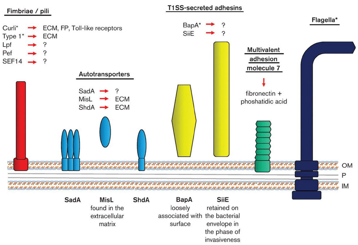
Figure 1. Schematic drawing of the cell envelope of S. enterica (OM, outer membrane; P, periplasm with peptidoglycan; IM, inner membrane) with symbolized bacterial adhesion molecules including their receptors (?, unknown receptors; ECM, extracellular matrix proteins; FP, fibrinolytic proteins). *Mediates adhesion to abiotic surfaces and biofilm formation. The structures depicted do not reflect the real macromolecule structures. Figure obtained after permission from Jaglic et al. (2014), Copyright Society of General Microbiology© 2015.
Pathogenicity islands typically accommodate large clusters of genes that contribute to a particular virulence phenotype, with S. Typhimurium possessing at least five such pathogenicity islands (SPIs; Marcus et al., 2000). Among these, SPI1 is primarily required for bacterial penetration of the epithelial cells of the intestine. Interestingly, Jennings et al. (2012) observed that S. Typhimurium cultures containing cloned SPI1 secretion system displayed an adherent biofilm and cell clumps in the media. This bacterial aggregation phenotype was associated with hyper-expression of SPI-1 type III secretion functions. With respect to mobile genetic elements, Salmonella genomic island 1 (SGI1) is associated with the multiple-drug-resistance (MDR) of S. Typhimurium DT104 strain (Boyd et al., 2001), while Malcova et al. (2008) has also demonstrated the additional positive effect of SGI1 on biofilm formation. In accordance, in recent years some other studies have revealed the role of multidrug efflux pumps in the ability of Salmonella spp. to produce biofilm (Baugh et al., 2012). Thus, resistance, biofilm production and fitness seem to be interrelated (Fàbrega et al., 2014). Noteworthily, eDNA has been found to inhibit biofilm development by S. Typhimurium and S. Typhi on abiotic surfaces (Wang et al., 2014).
Interspecies Interactions
Several studies have reported that biofilm production of Salmonella spp. may be promoted by the presence of other bacteria. In dual-species biofilms on stainless steel in drip-flow reactors, Staphylococcus piscifermentans and Pseudomonas sp. isolated from the feed industry increased biofilm growth of S. Agona compared to what was found in single species biofilms, with about threefold increases of the biovolume (Habimana et al., 2010b). In another flow system, with drinking water with acetate, S. Typhimurium did only form very thin-layered biofilm in monoculture on silicone tubes, but formed larger microcolonies when cultured together with a mixture of three strains of Paenibacillus sp., one Bacillus sp. and one strain of Enterococcus (Schaefer et al., 2013). The mechanism behind increased biofilm production was not investigated in these studies. However, in another study with a laboratory reactor simulating flow in water pipes, a much higher proportion of metabolically active cells were found in mixed biofilms than in single species biofilms. Also, the level of attachment of S. Enteritidis was seven times higher after 72 h in a dual biofilm with Klebsiella pneumoniae than in a single species biofilm (Jones and Bradshaw, 1997). Curli are important for biofilm formation of Salmonella. In a study of E. coli and S. Typhimurium, it was found that biofilm negative Salmonella could utilize parts of curli from E. coli and form biofilm on agar and pellicles (Zhou et al., 2012). In mixed biofilms of S. Typhimurium and E. coli O157:H7 on microtiter plates, Salmonella were generally found in higher numbers than E. coli, and a curli and cellulose producing Salmonella competed better with E. coli than a curli/cellulose negative Salmonella (Wang et al., 2013). Salmonella spp. or E. coli strains with negative EPS expression obtained significantly enhanced resistance to sanitation with a quaternary ammonium compound or chlorine by forming mixed biofilms with an EPS-producing companion strain of the other species (Wang et al., 2013).
A number of studies have demonstrated that the presence of other species may inhibit biofilm production of Salmonella spp. S. aureus dominated (99%) over S. Typhimurium in a constant-depth film fermentor (Knowles et al., 2005). Salmonella was primarily present in the top 40 μm of the biofilm and was not detected below 180 μm. This observation can be explained by that, in contrast to S. aureus, Salmonella require oxygen to produce biofilm and often form biofilm at liquid-air interphases (Scher et al., 2005). However, in another study where E. coli outcompeted S. enterica and L. monocytogenes in biofilms on different materials, S. enterica and L. monocytogenes were found close to the material surface, while E. coli was found in the biofilm top layer. The higher growth rate and exopolymer production ability of E. coli probably led this microorganism to outcompete the other two (Almeida et al., 2011). In a mixed species biofilm of S. simulans, Lactobacillus fermentum, Pseudomonas putida, L. monocytogenes, and S. Enteriditis, P. putida composed 98% of the population, while biofilm of S. Enteritidis was reduced by 1.5 log compared to the density of its population when this was grown as monoculture biofilm (Chorianopoulos et al., 2008).
Interbacterial communication signals like boronated-diester molecules (AI-2) and N-acyl homoserine lactones (AHLs) may be present in multi-species biofilms. Salmonella spp. can produce and respond to AI-2 and transcriptomics indicated an effect of AI-2 on biofilm formation in S. Typhimurium (Jesudhasan et al., 2010). However, Yoon and Sofos (2008a) found no differences in biofilm formation between AI-2 producing and non-producing S. Thompson. Brominated furanones which are believed to block QS signals have been shown to inhibit biofilm formation by Salmonella spp. (Janssens et al., 2008; Vestby et al., 2010, 2014). In the same way, grapefruit juice and its furocoumarins have been found to inhibit biofilm formation by E. coli O157:H7, S. Typhimurium and P. aeruginosa (Girennavar et al., 2008). These results suggest that targeting of microbial cell signaling processes could serve as a source to develop bacterial intervention strategies. Several types of bacteria have been reported to secrete compounds that inhibit biofilm of Salmonella spp. Heat stable compounds present in Hafnia alvei culture supernatant inhibited the early stage of biofilm development by S. Enteritidis on stainless steel. Although AHLs were detected in the supernatant, the role of AHLs could not be confirmed as synthetic AHLs did not affect the initial stage of biofilm formation by this pathogen (Chorianopoulos et al., 2010). The marine bacterium Pseudoalteromonas strongly inhibited adhesion of Salmonella spp. to glass through secretion of a compound of unknown identity (Dheilly et al., 2010). A probiotic Lb. plantarum strain led to 2-log decrease in biofilm forming colonies of Salmonella spp., compared to growing Salmonella spp. in monoculture. The effect was probably due to an unknown compound excreted by Lb. plantarum that also killed planktonic Salmonella spp. (Das et al., 2013a). Likewise, Woo and Ahn (2013) suggested that probiotic strains can be used as an alternative strategy to effectively reduce the biofilm formation in pathogenic bacteria through competition, exclusion, and displacement.
It has also been reported that Salmonella spp. can have an effect on biofilm of other bacteria. Thus, S. Typhimurium was found to outgrow and displace E. coli when it formed a biofilm on HEp-2 cells in a flow-through continuous culture system (Esteves et al., 2005). In biofilms on stainless steel, S. enterica influenced the intraspecies distribution of three L. monocytogenes strains in multi-species biofilms (Kostaki et al., 2012). The mechanism behind this interaction was not revealed. Most biofilm studies are on mixed cultures of bacteria, but two studies have also demonstrated interactions between Salmonella spp. and fungi. Tampakakis et al. (2009) showed that S. Typhimurium was able to secrete a heat stable substance that inhibited filamentation and biofilm formation of Candida albicans on silicone pads. In another study, S. Typhimurium was reported to rapidly attach to and forms biofilms on the hyphae of the fungus, Aspergillus niger. Interactions between cellulose produced by S. Typhimurium and chitin of A. niger was required for the production of the mixed biofilms (Brandl et al., 2011).
Intercellular Interactions in Biofilms of Salmonella: Current Knowledge and Concepts for Future Research
The persistence of Salmonella within the food chain is a major health concern, with its ability to form biofilms in food processing environments to serve as a potential reservoir for the contamination of food products. Undoubtedly, this ability contributes to its survival in non-host environments, stress hardening and its transmission to new hosts (Giaouris and Nesse, 2015). Like other bacterial pathogens, Salmonella is able to produce (depending on the surrounding conditions) various cell surface structures (especially of proteinaceous and carbohydrate nature) that all may result in the efficient coaggregation of its own cells with each other, as well as with cells of other species, facilitating thereby the formation of either mono- or multi-species biofilm communities. For instance, the co-expression of two of these components, curli fimbriae and cellulose, was shown to lead in the formation of a highly hydrophobic network with tightly packed cells aligned in parallel in a rigid matrix (Zogaj et al., 2001). Interestingly, White et al. (2010) showed by comparing extracellular matrix-embedded, wild-type S. Typhimurium and the matrix-deficient csgD mutant that the two populations present distinct metabolite and gene expression patterns, with wild-type cells expressing genes mainly involved in gluconeogenesis and stress-resistance pathways. Noteworthily, the effect of the simultaneous presence of other bacteria on the ability of Salmonella to form biofilms seems to greatly vary depending on the environmental conditions and bacteria tested. Thus, in laboratory studies, Salmonella spp. may be inhibited, promoted, or apparently do not respond to the presence of other bacteria in a biofilm. However, to the best of our knowledge, nothing is yet known on whether these multi-species communities could influence the pathogenicity of Salmonella (possibly by up-regulating the expression of virulence genes), while very little is still known on the effect of QS on Salmonella biofilm formation, especially in multi-species environments (Boyen et al., 2009). Targeting the QS mechanisms may provide a promising strategy for combatting biofilms and their associated problems.
Listeria monocytogenes
Listeria monocytogenes is a Gram-positive opportunistic and facultative intracellular bacterial pathogen that has served as a model organism for many virulence and biofilm related studies (Chavant et al., 2002; Møretrø and Langsrud, 2004; Cossart, 2011; Renier et al., 2011; Huang et al., 2012; da Silva and De Martinis, 2013). This is one of the major concerns in the food industry given that it can survive and even multiply in the harsh environmental conditions that exist in the production, processing, and storage of food products (Gandhi and Chikindas, 2007; Carpentier and Cerf, 2011). In addition, many L. monocytogenes strains can attach to various surfaces and form biofilms. However, strain and serotype differences occur, while conflicting results have been reported regarding correlations between biofilm forming abilities and persistence or phylogeny/lineage (Norwood and Gilmour, 2001; Djordjevic et al., 2002; Borucki et al., 2003; Di Bonaventura et al., 2008; Kadam et al., 2013; Valderrama and Cutter, 2013).
Intraspecies Interactions
Concerning L. monocytogenes intracellular interactions, the involvement of PrfA, the transcriptional activator of virulence operons, in biofilm formation was initially reported by Lemon et al. (2010) and Zhou et al. (2011), while Travier et al. (2013) subsequently demonstrated that actA, one the PrfA-regulated genes, was responsible for this function. In fact, ActA mediates interactions between L. monocytogenes cells via direct ActA-ActA interactions resulting in bacterial aggregation and biofilm formation. This aggregation property of ActA favors long-term gut colonization and fecal shedding, playing a key role in persistence within the host and in transmission from the host back to the environment (Travier et al., 2013; Travier and Lecuit, 2014).
Another protein involved in cell-to-cell interactions in this bacterium is SecA2, which is a paralog of SecA, a peripheral ATPase essential for the passage of pre-proteins through the cytoplasmic membrane. The secA2 gene has been identified in several pathogenic Gram-positive bacteria such as L. monocytogenes in which its deletion results in cell morphotype changes from discrete cells forming smooth colonies in wild type strain to long-chain cells forming rough colonies (Lenz and Portnoy, 2002). This colony morphotype is also observed when cwhA and murA, two genes encoding extracellular cell wall hydrolases, are deleted (Lenz et al., 2003; Renier et al., 2013, 2014) or when the SecA2-dependent secretion of their gene products is reduced (Machata et al., 2005). It is noticeable that this morphotype has been already observed from clinical patients, food samples and environmental biofilms (Rowan et al., 2000; Monk et al., 2004). In fact, this morphotypic conversion of bacterial cells is reversible and may have strong consequences on the ability of L. monocytogenes to colonize surfaces. In liquid medium, the inactivation of the SecA2 pathway results in extensive cell aggregation and sedimentation (Renier et al., 2014). So, the morphotypic conversion provides a significant advantage in listerial surface colonization under environmental conditions (Monk et al., 2004). It could be considered as an important risk factor for food processing plants and food products contamination but also of potential significance for asymptomatic human or animal carriage. Moreover, considering that biofilms are generally multi-species rather than mono-species, this cell differentiation could have consequences on L. monocytogenes implantation and interaction with other microbial species in various ecological niches (Sasahara and Zottola, 1993).
L. monocytogenes is a mobile bacterium thanks to 4–6 peritrichous flagella composed of thousands of monomers of the FlaA protein. However, this mobility is temperature-dependent because the transcription of flaA is stopped above 30°C. While flagella are important for biofilm formation of numerous bacterial species, their involvement in L. monocytogenes biofilm formation is quite controversial and seems dependent of environmental factors, namely more particularly growth medium, pH and temperature (Tresse et al., 2006, 2009), dynamic or static conditions (Rieu et al., 2008a). Undoubtedly, mutagenesis approaches have revealed that flagella and their motility play a role in L. monocytogenes biofilm formation. (Chang et al., 2012; Ouyang et al., 2012). However, this role seems to be limited to the positive role afforded by motility on the initial steps of surface attachment, probably by increasing the likelihood of encountering a surface and overcoming the repelling electrostatic forces, and not by flagella acting as surface adhesins per se (Vatanyoopaisarn et al., 2000; Lemon et al., 2007; Kumar et al., 2009; Jaglic et al., 2014). On the contrary, both non-flagellated and non-motile L. monocytogenes mutants were impaired in initial attachment but subsequently were revealed as hyper-biofilm formers when grown in flow cells (Todhanakasem and Young, 2008).
Contrary to the extracellular matrixes of many microbial biofilms, the one of L. monocytogenes seems to lack exopolysaccharides (Renier et al., 2011). Some observations by electron microscopy have sometimes revealed the presence of putative fimbriae-like structures binding cells to each other or to the surface (Hefford et al., 2005; Zameer et al., 2010; Renier et al., 2011). However, it was suggested that these fibrils resulted from the complete dehydration of a polymeric matrix during the processing of sample preparation. In the same way, the use of ruthenium red, a carbohydrate-binding dye, showed that L. monocytogenes bind this dye (Borucki et al., 2003; Zameer et al., 2010), which is consistent with the presence of exopolysaccharides. Nevertheless, as this dye can also bind carbohydrates on the bacterial cell surface unrelated to exopolysaccharides (peptidoglycans, teichoic acids, etc), these observations are not conclusive. Finally, it may be generally accepted that L. monocytogenes is a poor exopolysaccharide-producer by comparison with other bacterial species and that the putative presence of an extracellular matrix is dependent on the strain and environmental conditions (Borucki et al., 2003; Marsh et al., 2003; Zameer et al., 2010). Recently, Chen et al. (2014) identified a new c-di-GMP-inducible exopolysaccharide which caused cell aggregation in minimal medium and also impaired bacterial migration in semi-solid agar. However, this polysaccharide was not found to promote biofilm formation on abiotic surfaces. Harmsen et al. (2010) showed that in L. monocytogenes eDNA may be the only central component of the biofilm matrix and that it is necessary for both initial attachment and early biofilm formation. DNase I treatment resulted in dispersal of biofilms, not only in microtiter tray assays but also in flow cell biofilm assays. A dispersal of pre-existing L. monocytogenes biofilms by DNase treatment has also been shown by Nguyen and Burrows (2014). In agreement, the disruption of a putative DNA translocase gene impaired biofilm formation of L. monocytogenes on abiotic surfaces (Chang et al., 2013).
Interspecies Interactions
In food industry settings, interactions of L. monocytogenes in multi-species communities are likely, with the bacterial flora in food processing plants to include bacteria with the potential to increase or decrease colonization and biofilm formation by L. monocytogenes (Fox et al., 2014). Strains providing enhanced colonization and biofilm formation could contribute to L. monocytogenes persistence in food industry premises and consist a potential food safety risk. Bacteria with such properties, e.g., Pseudomonas spp., are commonly found on food processing surfaces even after cleaning and disinfection. They are also found to be significant biofilm producers. For instance, Hassan et al. (2004) showed increased attachment of L. monocytogenes on wet surfaces pre-colonized with P. putida. The change in surface properties due to P. putida EPS production which enhanced attachment of L. monocytogenes was the most likely cause to explain increased biofilm formation. Other spoilage organisms including Flavobacterium sp. have been reported to enhance biofilm formation of L. monocytogenes (Bremer et al., 2001). In contrast, biofilm formation by L. monocytogenes was reduced in co-cultures with Pseudomonas fragi (Norwood and Gilmour, 2001). Co-cultures with various Gram-negatives including Serratia spp., Aeromonas sp., and P. fluorescens have shown similar reductions (Gudmundsdottir et al., 2005; Daneshvar Alavi and Truelstrup Hansen, 2013). In less humid environments, staphylococci and other Gram-positives are regularly present. Co-culture biofilms with L. monocytogenes and S. aureus showed strain dependence and either increased, decreased or no effect on L. monocytogenes biofilm formation (Rieu et al., 2008b). The stimulated biofilm formation seemed to be caused by a S. aureus-excreted peptide. Biofilm formation of L. monocytogenes was decreased in the presence of S. sciuri where nutrient competition and extracellular substances produced by S. sciuiri explained the decreased adhesion and biofilm formation (Leriche and Carpentier, 2000). In another relevant study, Weiler et al. (2013) analyzed whether different L. monocytogenes strains are interacting with the microbial community of raw milk in terms of biofilm formation and found that the addition of individual L. monocytogenes strains to raw milk caused significant shifts in the biofilm biomass, in the chemical, as well as in the bacterial community composition. However, the added L. monocytogenes strains were not abundant, since mainly members of the genera Citrobacter and Lactococcus dominated the mixed culture bacterial biofilm community. In a study of 29 Gram-negative and Gram-positive isolates from food processing plants, 13% of the strains increased L. monocytogenes counts in the biofilms, while 53 and 34% had a negative and no effect on L. monocytogenes populations, respectively (Carpentier and Chassaing, 2004). Almeida et al. (2011) employed peptide nucleic acid fluorescence in situ hybridization (PNA FISH) to characterize S. enterica/L. monocytogenes/E. coli tri-species biofilm and identified two well-defined layers: the top one with E. coli, and the bottom one with mixed regions of L. monocytogenes and S. enterica. The higher growth rate and exopolymer production ability of E. coli probably led this microorganism to outcompete the other two. The competition of L. monocytogenes serotype 1/2a and 4b strains in mixed-culture biofilms has been demonstrated by Pan et al. (2009), with the serotype 1/2a strains tested to be generally more efficient at forming biofilms and to predominate in the mixed-culture biofilms.
Bacterial biofilms formed by useful technological bacteria, such as lactic acid bacteria (LAB) have the potential to control the development of L. monocytogenes through competitive exclusion and the synthesis of organic acids and bacteriocins (Guillier et al., 2008; Minei et al., 2008; Habimana et al., 2009, 2011; Woo and Ahn, 2013; Perez Ibarreche et al., 2014). For instance, Zhao et al. (2004) found 2 out of 413 microbial isolates from drains in food processing facilities to have significant anti-listerial activities. Follow-up studies have shown the potential of these isolates (Enterococcus durans and Lactococcus lactis) to control and eliminate L. monocytogenes from drains in the meat and poultry processing industry (Zhao et al., 2006, 2013). Although the mechanisms are not clear, both strains produce anti-listerial metabolites. Other studies have revealed nutrient competition as the principal mechanism behind the inhibition of L. monocytogenes in presence of multi-species biofilm microflora where L. monocytogenes often represent a minor part of the biofilm bacterial population (Chorianopoulos et al., 2008; Guillier et al., 2008), although a range of many other growth associated parameters may affect this distribution.
Once adhered to surfaces, L. monocytogenes often show enhanced survival and tolerance to food associated stresses which may even be enhanced by biofilm formation (Chavant et al., 2004; Pan et al., 2006; Carpentier and Cerf, 2011; Yun et al., 2012). Tolerance of L. monocytogenes to sanitizers and disinfectants has been reported to increase in dual culture biofilms as shown with L. monocytogenes and Lb. plantarum dual biofilms (van der Veen and Abee, 2011). Other studies have reported increased disinfectant tolerance of L. monocytogenes in mixed species biofilms. However, the protecting effects of the secondary species have been difficult to judge due to no results provided on single species biofilm resistance (Fatemi and Frank, 1999; Norwood and Gilmour, 2000). In dual culture biofilms of L. monocytogenes and S. enterica, the interspecies interactions did not influence either the biofilm forming ability or the resistance of each species to commonly used disinfectants. However, the intra- and interspecies interactions encountered in the biofilms had effect on the population dynamics and the resistance pattern of each L. monocytogenes strain present in the biofilm (Kostaki et al., 2012). Interactions between L. monocytogenes and P. putida provided increased tolerance to P. putida biofilm cells to benzalkonium chloride while L. monocytogenes tolerance remained unaffected in dual- or mono-species biofilms (Giaouris et al., 2013). This contrast to the study of Saa Ibusquiza et al. (2012) where increased tolerance of L. monocytogenes in dual L. monocytogenes/P. putida biofilms was reported.
Cell-to-cell signaling systems based on QS appear to be involved in bacterial biofilm formation although their exact role is still awaited (Li and Tian, 2012). In L. monocytogenes, there are two QS systems: the AI-2 signal system reported in both Gram-negative and Gram-positive bacteria and proposed as a universal, interspecies communication system, and the peptide-mediated QS system Agr, characteristic for Gram-positive bacteria (Waters and Bassler, 2005). Another communication system present in Gram-positive bacteria, involved in the development of competence in Bacillus subtilis, is associated with the autoinducer ComX. However, the main genes of this system are absent in the genome of L. monocytogenes for which, moreover, natural genetic transformation has never been observed (Borezee et al., 2000). Although L. monocytogenes can produce AI-2, experimental evidence indicates that L. monocytogenes lack receptors of AI-2. Thus, this suggests that AI-2 is not a communication signal in L. monocytogenes (Challan Belval et al., 2006; Garmyn et al., 2009). Concerning the agr system described in L. monocytogenes, it appears that this regulates major adaptive responses, such as the promotion of biofilm formation, expression of adhesion factors and internalins (Rieu et al., 2007, 2008a; Riedel et al., 2009). It has been shown that agrA or agrD mutant strains, two of the four genes composing the agr locus, were affected in adhesion and the first stage of biofilm formation. However, the agrA or agrD gene products, a response regulator of a two-component system and a precursor peptide respectively, are parts of a complex signaling system probably involved in multiple physiological regulation. Thus, there is no demonstration to date of the mechanism by which they intervene in biofilm formation.
Intercellular Interactions in Biofilms of L. monocytogenes: Current Knowledge and Concepts for Future Research
Listeria monocytogenes is considered as an environmental pathogen because it is capable of saprophytic life in the outside environment while also maintaining the ability to invade and replicate within mammalian cells (Xayarath and Freitag, 2012). This pathogen is capable of forming biofilms which considerably increase its resistance to harsh physicochemical conditions and particularly to cleaning and disinfection treatments. Thus, in this sessile mode of growth, bacteria may persist in production lines and constitute a permanent risk of contamination of food products. Literature has shown that L. monocytogenes biofilm formation, maturation, and structure depends on a multitude of external and internal factors, where both intra- and inter-species interactions seem to play an important role (Renier et al., 2011). However, their exact and respective underlying mechanisms are not always characterized, emphasizing that much remains to be investigated. Remarkably, L. monocytogenes may display increased disinfection tolerance when this forms biofilms with other species. This is certainly something which is worth to be further studied. Besides such an increase in disinfection resistance, the influence of various biofilm forming conditions on the virulence properties of these bacteria is still largely unexplored. Future research on this area should include both mono and multi-species biofilms and focus on the underlying molecular mechanisms hidden behind any observation. Finally, while peptide sensing is able to promote L. monocytogenes biofilm formation (Rieu et al., 2007, 2008a; Riedel et al., 2009), how this is achieved is not yet recognized.
Escherichia coli
Escherichia coli is primarily a commensal species which however also contains important pathogenic strains (Kaper et al., 2004), with the foodborne pathogenic E. coli to be diarrheagenic strains (Nataro and Kaper, 1998; Croxen et al., 2013). Those intestinal pathogenic E. coli (InPEC) can be subdivided into the following pathotypes, namely ETEC (enterotoxigenic E. coli), EAEC (enteroaggregative E. coli), DAEC (diffusively adherent E. coli), EIEC (enteroinvasive E. coli), EPEC (enteropathogenic E. coli), and EHEC (enterohemorrhagic E. coli), with the recently described subgroup of EAHEC (enteroaggregative and haemorrhagic E. coli, Brzuszkiewicz et al., 2011). By definition, EHEC are clinical strains of InPEC and belong to the larger group of Shiga toxin producing E. coli (STEC), which pathogenesis is not ascertained.
Intraspecies Interactions
Various cell surface molecules and structures have been implicated in biofilm formation in E. coli (Van Houdt and Michiels, 2005). In this bacterium, a major cell–cell interaction involves the autotransporter Ag43 (Antigen 43; van der Woude and Henderson, 2008), a protein of the Type Va secretion system (T5aSS; Desvaux et al., 2004b). This protein is found encoded in a majority of E. coli genomes, from domesticated K-12, commensal to pathogenic strains (van der Woude and Henderson, 2008). While Ag43 was known to be a self-recognizing protein promoting autoaggregation and consequently biofilm formation (Danese et al., 2000; Klemm et al., 2004), it was recently uncovered that its functional domain displays a twisted L-shaped β-helical structure firmly stabilized by a 3D hydrogen-bonded scaffold which facilitates self-association and cell aggregation via a mechanism described as a Velcro-like handshake (Heras et al., 2014). Very interestingly, the expression of the Ag43 is subjected to phase variation (Diderichsen, 1980; Henderson et al., 1997, 1999; Henderson and Owen, 1999). It was recently demonstrated that biofilm formation did not influence the frequency of switch between bacterial cells expressing the Ag43 (phase ON) and those which do not expressed it (phase OFF; Chauhan et al., 2013). Because of the autoaggregation phenotype, the phase ON E. coli cells were physically selected and prominent within the biofilm under dynamic flow conditions.
Of note, several other autotransporters can also be involved in cell aggregation, namely AIDA (Sherlock et al., 2004; Girard et al., 2010) and TibA (Sherlock et al., 2005), i.e., SAATs (self-associating autotransporters; Klemm et al., 2006), but also EhaA (Wells et al., 2008), at least when overexpressed. In E. coli O157:H7, Cah is a calcium-binding and heat-extractable autotransporter protein homologous to Ag43 and AIDA (Torres et al., 2002). Besides, TolC was supposedly also involved in E. coli aggregation (Imuta et al., 2008), as well as curli and Type 1 pili (Barnhart and Chapman, 2006; Tree et al., 2007; Ulett et al., 2007). Expression of Type 1 pili and polar localization of Ag43 can further influence bacterial cell chain formation in biofilm (Vejborg and Klemm, 2009). Within the T5SS (Desvaux et al., 2004a; Henderson et al., 2004), some proteins of the subfamily b (T5bSS) of the two-partner secretion (TPS) are involved in contact-dependent inhibition (CDI; Hayes et al., 2014). This system allows regulating bacterial growth in response to changing environmental conditions upon direct cell–cell interactions (Aoki et al., 2005; Hayes and Low, 2009; Hayes et al., 2010). The CDI activity is very specific since it is limited to target cells of the same species. So far, the role of CDI in the course of biofilm formation has not been investigated in the scientific literature.
Pili are cell-surface supramolecular protein complexes involved in cell-to-cell interactions (Lasaro et al., 2009; Hernandes et al., 2013), which have also been termed fimbriae or curli based on some morphological differences. Curli production occurs via the Type VIII secretion system and is dependent on the CsgD transcription activator, which also promotes cellulose biosynthesis (Brombacher et al., 2006). In E. coli, the expression of the curli-specific genes (csg) which are clustered in the csgBA and csgDEFG operons is dependent on a combination of environmental parameters such as low growth temperature and low osmolarity (Hammar et al., 1995; Römling et al., 1998). Previous studies revealed a positive correlation between curli expression, exopolysaccharides production (such as cellulose), and autoaggregation by E. coli (Ryu et al., 2004a,b; Uhlich et al., 2006; Tree et al., 2007; Gualdi et al., 2008; Saldaña et al., 2009; Goulter et al., 2010). The E. coli common pilus (ECP) represents a remarkable family of extracellular fibers and plays a dual role in early-stage biofilm development and host cell recognition (Garnett et al., 2012). The microcolony formation on biotic surfaces in EPEC is mediated by several adhesins including the type IV bundle-forming pilus (BFP) and the EspA filament, which are also involved in bacterial aggregation during biofilm formation on abiotic surfaces together with the type 1 fimbriae and the antigen Ag43 (Moreira et al., 2006). EAEC forms thick biofilms on the intestinal mucosa by virtue of a plasmid-encoded fimbrial adhesin designated aggregative adherence fimbria I (AAF/I; Czeczulin et al., 1997). EHEC factor for adherence Efa1 confers haemagglutination, adherence to epithelial cells and autoaggregation (Nicholls et al., 2000). Recently, Gómez-Gómez and Amils (2014) characterized a novel E. coli sessile behavior termed “crowning” which is developed independently of the adhesiveness of the major components of E. coli’s EPS matrix, the function of chemotaxis sensory system, type 1 pili and the biofilm master regulator CsgD, but its formation is suppressed by flagella-driven motility and glucose.
The F episome encodes a Type IVb secretion system (T4bSS) responsible for bacterial conjugation, the so-called sexual pili (Chagnot et al., 2013). It was elegantly demonstrated that the F episome in E. coli was not only involved in the horizontal transfer of genetic information to F-recipient cells but actively contributed to biofilm formation (Ghigo, 2001). Conjugation further decreased motility, which increased the biofilm formation in E. coli (Barrios et al., 2006; Reisner et al., 2006). As promoters of bacterial colonization and development of mature biofilms by providing aggregative properties, the conjugative plasmids in general should be more systematically considered as a risk factor among foodborne pathogenic bacteria (Dudley et al., 2006; Mliji el et al., 2007; May and Okabe, 2008; Norman et al., 2008); potentially, they cannot only promote intra- but also inter-species genetic transfer even between Gram-positive and Gram-negative bacteria (Goessweiner-Mohr et al., 2013).
Zhang et al. (2007) demonstrated, using DNA microarrays, that the expression of ycfR, which encodes the putative outer membrane protein YcfR (BhsA), is significantly induced in E. coli biofilms. This protein seems to be involved in the regulation of biofilm formation by decreasing cell aggregation and cell surface adhesion, by influencing the concentration of signal molecules, and by interfering with stress responses. Critical to the development of a biofilm is the elaboration of exo-polysaccharide that contributes to substrate and intercellular adhesion. Indeed, one way to identify the environmental cues that cause a given bacterial species to switch to the biofilm mode of growth is to monitor exo-polysaccharide elaboration in vitro (Jefferson and Cerca, 2006). Lipopolysaccharide (LPS) is the major component of the surface of Gram-negative bacteria and its polysaccharide portion is situated at the outermost region. Mutations known to affect the composition of E. coli LPS core oligosaccharide affected the biofilm formation which was associated with eDNA (Nakao et al., 2012). The spatial periodicity of E. coli cells within a biofilm has been associated to the secretion of the polysaccharide adhesin β-1,6-N-acetyl-d-glucosamine PGA (Agladze et al., 2005). Colanic acid also contributes to the biofilm architecture and allows for the formation of voluminous biofilms (Prigent-Combaret et al., 2000).
Quorum sensing has been shown to play a significant role on the surface chemistry and electrokinetic properties of E. coli cells, possibly through the regulation of outer membrane macromolecules (Eboigbodin et al., 2006). In addition, E. coli cells cultivated with an additional supplement of glucose, displayed a higher concentration of bacterial surface functional groups and a variation in outer membrane proteins, which consequently reduced the tendency for cell-to-cell attachment (Eboigbodin et al., 2007). AI-2 takes part both in intra- and inter-species interactions in E. coli and is involved in multiple physiological processes, including biofilm formation, exopolysaccharides production, and determination of cell surface properties via the regulation of the genes encoding outer membrane proteins and putative adhesins (DeLisa et al., 2001). The interactions between E. coli cell clusters involving particularly the AI-2 mediated cell-to-cell signaling have been found to play an important role in the spatial organization of the cell clusters in biofilms of E. coli (Gu et al., 2013). The two component signal-transduction system Cpx is also believed to act as a strategic signaling pathway for confronting adverse conditions and for settling biofilm communities by activating genes encoding periplasmic-protein-folding and degrading factors (Dorel et al., 2006). Interestingly, in recent years, plant phytochemicals have appeared as a novel promising strategy for controlling biofilm formation and virulence in E. coli and other pathogens through interfering with the bacterial cell–cell signaling pathways and the expression of cell surface adhesins (Vikram et al., 2010a,b; Lee et al., 2014a).
Interspecies Interactions
Escherichia coli interacts with other microorganisms and is able to form multi-species biofilms with many of the most common bacterial genera occurring on food processing surfaces including both Gram negatives (e.g., Pseudomonas, Acinetobacter) and Gram positives (e.g., staphylococci, Bacillus, Castonguay et al., 2006; Marouani-Gadri et al., 2009; Habimana et al., 2010a; Kuznetsova et al., 2013; Liu et al., 2014). Strains isolated from water and food processing environments have shown to stimulate co-adhesion and biofilm formation of E. coli. For instance, Castonguay et al. (2006) showed that biofilm formation of non-adherent E. coli from drinking water reservoirs was stimulated in dual and other multiculture biofilms with biofilm proficient bacteria from the same environment. The mechanism of biofilm stimulation required direct cell-to-cell contact. All but one of 20 randomly collected bacterial isolates obtained after cleaning and disinfection from a beef processing plant increased the counts of adhered E. coli O157:H7 in dual-culture biofilms (Marouani-Gadri et al., 2009). Low-nutrient conditions conferred by growth of the resident strains and which favored E. coli biofilm formation was hypothesized as an explanation of the observed phenomenon. Microflora isolates capable of producing biofilms are widely distributed in fresh produce processing facilities, while the presence of persistent biofilm forming strains is also reported (Van Houdt et al., 2004; Liu et al., 2013). Strong biofilm forming plant-associated bacteria promoted the incorporation of E. coli O157:H7 in biofilms at 30°C (Liu et al., 2014). Carter et al. (2012) examined the interaction of E. coli O157:H7 with spinach leaf indigenous microorganisms during co-colonization and establishment of a mixed biofilm on a stainless steel surface, by using a metagenomics analysis, and revealed competition for essential macronutrients as the primary interaction. Under flow conditions adhesion to glass surfaces of Acinetobacter calcoaceticus (Habimana et al., 2010a) or P. aeruginosa (Klayman et al., 2009) stimulated adhesion of E. coli O157:H7 which was not able to form biofilm in monoculture under dynamic flow conditions. Although the detailed mechanisms are not known, it is likely that the early colonizer provide surface structures or surfactants promoting co-adhesion of E. coli. Cell-to-cell dependent interactions promoting retention of E. coli O157:H7 in biofilms have also been shown by others (Uhlich et al., 2010). Bacteria affecting biofilm formation of E. coli through secreted metabolites have also been reported (Cabellos-Avelar et al., 2006; Lopes et al., 2011; Vacheva et al., 2012).
The above studies show that bacterial interactions can promote pathogenic E. coli biofilm formation and even enable adherence deficient strains to form biofilms. This is of significant relevance and highlights that the control of environmental bacteria promoting adherence and biofilm formation of other bacteria can be an important measure to prevent establishment of pathogenic E. coli and other pathogens in food processing environments. On the other hand, Da Re et al. (2013) analyzed genetic responses induced in commensal E. coli upon entry of a diarrheagenic EAEC or an unrelated K. pneumoniae pathogen into a biofilm community and identified some genes involved in limiting colonization of incoming pathogens within commensal biofilm. Valle et al. (2006) demonstrated that all E. coli expressing group II capsules release into their environment a soluble polysaccharide that induces physicochemical surface alterations, which prevent biofilm formation by a wide range of Gram positive and Gram negative bacteria. Similarly, Rendueles et al. (2011) identified an E. coli biofilm-associated anti-adhesion polysaccharide which reduced susceptibility to invasion and provided rapid exclusion of S. aureus from mixed E. coli and S. aureus biofilms. These last findings identify bacterial interference via surface active compounds produced during competitive interactions as a new strategy to limit biofilm formation on surfaces.
As previously mentioned in the Salmonella part of this review, dual cultures of E. coli O157:H7 and S. Typhimurium showed that EPS-producing strains were able to establish themselves in mixed biofilms more efficiently but also enhanced E. coli O157:H7 biofilm formation (Wang et al., 2013). The protective role of an EPS producing strain of S. Typhimurium in providing increased resistance to a non-EPS producing E. coli O157:H7 strain toward two industrial sanitizers was also shown (Wang et al., 2013). This indicates that EPS-producing strains also may provide protection to sensitive companion strains in mixed species biofilms. E. coli and its LPS has been shown to modulate in vitro biofilm formation by Candida species (Bandara et al., 2009). Putative F pili expressed by EAEC strains boosted mixed biofilm formation when in the presence of aggregative Citrobacter freundii (Pereira et al., 2010). E. coli transformed with green fluorescent protein (GFP) and Serratia marcescens stably co-existed in biofilms but did not affect the growth of each other (Skillman et al., 1998).
Another interesting concern is the correlation between the biofilm forming ability of pathogenic E. coli and the presence of probiotic bacteria (Chapman et al., 2012, 2014). Decreased E. coli O157:H7 biofilm formation was observed in the presence of EPS produced by a probiotic Lb. acidophilus strain by interfering with the expression of E. coli surface adhesins (Kim et al., 2009). In the investigations conducted by Kim et al. (2012) cell extract of Bifidobacterium longum caused 36% reduction in biofilm formation by E. coli O157:H7. Significant inhibition in AI-2 QS activity in E. coli O157:H7 was also observed, while proteome analysis showed that seven proteins were differentially regulated in E. coli in the presence of Bifidobacterium cell extract. However, Miyazaki et al. (2010) observed no inhibitory effect of the culture supernatant of Bifidobacterium spp. against biofilm formation by EAEC. Andrzejewska and Sobieszczanska (2013) showed that Lb. casei inhibited the biofilm formation by EAEC. The reason for this was not explained. Obviously probiotic bacteria may reduce biofilm formation by E. coli, but further analysis is still required.
Interspecies interactions in multi-species biofilms also include HGT (Madsen et al., 2012; Van Meervenne et al., 2014). Conjugative transfer of a plasmid encoding a type 3 fimbriae rendered a non-biofilm producing uropathogenic E. coli strain to be a significant member in a mixed biofilm (Ong et al., 2009). Conjugative transmission of natural plasmids carried by the E. coli lead to biofilm expansion in mixed E. coli biofilms (Reisner et al., 2006). Another study showed HGT of shiga toxin encoding genes (stx) to occur by transduction in E. coli (Solheim et al., 2013). BdcA binds the ubiquitous bacterial signal c-di-GMP and has been found to control biofilm dispersal in P. aeruginosa and Rhizobium meliloti via conjugation from E. coli in mixed-species biofilms (Ma et al., 2011). These reports illustrate the potential of HGT to spread determinants involved in biofilm formation, dispersal and virulence which have relevance for food-associated E. coli. Surely, the impact of the transfer of conjugative plasmids on multi-species biofilm formation is dependent on both the type of the plasmid and the plasmid host (Røder et al., 2013).
So far, only a few studies indicated the important role of AI-2 QS system in biofilm formation by E. coli (González Barrios et al., 2006; Silagyi et al., 2009; Zhou et al., 2014). González Barrios et al. (2006) showed that the addition of AI-2 enhanced biofilm formation by E. coli by stimulating flagellar motion and motility. Silagyi et al. (2009) observed that E. coli O157:H7 produced maximum levels of AI-2 signals in 12 h of incubation in tested foods and next formed strong biofilm in 24 h of incubation. In other studies AI-2-based QS activity of E. coli O157 did not affect biofilm formation in monocultures (Yoon and Sofos, 2008b). Indole has been proved as an interspecies signal that decreases E. coli biofilms through SdiA and increases those of pseudomonads (Lee et al., 2007). E. coli also possess receptors for AHL which can be produced by other bacteria (Moons et al., 2006; Ren et al., 2013). The potential effect of AHL producing bacteria on E. coli biofilm formation should be studied in multiculture biofilms.
Intercellular Interactions in Biofilms of E. coli: Current Knowledge and Concepts for Future Research
Escherichia coli possesses a wealth of cell surface structures involved in intercellular interactions during biofilm development (mainly by provoking cell aggregation). Their respective contribution and regulation in the course of sessile growth still remain to be further elucidated at a global scale respective to crucial environmental conditions as a function of the pH, temperature, and/or available nutrients. Of note for instance, the mechanism, regulation and function of Ag43 are quite restricted to studies using the non-pathogenic and domesticated E. coli K12 strain but further investigations respective to the different subfamilies of Ag43 and pathogenic E. coli species are undoubtedly required (van der Woude and Henderson, 2008). As another example and in addition to bacterial growth inhibition, CDI system might have other but as yet uncovered physiological functions; its potential involvement in the course of sessile development is particularly relevant and intriguing, which would hopefully trigger intense research investigations in the near future. Worth mentioning is also the growing evidence that biofilms of E. coli and other microorganisms represent an ideal microenvironment for HGT. Natural conjugative plasmids have been shown to promote the development of mature E. coli biofilms by providing aggregative properties, promoting cell-surface interactions, and stimulating colanic acid and curli production (Ghigo, 2001). However, there is no data in about the HGT by transformation (described as uptake of free DNA from the surrounding environment) in multi-species biofilms formed with E. coli contribution. For foodborne pathogenic E. coli like EHEC, their extremely low infectious dose combined with their ability to form biofilms and associate in multi-species biofilms poses an extra challenge. Finally, the possible effect of AHLs produced by other food related bacteria on E. coli biofilm formation is for sure another intriguing case of future research.
Staphylococcus aureus
Staphylococcus aureus is a Gram-positive, ubiquitous bacterial species commonly found on the skin and hair, as well as in the noses and throats of people and animals. It is the causative agent of a wide spectrum of human infections (Otto, 2013) and is also often responsible for foodborne intoxications through the production of heat stable enterotoxins in a variety of food products (Hennekinne et al., 2012). S. aureus can produce a multilayered biofilm embedded within a glycocalyx or slime layer with heterogeneous protein expression throughout (O’Gara, 2007; Archer et al., 2011; Periasamy et al., 2012; Laverty et al., 2013). Especially S. aureus harbors a variety of proteinaceous and non-proteinaceous adhesins that mediate attachment to a multitude of host factors, such as extracellular matrix and plasma proteins and human host cells, or intercellular adhesion, which is essential for biofilm accumulation (Heilmann, 2011).
Intraspecies Interactions
The major constituents of staphylococcal biofilms are polysaccharides, such as the polysaccharide intercellular adhesin (PIA) or poly-N-acetyl glucosamine (PNAG), cell surface and secreted bacterial proteins, and eDNA (Izano et al., 2008). However, the exact composition of biofilms often varies considerably between different strains of staphylococci and between different sites of infection by the same strain. PIA is encoded by the icaADBC operon, yet details of its biosynthesis are limited (Atkin et al., 2014). Regulation of icaADBC is extremely complex, this is influenced by many environmental factors and involves an array of coordinated regulatory mechanisms which have yet to be fully elucidated (Mack et al., 2004; O’Gara, 2007; Cue et al., 2012). Biofilm-specific transcriptional regulators include Rbf, which mediates the induction of biofilm formation at the cell–cell interaction stage in response to glucose and osmotic stress (Lim et al., 2004; Cue et al., 2009), and IcaR and TcaR, both of which negatively regulate biofilm formation (Jefferson et al., 2004). Global transcriptional regulators include staphylococcal accessory regulator (SarA), which is required for biofilm formation (Valle et al., 2003; Trotonda et al., 2005; Sambanthamoorthy et al., 2008; Mrak et al., 2012), and the two-component regulator ArlRS, a repressor of biofilm formation (Toledo-Arana et al., 2005). Inactivation of a global regulator of the bacterial stress response, the alternative transcription factor sigma(B), resulted in a biofilm-negative phenotype and loss of salt-induced biofilm production (Rachid et al., 2000). However, Valle et al. (2003) demonstrated that complete deletion of sigmaB did not affect PIA/PNAG production and biofilm formation, although it slightly decreased ica operon transcription. You et al. (2014) identified a new operon, gbaAB (glucose induced biofilm accessory gene) that is involved in the regulation of the multicellular aggregation step of S. aureus biofilm formation in response to glucose and showed that this regulation may be mediated through the ica operon. Osmotic stress was found to induce biofilm formation in a S. aureus mucosal isolate (Rachid et al., 2000).
Undoubtedly, the genetic and molecular basis of biofilm formation in staphylococci is multifaceted (Götz, 2002). Beyond PIA/PNAG, evidence is now emerging for the existence of ica-independent biofilm mechanisms capable of mediating intercellular accumulation in both S. aureus and S. epidermidis (O’Gara, 2007). Thus, a number of biofilm-negative mutants have been isolated in which PIA production appears to be unaffected. Two of the characterized mutants were affected in the major autolysin (atlE, Biswas et al., 2006; Bose et al., 2012) and in D-alanine esterification of teichoic acids (dltA, Gross et al., 2001). Teichoic acids are anchored to the outer layer of the cytoplasmic membrane via a glycolipid (lipoteichoic acid, LTA) or covalently to the cell-wall peptidoglycan (wall teichoic acid, WTA). A S. aureus ypfP mutant with strongly reduced LTA content was completely unable to form biofilm on plastic (Fedtke et al., 2007). WTA-deficient S. aureus mutants showed a higher degree of cell aggregation, but had reduced initial adherence to abiotic surfaces and had a reduced capacity to form biofilms under both steady-state and flow conditions (Vergara-Irigaray et al., 2008). eDNA provides structure and stability in mature biofilms of S. aureus (Izano et al., 2008), and many other species (Das et al., 2013b). Secreted proteins may also influence S. aureus multicellular behavior. Caiazza and O’Toole (2003) showed that alpha-toxin (also known as α-hemolysin or Hla), a secreted, multimeric, hemolytic toxin encoded by the hla gene, plays an integral role in S. aureus biofilm formation and is required for cell-to-cell interactions. Similarly, Anderson et al. (2012) showed that alpha-toxin also promotes S. aureus mucosal biofilm formation.
The surface of S. aureus is “decorated” with proteins that are in most cases covalently anchored to the cell wall peptidoglycan via an LPxTG motif cleaved by sortase A (SrtA). Interestingly, the overexpression of SrtA has resulted in increased levels of biofilm formation in some S. aureus strains (Sibbald et al., 2012). Structural and functional analysis has identified four distinct classes of surface proteins in this bacterium, of which microbial surface component recognizing adhesive matrix molecules (MSCRAMMs) are the largest class. These surface proteins have numerous functions, including adhesion to and invasion of host cells and tissues, evasion of immune responses and biofilm formation (Foster et al., 2014; Speziale et al., 2014). Surface proteins Bap (biofilm-associated protein), Eap (extracellular adherence protein), or PSM (phenol soluble modulin) promote S. aureus adherence to host cells and surfaces, as well as cell aggregation (Cucarella et al., 2001; Hussain et al., 2008; Thompson et al., 2010; Schwartz et al., 2012). Interestingly, Arrizubieta et al. (2004) found that Bap binds Ca2+ with low affinity and that this binding renders the protein non-competent for biofilm formation and for intercellular adhesion, while Tormo et al. (2007) described a process of phase variation that affects the expression of Bap in S. aureus. Thus, it is possible that S. aureus can detach from a biofilm by switching to a Bap-negative state. Schwartz et al. (2012) showed that the PSMs’ polymerization and aggregation into amyloid fibers stabilize and promotes S. aureus biofilm integrity. Intercellular auto-aggregation is also favored by SasG (S. aureus surface protein G; Corrigan et al., 2007; Kuroda et al., 2008; Geoghegan et al., 2010) and SasC (Schroeder et al., 2009), but inhibited by the delta hemolysin (Hld or PSM; Vuong et al., 2003).
Other examples of important surface proteins identified to be involved in S. aureus biofilm formation include accumulation-associated protein (Aap), clumping factor A (ClfA), staphylococcal surface protein (SSP1; Foster et al., 2014), protein A (Spa; Merino et al., 2009), serine-aspartate repeat protein SdrC (Barbu et al., 2014), and SraP, a surface-exposed serine-rich repeat glycoprotein (SRRP; Sanchez et al., 2010). Aap and SasG are homologous proteins containing sequence repeats known as G5 domains, which self-associate in the presence of Zn2+ resulting in the formation of extensive adhesive contacts between cells (Geoghegan et al., 2010; Conrady et al., 2013). Surface proteins with tandem G5 domains are also found in other bacterial species, suggesting that this mechanism for intercellular adhesion in biofilms may be conserved among staphylococci and other Gram-positive bacteria (Conrady et al., 2008). Expression of SasG masked the ability of exponentially grown S. aureus cells expressing protein A (Spa), clumping factor B (ClfB) and the fibronectin binding proteins A and B (FnBPA and FnBPB) to bind to IgG, cytokeratin 10 and fibronectin, respectively (Corrigan et al., 2007). SasG-expressing strains formed peritrichous fibrils of varying density on the cell wall, and also formed biofilm independently of the PIA. It was concluded that the fibrillar nature of SasG explains its ability to mask binding of S. aureus MSCRAMMs to their ligands and to promote formation of biofilm. S. aureus mutant strains unable to express the FnBPA and FnBPB lacked the ability to adhere to fibronectin and to form biofilms (O’Neill et al., 2008; McCourt et al., 2014). On the contrary, the expression of these two proteins increased bacterial aggregation suggesting that fibronectin-binding proteins can promote the accumulation phase of biofilm. Loss of fibronectin-binding proteins reduced the initial adherence of bacteria, indicating that these proteins are also involved in primary attachment.
Noteworthy, bacterial variants of S. aureus called small colony variants (SCVs) originate by mutations in metabolic genes, resulting in emergence of auxotrophic bacterial subpopulations (Melter and Radojevič, 2010). Environmental pressure such as antibiotics, select for isogenic SCV cells that are frequently found coexisting with their parent wild-type strains in a mixed bacterial culture. Such a menadione-auxotrophic S. aureus SCV displayed an autoaggregative phenotype and formed highly structured biofilms, consisting of large microcolonies separated by channels, and contained more biomass as well as significantly more PIA than wild-type biofilms (Singh et al., 2010).
Cell population density-dependent regulation of gene expression is an important determinant of bacterial biofilm formation. Staphylococci have two QS systems: the accessory gene regulator (agr) which is genus specific and uses a post-translationally modified peptide as an autoinducing signal and luxS, which is found in a variety of Gram-positive and Gram-negative bacteria (Kong et al., 2006). Importantly, unlike many QS systems described in Gram-negative bacteria, Agr and LuxS of staphylococci reduce rather than induce biofilm formation and virulence during biofilm-associated infection. Agr enhances biofilm detachment by up-regulation of the expression of detergent-like peptides, whereas LuxS reduces cell-to-cell adhesion by down-regulating expression of biofilm exopolysaccharide via an icaR-activation pathway (Yu et al., 2012). However, the role of the Agr QS system appears to vary depending on the strain or growth conditions, as disruption of agr can inhibit, enhance, or have no effect on biofilm formation (Yarwood et al., 2004).
Interspecies Interactions
Studying microbial interactions and adaptive processes leading to co- or poly-bacterial infections is evidently important. P. aeruginosa and S. aureus are the most prevalent pathogens in airway infections of cystic fibrosis (CF) patients, while significant research has been conducted on how these pathogens coexist and interact with each other (Hoffman et al., 2006; Davies and Marques, 2009; Chew et al., 2014). Interestingly, wild-type P. aeruginosa PAO1 has been found to facilitate S. aureus microcolony formation when both are grown in co-culture biofilms in a flow-chamber system (Yang et al., 2011a). Further investigations revealed that eDNA behaves as an essential EPS material shared by both species in co-culture biofilms, which facilitates interspecies interactions and that P. aeruginosa type IV pili are required for this process, probably through their ability to bind to eDNA. In another recent study, Fugère et al. (2014) found that culture supernatants from 63 P. aeruginosa clinical isolates retrieved from CF adult patients triggered a wide range of biofilm-stimulatory activities when added to the culture of a control S. aureus strain. However, when studying co-isolated pairs of P. aeruginosa and S. aureus retrieved from patients showing both pathogens, P. aeruginosa supernatants stimulated less biofilm production by the S. aureus counterparts compared to that observed using the control S. aureus strain. This suggests that colonization of the CF lungs promotes some type of strain selection, or that co-existence requires specific adaptations by either or both pathogens. In the same context, Wollenberg et al. (2014) demonstrated that coproporphyrin III (CIII), a diffusible small molecule excreted by nostril- and skin-associated Propionibacterium spp., induces S. aureus aggregation in a manner dependent on dose, growth phase, and pH. In another study on a polymicrobial wound infection, the presence of P. aeruginosa resulted in induced expression of S. aureus virulence factors (Pastar et al., 2013).
Bacteria and fungi are found together in a myriad of environments and particularly in a biofilm, where adherent species interact through diverse mechanisms (Shirtliff et al., 2009). The fungal species C. albicans and S. aureus are responsible for a majority of hospital-acquired infections and are increasingly co-isolated from implant-associated polymicrobial infections creating an incremental health care problem (Harriott and Noverr, 2009). This polymicrobial biofilm formation and subsequent resistance seems to be a multifactorial process that may require a unique combination of fungal and/or bacterial adhesins (Harriott and Noverr, 2010; Peters et al., 2012b). Thus, synergistic effects between both species seem to facilitate the formation of dual-species biofilms (Millsap et al., 2001). Confocal laser scanning microscopic (CLSM) analyses of such biofilms have revealed a unique biofilm architecture where S. aureus is commonly associated with the hyphal elements of C. albicans (Peters et al., 2010). In addition, during such co-existence, S. aureus is known to present enhanced pathogenesis due to the differential regulation of specific virulence factors (Peters et al., 2010). Interestingly, Fehrmann et al. (2013) also revealed the protective role of S. aureus fibrinogen-binding proteins coagulase and Efb against the phagocytosis of Candida cells by granulocytes.
Besides synergistic interactions, antagonistic ones have also been observed in mixed culture biofilms of S. aureus and other species. Thus, Malic et al. (2011) studied in vitro biofilms generated either individually or in dual combinations by P. aeruginosa, S. aureus, Streptococcus oralis, and Micrococcus luteus and observed distinct species antagonism with apparent antagonism of pathogenic species over “commensal” ones. Iwase et al. (2010) showed that the serine protease Esp secreted by a subset of commensal S. epidermidis, inhibits biofilm formation and nasal colonization by S. aureus and also destroys pre-existing biofilms of this pathogen. Furthermore, Esp enhances the susceptibility of S. aureus in biofilms to immune system components. Similar results were also obtained by Sugimoto et al. (2013) who demonstrated that Esp inhibits S. aureus colonization and biofilm formation by degrading specific proteins that are crucial for biofilm construction and host-pathogen interaction. Likewise, Chen et al. (2013) showed that Esp cleaves autolysin (Atl)-derived murein hydrolases and prevents staphylococcal release of DNA, which serves as extracellular matrix in biofilms.
Environmental biofilms grown in seawater on agar containing spent S. aureus filtrate were more inhibitory to S. aureus, as compared to environmental biofilms grown on plain agar (without added spent S. aureus filtrate; Lafleur et al., 2013). Jiang et al. (2011) isolated a bacterial exopolysaccharide (A101) from the culture supernatant of the marine bacterium Vibrio sp. QY101 able to inhibit cell aggregates and cell-surface interactions in S. aureus. Similarly, Rendueles et al. (2011) also isolated a new type of released high-molecular-weight polysaccharide from in vitro mature biofilms formed by natural E. coli isolates, whose production reduced susceptibility to invasion and provided rapid exclusion of S. aureus from mixed E. coli and S. aureus biofilms. Sadowska et al. (2010) demonstrated competitive direct interactions due to the activity of antagonistic substances produced by P. aeruginosa and Lb. acidophilus against planktonic and sessile populations of S. aureus strains. Similarly, Walencka et al. (2008) showed the inhibitory activity of surfactants obtained from three Lb. acidophilus strains on the ability of S. aureus and S. epidermidis to form biofilms, while also demonstrated that the addition of surfactants to preformed mature staphylococci biofilms accelerated their dispersal.
The interactions in dual species biofilms between L. monocytogenes EGD-e and several strains of S. aureus have also been studied, with S. aureus sessile population to either be increased or decreased or remain unaffected in the presence of L. monocytogenes (Rieu et al., 2008b). Respectively, the population of L. monocytogenes in dual species biofilms was not affected by the presence of S. aureus isolates except for one strain (either when this was in situ present or just its supernatant). In an intriguing study, Houry et al. (2012) studied the integrity of biofilms formed by S. aureus when this was challenged using either of two motile Bacillus species (B. thuringiensis and B. subtilis) expressing the S. aureus-specific cell wall endopeptidase, lysostaphin. Biofilms that were untreated or treated with non-motile B. thuringiensis expressing lysostaphin remained essentially intact, and their biovolumes were not statistically different. Remarkably, motile B. thuringiensis producing lysostaphin eradicated S. aureus within 24 h (Figure 2). Experiments using corresponding B. subtilis strains gave results equivalent to those obtained with B. thuringiensis. Besides competitive interactions, these results clearly demonstrate that motility gives toxin-carrying bacteria access deep into the biofilm layers to eradicate a preexisting population.
Figure 2. Biocide-carrying motile B. thuringiensis eradicates and supplants S. aureus biofilms. Confocal observations of S. aureus RN4220 GFP biofilms after treatment with motile B. thuringiensis 407 (+Bt), motile Bt expressing lysostaphin (+Bt pLysost), and non-motile cells expressing lysostaphin (+Bt Δfla pLysost). In these experiments, S. aureus biofilms were 24 h old before being exposed to Bt. (Scale bar = 30 μm.) In the lower left-hand corner of each confocal image is the quantification of the residual biovolume (μm3) of S. aureus GFP biofilms after contact with B. thuringiensis; each value is the average of ∼18 measurements performed in at least nine independent wells. Figure obtained after permission from Houry et al. (2012).
Intercellular Interactions in Biofilms of S. aureus: Current Knowledge and Concepts for Future Research
The matrix in which S. aureus cells are encased in a biofilm often consists of the PIA. However, in recent years, many surface proteins capable of promoting biofilm development in the absence of PIA/PNAG have been described. Like in many other species, eDNA also seems to provide a stable architectural organization in S. aureus biofilms. While significant progress in elucidating the role of the icaADBC-encoded PIA in staphylococcal biofilm development has been made, our understanding of how the ica locus and PIA/PNAG biosynthesis are regulated is far from complete and many questions remain. Interestingly, recent results on dual-species S. aureus and P. aeruginosa biofilm formation have provided insights on bacterial interactions and support the emerging perspective of a co-adaptation and interspecies cooperation that is largely contrasting with studies focusing on the competitive/inhibitory interactions between both bacterial species (Hoffman et al., 2006). Staying in the clinical settings, S. aureus and Candida species are increasingly co-isolated from implant-associated biofilm infections with amplified virulence during co-infection to occur. However, nothing is yet known on such putative virulence enhancement in S. aureus cells forming polymicrobial biofilms in food processing environments. This should be studied since this may significantly compromise food safety. Like in other bacteria, inter-species interactions encountered within S. aureus biofilms may be cooperative, antagonistic or even neutral. These largely depend on the bacteria and environmental conditions employed. Special attention should thus be given when someone is trying to formulate general conclusions. Undoubtedly, future studies on multi-species biofilms formed under food relevant conditions will shed light on this fascinating research area. Such information could then serve as a basis for exogenously modulating the interactions between biofilm constituents, resulting in novel approaches for controlling biofilm activities.
Conclusions and Future Prospects
It is now well accepted that biofilms represent a microbial phenotype, characterized by an explicit organization level, wherein microorganisms are involved in complex intercellular interactions that occur both within and between species and can be either competitive or cooperative (Elias and Banin, 2012; Rendueles and Ghigo, 2012). Such interactions are surely important for the selection of a specific microflora in a given ecological niche (Diaz, 2012; Boon et al., 2014). Moreover, the expression of different cell surface adhesins, their cognate receptors, and exopolymeric components by individual cell types within a biofilm can also contribute to overall biofilm development (Jefferson and Cerca, 2006; Garnett and Matthews, 2012; Das et al., 2013b; Demuyser et al., 2014). Surely, biofilms represent the most frequent mode of growth for many microbes. While headway is being made in understanding their formation and development, we are still far from being able to describe all of these processes from a molecular perspective (Shirtliff et al., 2002). As further insights into this complicated life style are made available, new targets to be exploited will arise, giving us a much wider scope to address problematic biofilms. In this context, intercellular interactions have a profound influence on the formation, structure, and physiology of biofilms (Moons et al., 2009).
Interactions encountered at the stage of microbial adhesion determine the initial community structure (i.e., which species are present) of the developing biofilm. As biofilm accumulation subsequently proceeds, stabilizing interactions between species can lead to increased biofilm thickness and stability and influence biofilm architecture. In addition, physiological interactions between microbial populations increase both the genetic and metabolic flexibility of the community. However, not only the microbial participants but also the environmental conditions in the niche determine the shape and phenotype of a mixed biofilm. Although significant progress in understanding intercellular interactions and their role in microbial growth, survival and virulence have been obtained in recent years, the molecular mechanisms of the regulatory networks involved in sensing and responding to environmental stimuli remain to be elucidated. Moreover, studies on mixed biofilms are beginning to unravel the complexity of interspecies interactions and their impact in clinical, industrial, and environmental settings (Wuertz et al., 2004).
The food industry, authorities and legislation tend to focus on surveillance and control of pathogenic bacteria like Salmonella spp., L. monocytogenes, STEC, and S. aureus. However, the majority of microorganisms in the food processing environment are non-pathogenic. Undoubtedly, the fate of pathogens in sessile multi-species communities may be affected by the type of other bacteria present, thus knowledge of the composition of such communities is important to understand survival and growth of pathogenic bacteria in the food industry (Jahid and Ha, 2014). Thus, the fate of each pathogen in multiculture biofilms including tolerance to food associated stresses clearly depends on the characteristic of co-cultured bacterium. An obvious approach to inhibit the virulence of biofilms would be to prevent the incorporation of potentially pathogenic organisms into biofilms. Since the incorporation of some organisms into biofilms is dependent upon other antecedent biofilm residents, it may be possible to identify such dependencies for specific pathogens and target these antecedent organisms for elimination. Moreover, since many unwanted bacteria are secondary colonizers and adhere on a pre-existing biofilm by means of adhesins, increasing or blocking available binding sites can potentially shift a community toward being less harmful. Shifting nutrients can also be used for directing biofilm functionality (Stoodley et al., 1998). Additionally, hydrodynamic conditions can significantly influence many of the processes involved in biofilm development. However, the effects of these on QS and biofilm formation require further study (Purevdorj et al., 2002).
The properties of biofilms providing protection to environmental stresses and increased resistance to antimicrobial agents are a big challenge (Coenye, 2010; Bridier et al., 2015). Furthermore, resistance is usually enhanced when multiple bacterial species coexist in biofilms. These protective, synergistic effects are highly relevant in many aspects of risk assessment and should be taken into account in selection and evaluation of treatment regimes and cleaning procedures. Bacterial biofilms can endure high concentrations of biocides, and new strategies for controlling them must therefore replace or complement the use of standard disinfectants, for example, by targeting the extracellular matrix to cause dispersal or increased antimicrobial susceptibility. As an example, eDNA is a matrix component of most biofilms, and is therefore an attractive target (Okshevsky and Meyer, 2015). Thus, its enzymatic degradation can prevent, disperse, or sensitize biofilm to antimicrobials (Okshevsky et al., 2014). In addition, given the typical involvement of QS in biofilm development and virulence, QS inhibitors could be used to reduce these characteristics (Burt et al., 2014). These molecules act primarily by quenching the QS system (Defoirdt et al., 2013). In fact, many organisms produce such inhibitors (e.g., halogenated furanones, N-acyl homoserine lactonases and acylases; Du et al., 2014), while synthetic compounds have also been successfully developed (de Lima Pimenta et al., 2013). However, despite the capacity of such compounds to influence the formation and virulence of single species biofilms (Rasmussen and Givskov, 2006; Lazar, 2011), their effect on multi-species biofilms so far remains largely unexplored.
Undoubtedly, biofilms should be envisioned as continuously evolving dynamic entities that cannot merely be seen as the sum of all components therein (Yang et al., 2011b; Burmølle et al., 2014). It is now clear that the physiology and function of these complex bacterial communities vary much from that of the individual species when examined as monocultures, and in some cases the underlying mechanisms are known. Accordingly, it follows that insights gained from research based on planktonic cells or even single species biofilms cannot readily be extrapolated to multi-species consortia. Evidently, we are just beginning to understand the complexity of biofilms, but it is already clear from the above examples that much is to be gained in doing so. Different approaches need to be combined, by using high throughput and high resolution methods applied in combination, for better understanding of these complex communities. As an example, comparisons of transcriptomes and proteomes from mono- and multi-species biofilms may allow identification of genes and proteins of which expression is affected by the presence of other species. Therefore, both underlying mechanisms and consequences of co-culturing may be revealed. Continued expansion of such information in the near future will be dependent on the development of new technologies designed to simultaneously identify multiple properties within biofilms. Such advanced knowledge on the physiology of complex biofilms formed by foodborne pathogenic bacteria and the interactions therein will be indispensable for the development of methods for controlling them in food areas.
Conflict of Interest Statement
The authors declare that the research was conducted in the absence of any commercial or financial relationships that could be construed as a potential conflict of interest.
Acknowledgments
All authors are members of the EU COST Action FA1202 (CGA-FA1202): A European Network for Mitigating Bacterial Colonization and Persistence on Foods and Food Processing Environments (http://www.bacfoodnet.org/) and acknowledge this action for facilitating collaborative networking that assisted in the writing of this review.
References
Agladze, K., Wang, X., and Romeo, T. (2005). Spatial periodicity of Escherichia coli K-12 biofilm microstructure initiates during a reversible, polar attachment phase of development and requires the polysaccharide adhesin PGA. J. Bacteriol. 187, 8237–8246. doi: 10.1128/JB.187.24.8237-8246.2005
Almeida, C., Azevedo, N. F., Santos, S., Keevil, C. W., and Vieira, M. J. (2011). Discriminating multi-species populations in biofilms with peptide nucleic acid fluorescence in situ hybridization (PNA FISH). PLoS ONE 6:e14786. doi: 10.1371/journal.pone.0014786
Anderson, G. G., and O’Toole, G. A. (2008). Innate and induced resistance mechanisms of bacterial biofilms. Curr. Top. Microbiol. Immunol. 322, 85–105. doi: 10.1007/978-3-540-75418-3_5
Anderson, M. J., Lin, Y. C., Gillman, A. N., Parks, P. J., Schlievert, P. M., and Peterson, M. L. (2012). Alpha-toxin promotes Staphylococcus aureus mucosal biofilm formation. Front. Cell. Infect. Microbiol. 2:64. doi: 10.3389/fcimb.2012.00064
Andrzejewska, B., and Sobieszczanska, B. M. (2013). Influence of Lactobacillus casei on biofilm formation by enteroaggregative Escherichia coli strains isolated from irritable bowel syndrome (IBS). Med. Dosw. Mikrobiol. 65, 11–17.
Anriany, Y., Sahu, S. N., Wessels, K. R., McCann, L. M., and Joseph, S. W. (2006). Alteration of the rugose phenotype in waaG and ddhC mutants of Salmonella enterica serovar Typhimurium DT104 is associated with inverse production of curli and cellulose. Appl. Environ. Microbiol. 72, 5002–5012. doi: 10.1128/AEM.02868-05
Aoki, S. K., Pamma, R., Hernday, A. D., Bickham, J. E., Braaten, B. A., and Low, D. A. (2005). Contact-dependent inhibition of growth in Escherichia coli. Science 309, 1245–1248. doi: 10.1126/science.1115109
Archer, N. K., Mazaitis, M. J., Costerton, J. W., Leid, J. G., Powers, M. E., and Shirtliff, M. E. (2011). Staphylococcus aureus biofilms: properties, regulation, and roles in human disease. Virulence 2, 445–459. doi: 10.4161/viru.2.5.17724
Arrizubieta, M. J., Toledo-Arana, A., Amorena, B., Penadés, J. R., and Lasa, I. (2004). Calcium inhibits bap-dependent multicellular behavior in Staphylococcus aureus. J. Bacteriol. 186, 7490–7498. doi: 10.1128/JB.186.22.7490-7498.2004
Atkin, K. E., MacDonald, S. J., Brentnall, A. S., Potts, J. R., and Thomas, G. H. (2014). A different path: revealing the function of staphylococcal proteins in biofilm formation. FEBS Lett. 588, 1869–1872. doi: 10.1016/j.febslet.2014.04.002
Austin, J. W., Sanders, G., Kay, W. W., and Collinson, S. K. (1998). Thin aggregative fimbriae enhance Salmonella enteritidis biofilm formation. FEMS Microbiol. Lett. 162, 295–301. doi: 10.1111/j.1574-6968.1998.tb13012.x
Bandara, H. M., Yau, J. Y., Watt, R. M., Jin, L. J., and Samaranayake, L. P. (2009). Escherichia coli and its lipopolysaccharide modulate in vitro Candida biofilm formation. J. Med. Microbiol. 58, 1623–1631. doi: 10.1099/jmm.0.012989-0
Barbu, E. M., Mackenzie, C., Foster, T. J., and Höök, M. (2014). SdrC induces staphylococcal biofilm formation through a homophilic interaction. Mol. Microbiol. 94, 172–185. doi: 10.1111/mmi.12750
Barnhart, M. M., and Chapman, M. R. (2006). Curli biogenesis and function. Annu. Rev. Microbiol. 60, 131–147. doi: 10.1146/annurev.micro.60.080805.142106
Barrios, A. F., Zuo, R., Ren, D., and Wood, T. K. (2006). Hha, YbaJ, and OmpA regulate Escherichia coli K12 biofilm formation and conjugation plasmids abolish motility. Biotechnol. Bioeng. 93, 188–200. doi: 10.1002/bit.20681
Baugh, S., Ekanayaka, A. S., Piddock, L. J., and Webber, M. A. (2012). Loss of or inhibition of all multidrug resistance efflux pumps of Salmonella enterica serovar Typhimurium results in impaired ability to form a biofilm. J. Antimicrob. Chemother. 67, 2409–2417. doi: 10.1093/jac/dks228
Biswas, R., Voggu, L., Simon, U. K., Hentschel, P., Thumm, G., and Götz, F. (2006). Activity of the major staphylococcal autolysin Atl. FEMS Microbiol. Lett. 259, 260–268. doi: 10.1111/j.1574-6968.2006.00281.x
Boon, E., Meehan, C. J., Whidden, C., Wong, D. H., Langille, M. G., and Beiko, R. G. (2014). Interactions in the microbiome: communities of organisms and communities of genes. FEMS Microbiol. Rev. 38, 90–118. doi: 10.1111/1574-6976.12035
Borezee, E., Msadek, T., Durant, L., and Berche, P. (2000). Identification in Listeria monocytogenes of MecA, a homologue of the Bacillus subtilis competence regulatory protein. J. Bacteriol. 182, 5931–5934. doi: 10.1128/JB.182.20.5931-5934.2000
Borucki, M. K., Peppin, J. D., White, D., Loge, F., and Call, D. R. (2003). Variation in biofilm formation among strains of Listeria monocytogenes. Appl. Environ. Microbiol. 69, 7336–7342. doi: 10.1128/AEM.69.12.7336-7342.2003
Bose, J. L., Lehman, M. K., Fey, P. D., and Bayles, K. W. (2012). Contribution of the Staphylococcus aureus Atl AM and GL murein hydrolase activities in cell division, autolysis, and biofilm formation. PLoS ONE 7:e42244. doi: 10.1371/journal.pone.0042244
Boyd, D., Peters, G. A., Cloeckaert, A., Boumedine, K. S., Chaslus-Dancla, E., Imberechts, H., et al. (2001). Complete nucleotide sequence of a 43-kilobase genomic island associated with the multidrug resistance region of Salmonella enterica serovar Typhimurium DT104 and its identification in phage type DT120 and serovar Agona. J. Bacteriol. 183, 5725–5732. doi: 10.1128/JB.183.19.5725-5732.2001
Boyen, F., Eeckhaut, V., Van Immerseel, F., Pasmans, F., Ducatelle, R., and Haesebrouck, F. (2009). Quorum sensing in veterinary pathogens: mechanisms, clinical importance and future perspectives. Vet. Microbiol. 135, 187–195. doi: 10.1016/j.vetmic.2008.12.025
Brandl, M. T., Carter, M. Q., Parker, C. T., Chapman, M. R., Huynh, S., and Zhou, Y. (2011). Salmonella biofilm formation on Aspergillus niger involves cellulose—chitin interactions. PLoS ONE 6:e25553. doi: 10.1371/journal.pone.0025553
Bremer, P. J., Monk, I., and Osborne, C. M. (2001). Survival of Listeria monocytogenes attached to stainless steel surfaces in the presence or absence of Flavobacterium spp. J. Food Prot. 64, 1369–1376.
Brenner, K., and Arnold, F. H. (2011). Self-organization, layered structure, and aggregation enhance persistence of a synthetic biofilm consortium. PLoS ONE 6:e16791. doi: 10.1371/journal.pone.0016791
Bridier, A., Sanchez-Vizuete, P., Guilbaud, M., Piard, J. C., Naïtali, M., and Briandet, R. (2015). Biofilm-associated persistence of food-borne pathogens. Food Microbiol. 45, 167–178. doi: 10.1016/j.fm.2014.04.015
Brombacher, E., Baratto, A., Dorel, C., and Landini, P. (2006). Gene expression regulation by the curli activator CsgD protein: modulation of cellulose biosynthesis and control of negative determinants for microbial adhesion. J. Bacteriol. 188, 2027–2037. doi: 10.1128/JB.188.6.2027-2037.2006
Brzuszkiewicz, E., Thürmer, A., Schuldes, J., Leimbach, A., Liesegang, H., Meyer, F. D., et al. (2011). Genome sequence analyses of two isolates from the recent Escherichia coli outbreak in Germany reveal the emergence of a new pathotype: entero-Aggregative-Hemorrhagic Escherichia coli (EAHEC). Arch. Microbiol. 193, 883–891. doi: 10.1007/s00203-011-0725-6
Burmølle, M., Bahl, M. I., Jensen, L. B., Sørensen, S. J., and Hansen, L. H. (2008). Type 3 fimbriae, encoded by the conjugative plasmid pOLA52, enhance biofilm formation and transfer frequencies in Enterobacteriaceae strains. Microbiology 154, 187–195. doi: 10.1099/mic.0.2007/010454-0
Burmølle, M., Ren, D., Bjarnsholt, T., and Sorensen, S. J. (2014). Interactions in multispecies biofilms: do they actually matter? Trends Microbiol. 22, 84–91. doi: 10.1016/j.tim.2013.12.004
Burmølle, M., Webb, J. S., Rao, D., Hansen, L. H., Sørensen, S. J., and Kjelleberg, S. (2006). Enhanced biofilm formation and increased resistance to antimicrobial agents and bacterial invasion are caused by synergistic interactions in multispecies biofilms. Appl. Environ. Microbiol. 72, 3916–3923. doi: 10.1128/AEM.03022-05
Burt, S. A., Ojo-Fakunle, V. T., Woertman, J., and Veldhuizen, E. J. (2014). The natural antimicrobial carvacrol inhibits quorum sensing in Chromobacterium violaceum and reduces bacterial biofilm formation at sub-lethal concentrations. PLoS ONE 9:e93414. doi: 10.1371/journal.pone.0093414
Cabellos-Avelar, T., Souza, V., and Membrillo-Hernandez, J. (2006). Spent media from cultures of environmental isolates of Escherichia coli can suppress the deficiency of biofilm formation under anoxic conditions of laboratory E coli strains. FEMS Microbiol. Ecol. 58, 414–424. doi: 10.1111/j.1574-6941.2006.00186.x
Caiazza, N. C., and O’Toole, G. A. (2003). Alpha-toxin is required for biofilm formation by Staphylococcus aureus. J. Bacteriol. 185, 3214–3217. doi: 10.1128/JB.185.10.3214-3217.2003
Carpentier, B., and Cerf, O. (2011). Review—Persistence of Listeria monocytogenes in food industry equipment and premises. Int. J. Food Microbiol. 145, 1–8. doi: 10.1016/j.ijfoodmicro.2011.01.005
Carpentier, B., and Chassaing, D. (2004). Interactions in biofilms between Listeria monocytogenes and resident microorganisms from food industry premises. Int. J. Food Microbiol. 97, 111–122. doi: 10.1016/j.ijfoodmicro.2004.03.031
Carter, M. Q., Xue, K., Brandl, M. T., Liu, F., Wu, L., Louie, J. W., et al. (2012). Functional metagenomics of Escherichia coli O157:H7 interactions with spinach indigenous microorganisms during biofilm formation. PLoS ONE 7:e44186. doi: 10.1371/journal.pone.0044186
Castelijn, G. A., van der Veen, S., Zwietering, M. H., Moezelaar, R., and Abee, T. (2012). Diversity in biofilm formation and production of curli fimbriae and cellulose of Salmonella Typhimurium strains of different origin in high and low nutrient medium. Biofouling 28, 51–63. doi: 10.1080/08927014.2011.648927
Castonguay, M. H., van der Schaaf, S., Koester, W., Krooneman, J., van der Meer, W., Harmsen, H., et al. (2006). Biofilm formation by Escherichia coli is stimulated by synergistic interactions and co-adhesion mechanisms with adherence-proficient bacteria. Res. Microbiol. 157, 471–478. doi: 10.1016/j.resmic.2005.10.003
Chagnot, C., Zorgani, M. A., Astruc, T., and Desvaux, M. (2013). Proteinaceous determinants of surface colonization in bacteria: bacterial adhesion and biofilm formation from a protein secretion perspective. Front. Microbiol. 4:303. doi: 10.3389/fmicb.2013.00303
Challan Belval, S., Gal, L., Margiewes, S., Garmyn, D., Piveteau, P., and Guzzo, J. (2006). Assessment of the roles of LusS, S-ribosyl homocysteine, and autoinducer 2 in cell attachment during biofilm formation by Listeria monocytogenes. Appl. Environ. Microbiol. 72, 2644–2650. doi: 10.1128/AEM.72.4.2644-2650.2006
Chang, Y., Gu, W., Fischer, N., and McLandsborough, L. (2012). Identification of genes involved in Listeria monocytogenes biofilm formation by mariner-based transposon mutagenesis. Appl. Microbiol. Biotechnol. 93, 2051–2062. doi: 10.1007/s00253-011-3719-z
Chang, Y., Gu, W., Zhang, F., and McLandsborough, L. (2013). Disruption of lmo1386, a putative DNA translocase gene, affects biofilm formation of Listeria monocytogenes on abiotic surfaces. Int. J. Food Microbiol. 161, 158–163. doi: 10.1016/j.ijfoodmicro.2012.12.003
Chapman, C. M. C., Gibson, G. R., and Rowland, I. (2012). In vitro evaluation of single- and multi-strain probiotics: interspecies inhibition between probiotic strains, and inhibition of pathogens. Anaerobe 18, 405–413. doi: 10.1016/j.anaerobe.2012.05.004
Chapman, C. M. C., Gibson, G. R., and Rowland, I. (2014). Effects of single- and multi-strain probiotics on biofilm formation and in vitro adhesion to bladder cells by urinary tract pathogens. Anaerobe 27, 71–76. doi: 10.1016/j.anaerobe.2014.02.001
Chauhan, A., Sakamoto, C., Ghigo, J. M., and Beloin, C. (2013). Did I pick the right colony? Pitfalls in the study of regulation of the phase variable antigen 43 adhesin. PLoS ONE 8:e73568. doi: 10.1371/journal.pone.0073568
Chavant, P., Gaillard-Martinie, B., and Hébraud, M. (2004). Antimicrobial effects of sanitizers against planktonic and sessile Listeria monocytogenes cells according to the growth phase. FEMS Microbiol. Lett. 236, 241–248. doi: 10.1111/j.1574-6968.2004.tb09653.x
Chavant, P., Martinie, B., Meylheuc, T., Bellon-Fontaine, M.-N., and Hebraud, M. (2002). Listeria monocytogenes L028: surface physicochemical properties and ability to form biofilms at different temperatures and growth phases. Appl. Environ. Microbiol. 68, 728–737. doi: 10.1128/AEM.68.2.728-737.2002
Chen, C., Krishnan, V., Macon, K., Manne, K., Narayana, S. V., and Schneewind, O. (2013). Secreted proteases control autolysin-mediated biofilm growth of Staphylococcus aureus. J. Biol. Chem. 288, 29440–29452. doi: 10.1074/jbc.M113.502039
Chen, L. H., Köseoğlu, V. K., Guvener, Z. T., Myers-Morales, T., Reed, J. M., D’Orazio, S. E., et al. (2014). Cyclic di-GMP-dependent signaling pathways in the pathogenic Firmicute Listeria monocytogenes. PLoS Pathog. 10:e1004301. doi: 10.1371/journal.ppat.1004301
Chew, S. C., Kundukad, B., Seviour, T., van der Maarel, J. R., Yang, L., Rice, S. A., et al. (2014). Dynamic remodeling of microbial biofilms by functionally distinct exopolysaccharides. MBio. 5:e01536–14. doi: 10.1128/mBio.01536-14
Chmielewski, R. A., and Frank, J. F. (2003). Biofilm formation and control in food processing facilities. Compr. Rev. Food Sci. Food Saf. 2, 22–32. doi: 10.1111/j.1541-4337.2003.tb00012.x
Chorianopoulos, N. G., Giaouris, E. D., Kourkoutas, Y., and Nychas, G.-J. (2010). Inhibition of the early stage of Salmonella enterica serovar Enteritidis biofilm development on stainless steel by cell-free supernatant of a Hafnia alvei culture. Appl. Environ. Microbiol. 76, 2018–2022. doi: 10.1128/AEM.02093-09
Chorianopoulos, N. G., Giaouris, E. D., Skandamis, P. N., Haroutounian, S. A., and Nychas, G.-J. (2008). Disinfectant test against monoculture and mixed-culture biofilms composed of technological, spoilage and pathogenic bacteria: bactericidal effect of essential oil and hydrosol of Satureja thymbra and comparison with standard acid-base sanitizers. J. Appl. Microbiol. 104, 1586–1596. doi: 10.1111/j.1365-2672.2007.03694.x
Christensen, B. B., Sternberg, C., Andersen, J. B., Eberl, L., Moller, S., Givskov, M., et al. (1998). Establishment of new genetic traits in a microbial biofilm community. Appl. Environ. Microbiol. 64, 2247–2255.
Coenye, T. (2010). Response of sessile cells to stress: from changes in gene expression to phenotypic adaptation. FEMS Immunol. Med. Microbiol. 59, 239–252. doi: 10.1111/j.1574-695X.2010.00682.x
Conrady, D. G., Brescia, C. C., Horii, K., Weiss, A. A., Hassett, D. J., and Herr, A. B. (2008). A zinc-dependent adhesion module is responsible for intercellular adhesion in staphylococcal biofilms. Proc. Natl. Acad. Sci. U.S.A. 105, 19456–19461. doi: 10.1073/pnas.0807717105
Conrady, D. G., Wilson, J. J., and Herr, A. B. (2013). Structural basis for Zn2+-dependent intercellular adhesion in staphylococcal biofilms. Proc. Natl. Acad. Sci. U.S.A. 110, E202–E211. doi: 10.1073/pnas.1208134110
Corrigan, R. M., Rigby, D., Handley, P., and Foster, T. J. (2007). The role of Staphylococcus aureus surface protein SasG in adherence and biofilm formation. Microbiology 153, 2435–2446. doi: 10.1099/mic.0.2007/006676-0
Cossart, P. (2011). Illuminating the landscape of host-pathogen interactions with the bacterium Listeria monocytogenes. Proc. Natl. Acad. Sci. U.S.A. 108, 19484–19491. doi: 10.1073/pnas.1112371108
Crawford, R. W., Gibson, D. L., Kay, W. W., and Gunn, J. S. (2008). Identification of a bile-induced exopolysaccharide required for Salmonella biofilm formation on gallstone surfaces. Infect. Immun. 76, 5341–5349. doi: 10.1128/IAI.00786-08
Croxen, M. A., Law, R. J., Scholz, R., Keeney, K. M., Wlodarska, M., and Finlay, B. B. (2013). Recent advances in understanding enteric pathogenic Escherichia coli. Clin. Microbiol. Rev. 26, 822–880. doi: 10.1128/CMR.00022-13
Cucarella, C., Solano, C., Valle, J., Amorena, B., Lasa, I., and Penadés, J. R. (2001). Bap, a Staphylococcus aureus surface protein involved in biofilm formation. J. Bacteriol. 183, 2888–2896. doi: 10.1128/JB.183.9.2888-2896.2001
Cue, D., Lei, M. G., and Lee, C. Y. (2012). Genetic regulation of the intercellular adhesion locus in staphylococci. Front. Cell. Infect. Microbiol. 2:38. doi: 10.3389/fcimb.2012.00038
Cue, D., Lei, M. G., Luong, T. T., Kuechenmeister, L., Dunman, P. M., O’Donnell, S., et al. (2009). Rbf promotes biofilm formation by Staphylococcus aureus via repression of icaR, a negative regulator of icaADBC. J. Bacteriol. 191, 6363–6373. doi: 10.1128/JB.00913-09
Czeczulin, J. R., Balepur, S., Hicks, S., Phillips, A., Hall, R., Kothary, M. H., et al. (1997). Aggregative adherence fimbria II, a second fimbrial antigen mediating aggregative adherence in enteroaggregative Escherichia coli. Infect. Immun. 65, 4135–4145.
Danese, P. N., Pratt, L. A., Dove, S. L., and Kolter, R. (2000). The outer membrane protein, antigen 43, mediates cell-to-cell interactions within Escherichia coli biofilms. Mol. Microbiol. 37, 424–432. doi: 10.1046/j.1365-2958.2000.02008.x
Daneshvar Alavi, H. E., and Truelstrup Hansen, L. (2013). Kinetics of biofilm formation and desiccation survival of Listeria monocytogenes in single and dual species biofilms with Pseudomonas fluorescens, Serratia proteamaculans, or Shewanella baltica on food-grade stainless steel surfaces. Biofouling 29, 1253–1268. doi: 10.1080/08927014.2013.835805
Da Re, S., Valle, J., Charbonnel, N., Beloin, C., Latour-Lambert, P., Faure, P., et al. (2013). Identification of commensal Escherichia coli genes involved in biofilm resistance to pathogen colonization. PLoS ONE 8:e61628. doi: 10.1371/journal.pone.0061628
da Silva, E. P., and De Martinis, E. C. (2013). Current knowledge and perspectives on biofilm formation: the case of Listeria monocytogenes. Appl. Microbiol. Biotechnol. 97, 957–968. doi: 10.1007/s00253-012-4611-1
Das, J. K., Mishra, D., Ray, P., Tripathy, P., Beuria, T. K., Singh, N., et al. (2013a). In vitro evaluation of anti-infective activity of a Lactobacillus plantarum strain against Salmonella enterica serovar Enteritidis. Gut Pathog. 5, 11. doi: 10.1186/1757-4749-5-11
Das, T., Sehar, S., and Manefield, M. (2013b). The roles of extracellular DNA in the structural integrity of extracellular polymeric substance and bacterial biofilm development. Environ. Microbiol. Rep. 5, 778–786. doi: 10.1111/1758-2229.12085
Davies, D. G., and Marques, C. N. (2009). A fatty acid messenger is responsible for inducing dispersion in microbial biofilms. J. Bacteriol. 191, 1393–1403. doi: 10.1128/JB.01214-08
Davies, D. G., Parsek, M. R., Pearson, J. P., Iglewski, B. H., Costerton, J. W., and Greenberg, E. P. (1998). The involvement of cell-to-cell signals in the development of a bacterial biofilm. Science 280, 295–298. doi: 10.1126/science.280.5361.295
Defoirdt, T., Brackman, G., and Coenye, T. (2013). Quorum sensing inhibitors: how strong is the evidence? Trends Microbiol. 21, 619–624. doi: 10.1016/j.tim.2013.09.006
de Lima Pimenta, A., Chiaradia-Delatorre, L. D., Mascarello, A., de Oliveira, K. A., Leal, P. C., Yunes, R. A., et al. (2013). Synthetic organic compounds with potential for bacterial biofilm inhibition, a path for the identification of compounds interfering with quorum sensing. Int. J. Antimicrob. Agents 42, 519–523. doi: 10.1016/j.ijantimicag.2013.07.006
DeLisa, M. P., Wu, C. F., Wang, L., Valdes, J. J., and Bentley, W. E. (2001). DNA microarray-based identification of genes controlled by autoinducer 2-stimulated quorum sensing in Escherichia coli. J. Bacteriol. 183, 5239–5247. doi: 10.1128/JB.183.18.5239-5247.2001
Demuyser, L., Jabra-Rizk, M. A., and Van Dijck, P. (2014). Microbial cell surface proteins and secreted metabolites involved in multispecies biofilms. Pathog. Dis. 70, 219–230. doi: 10.1111/2049-632X.12123
Desvaux, M., Parham, N. J., and Henderson, I. R. (2004a). The autotransporter secretion system. Res. Microbiol. 155, 53–60. doi: 10.1016/j.resmic.2003.10.002
Desvaux, M., Parham, N. J., and Henderson, I. R. (2004b). Type V protein secretion: simplicity gone away? Curr. Issues Mol. Biol. 6, 111–124.
Dheilly, A., Soum-Soutéra, E., Klein, G. L., Bazire, A., Compère, C., Haras, D., et al. (2010). Antibiofilm activity of the marine bacterium Pseudoalteromonas sp. strain 3J6. Appl. Environ. Microbiol. 76, 3452–3461. doi: 10.1128/AEM.02632-09
Diaz, P. I. (2012). Microbial diversity and interactions in subgingival biofilm communities. Front. Oral Biol. 15:17–40. doi: 10.1159/000329669
Di Bonaventura, G., Piccolomini, R., Paludi, D., D’Orio, V., Vergara, A., Conter, M., et al. (2008). Influence of temperature on biofilm formation by Listeria monocytogenes on various food-contact surfaces: relationship with motility and cell surface hydrophobicity. J. Appl. Microbiol. 104, 1552–1561. doi: 10.1111/j.1365-2672.2007.03688.x
Diderichsen, B. (1980). flu, a metastable gene controlling surface properties of Escherichia coli. J. Bacteriol. 141, 858–867.
Djordjevic, D., Wiedmann, A., and McLandsborough, L. A. (2002). Microtiter plate assay for assessment of Listeria monocytogenes biofilm formation. Appl. Environ. Microbiol. 68, 2950–2958. doi: 10.1128/AEM.68.6.2950-2958.2002
Dorel, C., Lejeune, P., and Rodrigue, A. (2006). The Cpx system of Escherichia coli, a strategic signaling pathway for confronting adverse conditions and for settling biofilm communities? Res. Microbiol. 157, 306–314. doi: 10.1016/j.resmic.2005.12.003
Dudley, E. G., Abe, C., Ghigo, J. M., Latour-Lambert, P., Hormazabal, J. C., and Nataro, J. P. (2006). An IncI1 plasmid contributes to the adherence of the atypical enteroaggregative Escherichia coli strain C1096 to cultured cells and abiotic surfaces. Infect. Immun. 74, 2102–2114. doi: 10.1128/IAI.74.4.2102-2114.2006
Dunny, G. M., Brickman, T. J., and Dworkin, M. (2008). Multicellular behavior in bacteria: communication, cooperation, competition and cheating. Bioessays 30, 296–298. doi: 10.1002/bies.20740
Du, Y., Li, T., Wan, Y., Long, Q., and Liao, P. (2014). Signal molecule-dependent quorum-sensing and quorum-quenching enzymes in bacteria. Crit. Rev. Eukaryot. Gene Expr. 24, 117–132. doi: 10.1615/CritRevEukaryotGeneExpr.2014008034
Eboigbodin, K. E., Newton, J. R., Routh, A. F., and Biggs, C. A. (2006). Bacterial quorum sensing and cell surface electrokinetic properties. Appl. Microbiol. Biotechnol. 73, 669–675. doi: 10.1007/s00253-006-0505-4
Eboigbodin, K. E., Ojeda, J. J., and Biggs, C. A. (2007). Investigating the surface properties of Escherichia coli under glucose controlled conditions and its effect on aggregation. Langmuir 23, 6691–6697. doi: 10.1021/la063404z
Elias, S., and Banin, E. (2012). Multi-species biofilms: living with friendly neighbors. FEMS Microbiol. Rev. 36, 990–1004. doi: 10.1111/j.1574-6976.2012.00325.x
Esteves, C. L., Jones, B. D., and Clegg, S. (2005). Biofilm formation by Salmonella enterica serovar Typhimurium and Escherichia coli on epithelial cells following mixed inoculations. Infect. Immun. 73, 5198–5203. doi: 10.1128/IAI.73.8.5198-5203.2005
Fàbrega, A., Soto, S. M., Ballesté-Delpierre, C., Fernández-Orth, D., Jiménez de Anta, M. T., and Vila, J. (2014). Impact of quinolone-resistance acquisition on biofilm production and fitness in Salmonella enterica. J. Antimicrob. Chemother. 69, 1815–1824. doi: 10.1093/jac/dku078
Fatemi, P., and Frank, J. F. (1999). Inactivation of Listeria monocytogenes/Pseudomonas biofilms by peracid sanitizers. J. Food Prot. 62, 761–765.
Fedtke, I., Mader, D., Kohler, T., Moll, H., Nicholson, G., Biswas, R., et al. (2007). A Staphylococcus aureus ypfP mutant with strongly reduced lipoteichoic acid (LTA) content: LTA governs bacterial surface properties and autolysin activity. Mol. Microbiol. 65, 1078–1091. doi: 10.1111/j.1365-2958.2007.05854.x
Fehrmann, C., Jurk, K., Bertling, A., Seidel, G., Fegeler, W., Kehrel, B. E., et al. (2013). Role for the fibrinogen-binding proteins coagulase and Efb in the Staphylococcus aureus-Candida interaction. Int. J. Med. Microbiol. 303, 230–238. doi: 10.1016/j.ijmm.2013.02.011
Foley, S. L., Johnson, T. J., Ricke, S. C., Nayak, R., and Danzeisen, J. (2013). Salmonella pathogenicity and host adaptation in chicken-associated serovars. Microbiol. Mol. Biol. Rev. 77, 582–607. doi: 10.1128/MMBR.00015-13
Foster, T. J., Geoghegan, J. A., Ganesh, V. K., and Höök, M. (2014). Adhesion, invasion and evasion: the many functions of the surface proteins of Staphylococcus aureus. Nat. Rev. Microbiol. 12, 49–62. doi: 10.1038/nrmicro3161
Fox, E. M., Solomon, K., Moore, J. E., Wall, P. G., and Fanning, S. (2014). Phylogenetic profiles of in-house microflora in drains at a food production facility: comparison and biocontrol implications of Listeria-positive and -negative bacterial populations. Appl. Environ. Microbiol. 80, 3369–3374. doi: 10.1128/AEM.00468-14
Fugère, A., Lalonde Séguin, D., Mitchell, G., Déziel, E., Dekimpe, V., Cantin, A. M., et al. (2014). Interspecific small molecule interactions between clinical isolates of Pseudomonas aeruginosa and Staphylococcus aureus from adult cystic fibrosis patients. PLoS ONE 9:e86705. doi: 10.1371/journal.pone.0086705
Galkina, S. I., Romanova, J. M., Bragina, E. E., Tiganova, I. G., Stadnichuk, V. I., Alekseeva, N. V., et al. (2011). Membrane tubules attach Salmonella Typhimurium to eukaryotic cells and bacteria. FEMS Immunol. Med. Microbiol. 61, 114–124. doi: 10.1111/j.1574-695X.2010.00754.x
Gandhi, M., and Chikindas, M. L. (2007). Listeria: a foodborne pathogen that knows how to survive. Int. J. Food Microbiol. 113, 1–15. doi: 10.1016/j.ijfoodmicro.2006.07.008
Garmyn, D., Gal, L., Lemaitre, J.-P., Hartmann, A., and Piveteau, P. (2009). Communication and autoinduction in the species Listeria monocytogenes: a central role for the agr system. Commun. Integr. Biol. 2, 371–374. doi: 10.4161/cib.2.4.8610
Garnett, J. A., Martinez-Santos, V. I., Saldana, Z., Pape, T., Hawthorne, W., Chan, J., et al. (2012). Structural insights into the biogenesis and biofilm formation by the Escherichia coli common pilus. Proc. Natl. Acad. Sci. U.S.A. 109, 3950–3955. doi: 10.1073/pnas.1106733109
Garnett, J. A., and Matthews, S. (2012). Interactions in bacterial biofilm development: a structural perspective. Curr. Protein Pept. Sci. 13, 739–755. doi: 10.2174/1389203711213080004
Geoghegan, J. A., Corrigan, R. M., Gruszka, D. T., Speziale, P., O’Gara, J. P., Potts, J. R., et al. (2010). Role of surface protein SasG in biofilm formation by Staphylococcus aureus. J. Bacteriol. 192, 5663–5673. doi: 10.1128/JB.00628-10
Gerstel, U., and Römling, U. (2003). The csgD promoter, a control unit for biofilm formation in Salmonella Typhimurium. Res. Microbiol. 154, 659–667. doi: 10.1016/j.resmic.2003.08.005
Ghigo, J. M. (2001). Natural conjugative plasmids induce bacterial biofilm development. Nature 412, 442–445. doi: 10.1038/35086581
Giaouris, E., Chorianopoulos, N., Doulgeraki, A., and Nychas, G.-J. (2013). Co-culture with Listeria monocytogenes within a dual-species biofilm community strongly increases resistance of Pseudomonas putida to benzalkonium chloride. PLoS ONE 8:e77276. doi: 10.1371/journal.pone.0077276
Giaouris, E., and Nesse, L. L. (2015). “Attachment of Salmonella spp. to food contact and product surfaces and biofilm formation on them as stress adaptation and survival strategies,” in Salmonella: Prevalence, Risk Factors and Treatment Options, ed. C. B. Hackett (New York, NY: Nova Science Publishers, Inc.), 111–136.
Girard, V., Cote, J. P., Charbonneau, M. E., Campos, M., Berthiaume, F., Hancock, M. A., et al. (2010). Conformation change in a self-recognizing autotransporter modulates bacterial cell-cell interaction. J. Biol. Chem. 285, 10616–10626. doi: 10.1074/jbc.M109.069070
Girennavar, B., Cepeda, M. L., Soni, K. A., Vikram, A., Jesudhasan, P., Jayaprakasha, G. K., et al. (2008). Grapefruit juice and its furocoumarins inhibits autoinducer signaling and biofilm formation in bacteria. Int. J. Food Microbiol. 125, 204–208. doi: 10.1016/j.ijfoodmicro.2008.03.028
Goessweiner-Mohr, N., Arends, K., Keller, W., and Grohmann, E. (2013). Conjugative type IV secretion systems in Gram-positive bacteria. Plasmid 70, 289–302. doi: 10.1016/j.plasmid.2013.09.005
Gómez-Gómez, J. M., and Amils, R. (2014). Crowning: a novel Escherichia coli colonizing behaviour generating a self-organized corona. BMC Res. Notes 7:108. doi: 10.1186/1756-0500-7-108
González Barrios, A. F., Zuo, R., Hashimoto, Y., Yang, L., Bentley, W. E., and Wood, T. K. (2006). Autoinducer 2 controls biofilm formation in Escherichia coli through a novel motility quorum-sensing regulator (MqsR, B3022). J. Bacteriol. 188, 305–316. doi: 10.1128/JB.188.1.305-316.2006
Götz, F. (2002). Staphylococcus and biofilms. Mol. Microbiol. 43, 1367–1378. doi: 10.1046/j.1365-2958.2002.02827.x
Goulter, R. M., Gentle, I. R., and Dykes, G. A. (2010). Characterisation of curli production, cell surface hydrophobicity, autoaggregation and attachment behaviour of Escherichia coli O157. Curr. Microbiol. 61, 157–162. doi: 10.1007/s00284-010-9589-2
Gross, M., Cramton, S. E., Götz, F., and Peschel, A. (2001). Key role of teichoic acid net charge in Staphylococcus aureus colonization of artificial surfaces. Infect. Immun. 69, 3423–3426. doi: 10.1128/IAI.69.5.3423-3426.2001
Gu, H., Hou, S., Yongyat, C., De Tore, S., and Ren, D. (2013). Patterned biofilm formation reveals a mechanism for structural heterogeneity in bacterial biofilms. Langmuir 29, 11145–11153. doi: 10.1021/la402608z
Gualdi, L., Tagliabue, L., Bertagnoli, S., Ierano, T., De Castro, C., and Landini, P. (2008). Cellulose modulates biofilm formation by counteracting curli-mediated colonization of solid surfaces in Escherichia coli. Microbiology 154, 2017–2024. doi: 10.1099/mic.0.2008/018093-0
Gudmundsdottir, S., Gudbjornsdottir, B., Lauzon, H. L., Einarsson, H., Kristinsson, K. G., and Kristjansson, M. (2005). Tracing Listeria monocytogenes isolates from cold-smoked salmon and its processing environment in Iceland using pulsed-field gel electrophoresis. Int. J. Food Microbiol. 101, 41–51. doi: 10.1016/j.ijfoodmicro.2004.08.023
Guillier, L., Stahl, V., Hezard, B., Notz, E., and Briandet, R. (2008). Modelling the competitive growth between Listeria monocytogenes and biofilm microflora of smear cheese wooden shelves. Int. J. Food Microbiol. 128, 51–57. doi: 10.1016/j.ijfoodmicro.2008.06.028
Guo, L., He, X., and Shi, W. (2014). Intercellular communications in multispecies oral microbial communities. Front. Microbiol. 5:328. doi: 10.3389/fmicb.2014.00328
Habimana, O., Guillier, L., Kulakauskas, S., and Briandet, R. (2011). Spatial competition with Lactococcus lactis in mixed-species continuous-flow biofilms inhibits Listeria monocytogenes growth. Biofouling 27, 1065–1072. doi: 10.1080/08927014.2011.626124
Habimana, O., Heir, E., Langsrud, S., Asli, A. W., and Moretro, T. (2010a). Enhanced surface colonization by Escherichia coli O157:H7 in biofilms formed by an Acinetobacter calcoaceticus isolate from meat-processing environments. Appl. Environ. Microbiol. 76, 4557–4559. doi: 10.1128/AEM.02707-09
Habimana, O., Møretrø, T., Langsrud, S., Vestby, L. K., Nesse, L. L., and Heir, E. (2010b). Micro ecosystems from feed industry surfaces: a survival and biofilm study of Salmonella versus host resident flora strains. BMC Vet. Res. 6:48. doi: 10.1186/1746-6148-6-48
Habimana, O., Meyrand, M., Meylheuc, T., Kulakauskas, S., and Briandet, R. (2009). Genetic features of resident biofilms determine attachment of Listeria monocytogenes. Appl. Environ. Microbiol. 75, 7814–7821. doi: 10.1128/AEM.01333-09
Hall-Stoodley, L., Costerton, J. W., and Stoodley, P. (2004). Bacterial biofilms: from the natural environment to infectious diseases. Nat. Rev. Microbiol. 2, 95–108. doi: 10.1038/nrmicro821
Hammar, M., Arnqvist, A., Bian, Z., Olsén, A., and Normark, S. (1995). Expression of two csg operons is required for production of fibronectin- and congo red-binding curli polymers in Escherichia coli K-12. Mol. Microbiol. 18, 661–670. doi: 10.1111/j.1365-2958.1995.mmi_18040661.x
Harmsen, M., Lappann, M., Knøchel, S., and Molin, S. (2010). Role of extracellular DNA during biofilm formation by Listeria monocytogenes. Appl. Environ. Microbiol. 76, 2271–2279. doi: 10.1128/AEM.02361-09
Harriott, M. M., and Noverr, M. C. (2009). Candida albicans and Staphylococcus aureus form polymicrobial biofilms: effects on antimicrobial resistance. Antimicrob. Agents Chemother. 53, 3914–3922. doi: 10.1128/AAC.00657-09
Harriott, M. M., and Noverr, M. C. (2010). Ability of Candida albicans mutants to induce Staphylococcus aureus vancomycin resistance during polymicrobial biofilm formation. Antimicrob. Agents Chemother. 54, 3746–3755. doi: 10.1128/AAC.00573-10
Hassan, A. N., Birt, D. M., and Frank, J. F. (2004). Behavior of Listeria monocytogenes in a Pseudomonas putida biofilm on a condensate-forming surface. J. Food Prot. 67, 322–327.
Hayes, C. S., Aoki, S. K., and Low, D. A. (2010). Bacterial contact-dependent delivery systems. Annu. Rev. Genet. 44, 71–90. doi: 10.1146/annurev.genet.42.110807.091449
Hayes, C. S., Koskiniemi, S., Ruhe, Z. C., Poole, S. J., and Low, D. A. (2014). Mechanisms and biological roles of contact-dependent growth inhibition systems. Cold Spring Harb. Perspect. Med. 4, pii: a010025. doi: 10.1101/cshperspect.a010025
Hayes, C. S., and Low, D. A. (2009). Signals of growth regulation in bacteria. Curr. Opin. Microbiol. 12, 667–673. doi: 10.1016/j.mib.2009.09.006
Hefford, M. A., D’Aoust, S., Cyr, T. D., Austin, J. W., Sanders, G., Kheradpir, E., et al. (2005). Proteomic and microscopic analysis of biofilms formed by Listeria monocytogenes 568. Can. J. Microbiol. 51, 197–208. doi: 10.1139/w04-129
Heilmann, C. (2011). Adhesion mechanisms of staphylococci. Adv. Exp. Med. Biol. 715, 105–123. doi: 10.1007/978-94-007-0940-9_7
Henderson, I. R., Meehan, M., and Owen, P. (1997). Antigen 43, a phase-variable bipartite outer membrane protein, determines colony morphology and autoaggregation in Escherichia coli K-12. FEMS Microbiol. Lett. 149, 115–120. doi: 10.1111/j.1574-6968.1997.tb10317.x
Henderson, I. R., Navarro-Garcia, F., Desvaux, M., Fernandez, R. C., and Ala’Aldeen, D. (2004). Type V protein secretion pathway: the autotransporter story. Microbiol. Mol. Biol. Rev. 68, 692–744. doi: 10.1128/MMBR.68.4.692-744.2004
Henderson, I. R., and Owen, P. (1999). The major phase-variable outer membrane protein of Escherichia coli structurally resembles the immunoglobulin A1 protease class of exported protein and is regulated by a novel mechanism involving Dam and oxyR. J. Bacteriol. 181, 2132–2141.
Henderson, I. R., Owen, P., and Nataro, J. P. (1999). Molecular switches: the ON and OFF of bacterial phase variation. Mol. Microbiol. 33, 919–932. doi: 10.1046/j.1365-2958.1999.01555.x
Hennekinne, J. A., De Buyser, M. L., and Dragacci, S. (2012). Staphylococcus aureus and its food poisoning toxins: characterization and outbreak investigation. FEMS Microbiol. Rev. 36, 815–836. doi: 10.1111/j.1574-6976.2011.00311.x
Heras, B., Totsika, M., Peters, K. M., Paxman, J. J., Gee, C. L., Jarrott, R. J., et al. (2014). The antigen 43 structure reveals a molecular Velcro-like mechanism of autotransporter-mediated bacterial clumping. Proc. Natl. Acad. Sci. U.S.A. 111, 457–462. doi: 10.1073/pnas.1311592111
Hernandes, R. T., De la Cruz, M. A., Yamamoto, D., Girón, J. A., and Gomes, T. A. (2013). Dissection of the role of pili and type 2 and 3 secretion systems in adherence and biofilm formation of an atypical enteropathogenic Escherichia coli strain. Infect. Immun. 81, 3793–3802. doi: 10.1128/IAI.00620-13
Hoffman, L. R., Déziel, E., D’Argenio, D. A., Lépine, F., Emerson, J., McNamara, S., et al. (2006). Selection for Staphylococcus aureus small-colony variants due to growth in the presence of Pseudomonas aeruginosa. Proc. Natl. Acad. Sci. U.S.A. 103, 19890–19895. doi: 10.1073/pnas.0606756104
Hojo, K., Nagaoka, S., Ohshima, T., and Maeda, N. (2009). Bacterial interactions in dental biofilm development. J. Dent. Res. 88, 982–990. doi: 10.1177/0022034509346811
Houry, A., Gohar, M., Deschamps, J., Tischenko, E., Aymerich, S., Gruss, A., et al. (2012). Bacterial swimmers that infiltrate and take over the biofilm matrix. Proc. Natl. Acad. Sci. U.S.A. 109, 13088–13093. doi: 10.1073/pnas.1200791109
Huang, R., Li, M., and Gregory, R. L. (2011). Bacterial interactions in dental biofilm. Virulence 2, 435–444. doi: 10.4161/viru.2.5.16140
Huang, Y., Shi, C., Yu, S., Li, K., and Shi, X. (2012). A putative MerR family regulator involved in biofilm formation in Listeria monocytogenes 4b G. Foodborne Pathog. Dis. 9, 767–772. doi: 10.1089/fpd.2011.1101
Hussain, M., Haggar, A., Peters, G., Chhatwal, G. S., Herrmann, M., Flock, J. I., et al. (2008). More than one tandem repeat domain of the extracellular adherence protein of Staphylococcus aureus is required for aggregation, adherence, and host cell invasion but not for leukocyte activation. Infect. Immun. 76, 5615–5623. doi: 10.1128/IAI.00480-08
Imuta, N., Nishi, J., Tokuda, K., Fujiyama, R., Manago, K., Iwashita, M., et al. (2008). The Escherichia coli efflux pump TolC promotes aggregation of enteroaggregative E. coli 042. Infect. Immun. 76, 1247–1256. doi: 10.1128/IAI.00758-07
Irie, Y., and Parsek, M. R. (2008). Quorum sensing and microbial biofilms. Curr. Top. Microbiol. Immunol. 322, 67–84. doi: 10.1007/978-3-540-75418-3_4
Iwase, T., Uehara, Y., Shinji, H., Tajima, A., Seo, H., Takada, K., et al. (2010). Staphylococcus epidermidis Esp inhibits Staphylococcus aureus biofilm formation and nasal colonization. Nature 465, 346–349. doi: 10.1038/nature09074
Izano, E. A., Amarante, M. A., Kher, W. B., and Kaplan, J. B. (2008). Differential roles of poly-N-acetylglucosamine surface polysaccharide and extracellular DNA in Staphylococcus aureus and Staphylococcus epidermidis biofilms. Appl. Environ. Microbiol. 74, 470–476. doi: 10.1128/AEM.02073-07
Jaglic, Z., Desvaux, M., Weiss, A., Nesse, L. L., Meyer, R. L., Demnerova, K., et al. (2014). Surface adhesins and exopolymers of selected foodborne pathogens. Microbiology 160, 2561–2582. doi: 10.1099/mic.0.075887-0
Jahid, I. K., and Ha, S.-D. (2014). The paradox of mixed-species biofilms in the context of food safety. Compr. Rev. Food Sci. Food Saf. 13, 990–1011. doi: 10.1111/1541-4337.12087
Jain, S., and Chen, J. (2007). Attachment and biofilm formation by various serotypes of Salmonella as influenced by cellulose production and thin aggregative fimbriae biosynthesis. J. Food Prot. 70, 2473–2479.
James, G. A., Beaudette, L., and Costerton, J. W. (1995). Interspecies bacterial interactions in biofilms. J. Ind. Microbiol. 15, 257–262. doi: 10.1007/BF01569978
Janssens, J. C., Steenackers, H., Robijns, S., Gellens, E., Levin, J., Zhao, H., et al. (2008). Brominated furanones inhibit biofilm formation by Salmonella enterica serovar Typhimurium. Appl. Environ. Microbiol. 74, 6639–6648. doi: 10.1128/AEM.01262-08
Jefferson, K. K., and Cerca, N. (2006). Bacterial-bacterial cell interactions in biofilms: detection of polysaccharide intercellular adhesins by blotting and confocal microscopy. Methods Mol. Biol. 341, 119–126. doi: 10.1385/1-59745-113-4:119
Jefferson, K. K., Pier, D. B., Goldmann, D. A., and Pier, G. B. (2004). The teicoplanin-associated locus regulator (TcaR) and the intercellular adhesin locus regulator (IcaR) are transcriptional inhibitors of the ica locus in Staphylococcus aureus. J. Bacteriol. 186, 2449–2456. doi: 10.1128/JB.186.8.2449-2456.2004
Jennings, M. E., Quick, L. N., Ubol, N., Shrom, S., Dollahon, N., and Wilson, J. W. (2012). Characterization of Salmonella type III secretion hyper-activity which results in biofilm-like cell aggregation. PLoS ONE 7:e33080. doi: 10.1371/journal.pone.0033080
Jesudhasan, P. R., Cepeda, M. L., Widmer, K., Dowd, S. E., Soni, K. A., Hume, M. E., et al. (2010). Transcriptome analysis of genes controlled by luxS/autoinducer-2 in Salmonella enterica serovar Typhimurium. Foodborne Pathog. Dis. 7, 399–410. doi: 10.1089/fpd.2009.0372
Jiang, P., Li, J., Han, F., Duan, G., Lu, X., Gu, Y., et al. (2011). Antibiofilm activity of an exopolysaccharide from marine bacterium Vibrio sp. QY101. PLoS ONE 6:e18514. doi: 10.1371/journal.pone.0018514
Jonas, K., Tomenius, H., Kader, A., Normark, S., Römling, U., Belova, L. M., et al. (2007). Roles of curli, cellulose and BapA in Salmonella biofilm morphology studied by atomic force microscopy. BMC Microbiol. 7:70. doi: 10.1186/1471-2180-7-70
Jones, K., and Bradshaw, S. B. (1997). Synergism in biofilm formation between Salmonella enteritidis and a nitrogen-fixing strain of Klebsiella pneumoniae. J. Appl. Microbiol. 82, 663–668. doi: 10.1111/j.1365-2672.1997.tb03600.x
Kadam, S. R., den Besten, H. M., van der Veen, S., Zwietering, M. H., Moezelaar, R., and Abee, T. (2013). Diversity assessment of Listeria monocytogenes biofilm formation: impact of growth condition, serotype and strain origin. Int. J. Food Microbiol. 165, 259–264. doi: 10.1016/j.ijfoodmicro.2013.05.025
Kaper, J. B., Nataro, J. P., and Mobley, H. L. (2004). Pathogenic Escherichia coli. Nat. Rev. Microbiol. 2, 123–140. doi: 10.1038/nrmicro818
Kim, Y., Lee, J. W., Kang, S. G., Oh, S., and Griffiths, M. W. (2012). Bifidobacterium spp. influences the production of autoinducer-2 and biofilm formation by Escherichia coli O157:H7. Anaerobe 18, 539–545. doi: 10.1016/j.anaerobe.2012.08.006
Kim, Y., Oh, S., and Kima, S. H. (2009). Released exopolysaccharide (r-EPS) produced from probiotic bacteria reduce biofilm formation of enterohemorrhagic Escherichia coli O157:H7. Biochem. Biophys. Res. Commun. 379, 324–329. doi: 10.1016/j.bbrc.2008.12.053
Kjelleberg, S., and Molin, S. (2002). Is there a role for quorum sensing signals in bacterial biofilms? Curr. Opin. Microbiol. 5, 254–258. doi: 10.1016/S1369-5274(02)00325-9
Klayman, B. J., Volden, P. A., Stewart, P. S., and Camper, A. K. (2009). Escherichia coli O157:H7 requires colonizing partner to adhere and persist in a capillary flow cell. Environ. Sci. Technol. 43, 2105–2111. doi: 10.1021/es802218q
Klemm, P., Hjerrild, L., Gjermansen, M., and Schembri, M. A. (2004). Structure-function analysis of the self-recognizing Antigen 43 autotransporter protein from Escherichia coli. Mol. Microbiol. 51, 283–296. doi: 10.1046/j.1365-2958.2003.03833.x
Klemm, P., Vejborg, R. M., and Sherlock, O. (2006). Self-associating autotransporters, SAATs: functional and structural similarities. Int. J. Med. Microbiol. 296, 187–195. doi: 10.1016/j.ijmm.2005.10.002
Knowles, J. R., Roller, S., Murray, D. B., and Naidu, A. S. (2005). Antimicrobial action of carvacrol at different stages of dual-species biofilm development by Staphylococcus aureus and Salmonella enterica serovar Typhimurium. Appl. Environ. Microbiol. 71, 797–803. doi: 10.1128/AEM.71.2.797-803.2005
Kolenbrander, P. E., Palmer, R. J., Jr., Rickard, A. H., Jakubovics, N. S., Chalmers, N. I., et al. (2006). Bacterial interactions and successions during plaque development. Periodontol. 2000 42, 47–79. doi: 10.1111/j.1600-0757.2006.00187.x
Kong, K. F., Vuong, C., and Otto, M. (2006). Staphylococcus quorum sensing in biofilm formation and infection. Int. J. Med. Microbiol. 296, 133–139. doi: 10.1016/j.ijmm.2006.01.042
Kostaki, M., Chorianopoulos, N., Braxou, E., Nychas, G.-J., and Giaouris, E. (2012). Differential biofilm formation and chemical disinfection resistance of sessile cells of Listeria monocytogenes strains under monospecies and dual-Species (with Salmonella enterica) conditions. Appl. Environ. Microbiol. 78, 2586–2595. doi: 10.1128/AEM.07099-11
Kumar, S., Parvathi, A., George, J., Krohne, G., Karunasagar, I., and Karunasagar, I. (2009). A study on the effects of some laboratory-derived genetic mutations on biofilm formation by Listeria monocytogenes. World J. Microbiol. Biotechnol. 25, 527–531. doi: 10.1007/s11274-008-9919-8
Kuramitsu, H. K., He, X., Lux, R., Anderson, M. H., and Shi, W. (2007). Interspecies interactions within oral microbial communities. Microbiol. Mol. Biol. Rev. 71, 653–670. doi: 10.1128/MMBR.00024-07
Kuroda, M., Ito, R., Tanaka, Y., Yao, M., Matoba, K., Saito, S., et al. (2008). Staphylococcus aureus surface protein SasG contributes to intercellular autoaggregation of Staphylococcus aureus. Biochem. Biophys. Res. Commun. 377, 1102–1106. doi: 10.1016/j.bbrc.2008.10.134
Kuznetsova, M. V., Maslennikova, I. L., Karpunina, T. I., Nesterova, L. Y., and Demakov, V. A. (2013). Interactions of Pseudomonas aeruginosa in predominant biofilm or planktonic forms of existence in mixed culture with Escherichia coli in vitro. Can. J. Microbiol. 59, 604–610. doi: 10.1139/cjm-2013-0168
Lafleur, J., Yasuda, M., and Shiaris, M. (2013). Induction of resistance to Staphylococcus aureus in an environmental marine biofilm. J. Microbiol. Methods. 93, 68–71. doi: 10.1016/j.mimet.2013.01.005
Lasaro, M. A., Salinger, N., Zhang, J., Wang, Y., Zhong, Z., Goulian, M., et al. (2009). F1C fimbriae play an important role in biofilm formation and intestinal colonization by the Escherichia coli commensal strain Nissle 1917. Appl. Environ. Microbiol. 75, 246–251. doi: 10.1128/AEM.01144-08
Latasa, C., Roux, A., Toledo-Arana, A., Ghigo, J. M., Gamazo, C., Penadés, J. R., et al. (2005). BapA, a large secreted protein required for biofilm formation and host colonization of Salmonella enterica serovar Enteritidis. Mol. Microbiol. 58, 1322–1339. doi: 10.1111/j.1365-2958.2005.04907.x
Laverty, G., Gorman, S. P., and Gilmore, B. F. (2013). Biomolecular mechanisms of staphylococcal biofilm formation. Future Microbiol. 8, 509–524. doi: 10.2217/fmb.13.7
Lazar, V. (2011). Quorum sensing in biofilms—how to destroy the bacterial citadels or their cohesion/power? Anaerobe 17, 280–285. doi: 10.1016/j.anaerobe.2011.03.023
Ledeboer, N. A., Frye, J. G., McClelland, M., and Jones, B. D. (2006). Salmonella enterica serovar Typhimurium requires the Lpf, Pef, and Tafi fimbriae for biofilm formation on HEp-2 tissue culture cells and chicken intestinal epithelium. Infect. Immun. 74, 3156–3169. doi: 10.1128/IAI.01428-05
Ledeboer, N. A., and Jones, B. D. (2005). Exopolysaccharide sugars contribute to biofilm formation by Salmonella enterica serovar typhimurium on HEp-2 cells and chicken intestinal epithelium. J. Bacteriol. 187, 3214–3226. doi: 10.1128/JB.187.9.3214-3226.2005
Lee, J., Jayaraman, A., and Wood, T. K. (2007). Indole is an inter-species biofilm signal mediated by SdiA. BMC Microbiol. 7:42. doi: 10.1186/1471-2180-7-42
Lee, J. H., Kim, Y. G., Cho, H. S., Ryu, S. Y., Cho, M. H., and Lee, J. (2014a). Coumarins reduce biofilm formation and the virulence of Escherichia coli O157:H7. Phytomedicine 21, 1037–1042. doi: 10.1016/j.phymed.2014.04.008
Lee, K. W., Periasamy, S., Mukherjee, M., Xie, C., Kjelleberg, S., and Rice, S. A. (2014b). Biofilm development and enhanced stress resistance of a model, mixed-species community biofilm. ISME J. 8, 894–907. doi: 10.1038/ismej.2013.194
Le Guyon, S., Simm, R., Rhen, M., and Römling, U. (2015). Dissecting the cyclic di-guanylate monophosphate signaling network regulating motility in Salmonella enterica serovar Typhimurium. Environ. Microbiol. 17, 1310–1320. doi: 10.1111/1462-2920.12580
Lemon, K. P., Freitag, N. E., and Kolter, R. (2010). The virulence regulator PrfA promotes biofilm formation by Listeria monocytogenes. J. Bacteriol. 192, 3969–3976. doi: 10.1128/JB.00179-10
Lemon, K. P., Higgins, D. E., and Kolter, R. (2007). Flagellar motility is critical for Listeria monocytogenes biofilm formation. J. Bacteriol. 189, 4418–4424. doi: 10.1128/JB.01967-06
Lenz, L. L., Mohammadi, S., Geissler, A., and Portnoy, D. A. (2003). SecA2-dependent secretion of autolytic enzymes promotes Listeria monocytogenes pathogenesis. Proc. Natl. Acad. Sci. U.S.A. 100, 12432–12437. doi: 10.1073/pnas.2133653100
Lenz, L. L., and Portnoy, D. A. (2002). Identification of a second Listeria secA gene associated with protein secretion and the rough phenotype. Mol. Microbiol. 45, 1043–1056. doi: 10.1046/j.1365-2958.2002.03072.x
Leriche, V., and Carpentier, B. (2000). Limitation of adhesion and growth of Listeria monocytogenes on stainless steel surfaces by Staphylococcus sciuri biofilms. J. Appl. Microbiol. 88, 594–605. doi: 10.1046/j.1365-2672.2000.01000.x
Lim, Y., Jana, M., Luong, T. T., and Lee, C. Y. (2004). Control of glucose- and NaCl-induced biofilm formation by rbf in Staphylococcus aureus. J. Bacteriol. 186, 722–729. doi: 10.1128/JB.186.3.722-729.2004
Liu, N. T., Lefcourt, A. M., Nou, X., Shelton, D. R., Zhang, G., and Lo, Y. M. (2013). Native microflora in fresh-cut produce processing plants and their potentials for biofilm formation. J. Food Prot. 76, 827–832. doi: 10.4315/0362-028X.JFP-12-433
Liu, N. T., Nou, X., Lefcourt, A. M., Shelton, D. R., and Lo, Y. M. (2014). Dual-species biofilm formation by Escherichia coli O157:H7 and environmental bacteria isolated from fresh-cut processing facilities. Int. J. Food Microbiol. 171, 15–20. doi: 10.1016/j.ijfoodmicro.2013.11.007
Li, Y. H., and Tian, X. (2012). Quorum sensing and bacterial social interactions in biofilms. Sensors (Basel) 12, 2519–2538. doi: 10.3390/s120302519
Lopes, S. P., Machado, I., and Pereira, M. O. (2011). Role of planktonic and sessile extracellular metabolic byproducts on Pseudomonas aeruginosa and Escherichia coli intra and interspecies relationships. J. Ind. Microbiol. Biotechnol. 38, 133–140. doi: 10.1007/s10295-010-0838-y
Ma, Q., Zhang, G., and Wood, T. K. (2011). Escherichia coli BdcA controls biofilm dispersal in Pseudomonas aeruginosa and Rhizobium meliloti. BMC Res. Notes 4:447. doi: 10.1186/1756-0500-4-447
Machata, S., Hain, T., Rohde, M., and Chakraborty, T. (2005). Simultaneous deficiency of both MurA and p60 proteins generates a rough phenotype in Listeria monocytogenes. J. Bacteriol. 187, 8385–8394. doi: 10.1128/JB.187.24.8385-8394.2005
Mack, D., Becker, P., Chatterjee, I., Dobinsky, S., Knobloch, J. K., Peters, G., et al. (2004). Mechanisms of biofilm formation in Staphylococcus epidermidis and Staphylococcus aureus: functional molecules, regulatory circuits, and adaptive responses. Int. J. Med. Microbiol. 294, 203–212. doi: 10.1016/j.ijmm.2004.06.015
Madsen, J. S, Burmølle, M., Hansen, L. H., and Sørensen, S. J. (2012). The interconnection between biofilm formation and horizontal gene transfer. FEMS Immunol. Med. Microbiol. 65, 183–195. doi: 10.1111/j.1574-695X.2012.00960.x
Malcova, M., Hradecka, H., Karpiskova, R., and Rychlik, I. (2008). Biofilm formation in field strains of Salmonella enterica serovar Typhimurium: identification of a new colony morphology type and the role of SGI1 in biofilm formation. Vet. Microbiol. 129, 360–366. doi: 10.1016/j.vetmic.2007.12.006
Malic, S., Hill, K. E., Playle, R., Thomas, D. W., and Williams, D. W. (2011). In vitro interaction of chronic wound bacteria in biofilms. J. Wound Care. 20, 569–70, 572, 574–577. doi: 10.12968/jowc.2011.20.12.569
Marcus, S. L., Brumell, J. H., Pfeifer, C. G., and Finlay, B. B. (2000). Salmonella pathogenicity islands: big virulence in small packages. Microbes Infect. 2, 145–156. doi: 10.1016/S1286-4579(00)00273-2
Marouani-Gadri, N., Augier, G., and Carpentier, B. (2009). Characterization of bacterial strains isolated from a beef-processing plant following cleaning and disinfection—Influence of isolated strains on biofilm formation by Sakai and EDL 933 E. coli O157:H7. Int. J. Food Microbiol. 133, 62–67. doi: 10.1016/j.ijfoodmicro.2009.04.028
Marsh, E. J., Luo, H., and Wang, H. (2003). A three-tiered approach to differentiate Listeria monocytogenes biofilm-forming abilities. FEMS Microbiol. Lett. 228, 203–210. doi: 10.1016/S0378-1097(03)00752-3
May, T., and Okabe, S. (2008). Escherichia coli harboring a natural IncF conjugative F plasmid develops complex mature biofilms by stimulating synthesis of colanic acid and curli. J. Bacteriol. 190, 7479–7490. doi: 10.1128/JB.00823-08
McCourt, J., O’Halloran, D. P., McCarthy, H., O’Gara, J. P., and Geoghegan, J. A. (2014). Fibronectin-binding proteins are required for biofilm formation by community-associated methicillin-resistant Staphylococcus aureus strain LAC. FEMS Microbiol. Lett. 353, 157–164. doi: 10.1111/1574-6968.12424
McLean, R. J., and Kakirde, K. S. (2013). Enhancing metagenomics investigations of microbial interactions with biofilm technology. Int. J. Mol. Sci. 14, 22246–22257. doi: 10.3390/ijms141122246
Melter, O., and Radojevič, B. (2010). Small colony variants of Staphylococcus aureus—review. Folia Microbiol. (Praha) 55, 548–558. doi: 10.1007/s12223-010-0089-3
Merino, N., Toledo-Arana, A., Vergara-Irigaray, M., Valle, J., Solano, C., Calvo, E., et al. (2009). Protein A-mediated multicellular behavior in Staphylococcus aureus. J. Bacteriol. 191, 832–843. doi: 10.1128/JB.01222-08
Millsap, K. W., Bos, R., van der Mei, H. C., and Busscher, H. J. (2001). Adhesive interactions between voice prosthetic yeast and bacteria on silicone rubber in the absence and presence of saliva. Antonie Van Leeuwenhoek 79, 337–343. doi: 10.1023/A:1012013101862
Minei, C. C., Gomes, B. C., Ratti, R. P., D’Angelis, C. E., and De Martinis, E. C. (2008). Influence of peroxyacetic acid and nisin and coculture with Enterococcus faecium on Listeria monocytogenes biofilm formation. J. Food Prot. 71, 634–638.
Mitri, S., Xavier, J. B., and Foster, K. R. (2011). Social evolution in multispecies biofilms. Proc. Natl. Acad. Sci. U.S.A. 108(Suppl. 2), 10839–10846. doi: 10.1073/pnas.1100292108
Miyazaki, Y., Kamiya, S., Hanawa, T., Fukuda, M., Kawakami, H., Takahashi, H., et al. (2010). Effect of probiotic bacterial strains of Lactobacillus, Bifidobacterium, and Enterococcus on enteroaggregative Escherichia coli. J. Infect. Chemother. 16, 10–18. doi: 10.1007/s10156-009-0007-2
Mliji el, M., Hamadi, F., Latrache, H., Cohen, N., El Ghmari, A., and Timinouni, M. (2007). Association between plasmid carrying an expanded-spectrum cephalosporin resistance and biofilm formation in Escherichia coli. New Microbiol. 30, 19–27.
Molin, S., and Tolker-Nielsen, T. (2003). Gene transfer occurs with enhanced efficiency in biofilms and induces enhanced stabilisation of the biofilm structure. Curr. Opin. Biotechnol. 14, 255–261. doi: 10.1016/S0958-1669(03)00036-3
Møller, S., Sternberg, C., Andersen, J. B., Christensen, B. B., Ramos, J. L., Givskov, M., et al. (1998). In situ gene expression in mixed-culture biofilms: evidence of metabolic interactions between community members. Appl. Environ. Microbiol. 64, 721–732.
Monk, I. R., Cook, G. M., Monk, B. C., and Bremer, P. J. (2004). Morphotypic conversion in Listeria monocytogenes biofilm formation: biological significance of rough colony isolates. Appl. Environ. Microbiol. 70, 6686–6694. doi: 10.1128/AEM.70.11.6686-6694.2004
Moons, P., Michiels, C. W., and Aertsen, A. (2009). Bacterial interactions in biofilms. Crit. Rev. Microbiol. 35, 157–168. doi: 10.1080/10408410902809431
Moons, P., Van Houdt, R., Aertsen, A., Vanoirbeek, K., Engelborghs, Y., and Michiels, C. W. (2006). Role of quorum sensing and antimicrobial component production by Serratia plymuthica in formation of biofilms, including mixed biofilms with Escherichia coli. Appl. Environ. Microbiol. 72, 7294–7300. doi: 10.1128/AEM.01708-06
Moreira, C. G., Palmer, K., Whiteley, M., Sircili, M. P., Trabulsi, L. R., Castro, A. F., et al. (2006). Bundle-forming pili and EspA are involved in biofilm formation by enteropathogenic Escherichia coli. J. Bacteriol. 188, 3952–3961. doi: 10.1128/JB.00177-06
Møretrø, T., and Langsrud, S. (2004). Listeria monocytogenes: biofilm formation and persistence in food processing environments. Biofilms 1, 107–121. doi: 10.1017/S1479050504001322
Mrak, L. N., Zielinska, A. K., Beenken, K. E., Mrak, I. N., Atwood, D. N., Griffin, L. M., et al. (2012). saeRS and sarA act synergistically to repress protease production and promote biofilm formation in Staphylococcus aureus. PLoS ONE 7:e38453. doi: 10.1371/journal.pone.0038453
Nadell, C. D., Xavier, J. B., and Foster, K. R. (2009). The sociobiology of biofilms. FEMS Microbiol. Rev. 33, 206–224. doi: 10.1111/j.1574-6976.2008.00150.x
Nakao, R., Ramstedt, M., Wai, S. N., and Uhlin, B. E. (2012). Enhanced biofilm formation by Escherichia coli LPS mutants defective in Hep biosynthesis. PLoS ONE 7:e51241. doi: 10.1371/journal.pone.0051241
Nataro, J. P., and Kaper, J. B. (1998). Diarrheagenic Escherichia coli. Clin. Microbiol. Rev. 11, 142–201.
Neu, T. R., Manz, B., Volke, F., Dynes, J. J., Hitchcock, A. P., and Lawrence, J. R. (2010). Advanced imaging techniques for assessment of structure, composition and function in biofilm systems. FEMS Microbiol. Ecol. 72, 1–21. doi: 10.1111/j.1574-6941.2010.00837.x
Nguyen, U. T., and Burrows, L. L. (2014). DNase I and proteinase K impair Listeria monocytogenes biofilm formation and induce dispersal of pre-existing biofilms. Int. J. Food Microbiol. 187, 26–32. doi: 10.1016/j.ijfoodmicro.2014.06.025
Nicholls, L., Grant, T. H., and Robins-Browne, R. M. (2000). Identification of a novel genetic locus that is required for in vitro adhesion of a clinical isolate of enterohaemorrhagic Escherichia coli to epithelial cells. Mol. Microbiol. 35, 275–288. doi: 10.1046/j.1365-2958.2000.01690.x
Norman, A., Hansen, L. H, She, Q., and Sørensen, S. J. (2008). Nucleotide sequence of pOLA52: a conjugative IncX1 plasmid from Escherichia coli which enables biofilm formation and multidrug efflux. Plasmid 60, 59–74. doi: 10.1016/j.plasmid.2008.03.003
Norwood, D. E., and Gilmour, A. (2000). The growth and resistance to sodium hypochlorite of Listeria monocytogenes in a steady-state multispecies biofilm. J. Appl. Microbiol. 88, 512–520. doi: 10.1046/j.1365-2672.2000.00990.x
Norwood, D. E., and Gilmour, A. (2001). The differential adherence capabilities of two Listeria monocytogenes strains in monoculture and multispecies biofilms as a function of temperature. Lett. Appl. Microbiol. 33, 320–324. doi: 10.1046/j.1472-765X.2001.01004.x
O’Gara, J. P. (2007). ica and beyond: biofilm mechanisms and regulation in Staphylococcus epidermidis and Staphylococcus aureus. FEMS Microbiol. Lett. 270, 179–188. doi: 10.1111/j.1574-6968.2007.00688.x
Okshevsky, M., and Meyer, R. L. (2015). The role of extracellular DNA in the establishment, maintenance and perpetuation of bacterial biofilms. Crit. Rev. Microbiol. 41, 341–352. doi: 10.3109/1040841X.2013.841639
Okshevsky, M., Regina, V. R., and Meyer, R. L. (2014). Extracellular DNA as a target for biofilm control. Curr. Opin. Biotechnol. 33, 73–80. doi: 10.1016/j.copbio.2014.12.002
O’Neill, E., Pozzi, C., Houston, P., Humphreys, H., Robinson, D. A., Loughman, A., et al. (2008). A novel Staphylococcus aureus biofilm phenotype mediated by the fibronectin-binding proteins, FnBPA and FnBPB. J. Bacteriol. 190, 3835–3850. doi: 10.1128/JB.00167-08
Ong, C.-L. Y., Beatson, S. A., McEwan, A. G., and Schembri, M. A. (2009). Conjugative plasmid transfer and adhesion dynamics in an Escherichia coli biofilm. Appl. Environ. Microbiol. 75, 6783–6791. doi: 10.1128/AEM.00974-09
Otto, M. (2013). Staphylococcal infections: mechanisms of biofilm maturation and detachment as critical determinants of pathogenicity. Annu. Rev. Med. 64, 175–188. doi: 10.1146/annurev-med-042711-140023
Ouyang, Y., Li, J., Dong, Y., Blakely, L. V., and Cao, M. (2012). Genome wide screening of genes required for Listeria monocytogenes biofilm formation. J. Biotech. Res. 4, 13–25.
Pan, Y., Breidt, F. Jr., and Kathariou, S. (2006). Resistance of Listeria monocytogenes biofilms to sanitizing agents in a simulated food processing environment. Appl. Environ. Microbiol. 72, 7711–7717. doi: 10.1128/AEM.01065-06
Pan, Y., Breidt, F. Jr., and Kathariou, S. (2009). Competition of Listeria monocytogenes serotype 1/2a and 4b strains in mixed-culture biofilms. Appl. Environ. Microbiol. 75, 5846–5852. doi: 10.1128/AEM.00816-09
Pastar, I., Nusbaum, A. G., Gil, J., Patel, S. B., Chen, J., Valdes, J., et al. (2013). Interactions of methicillin resistant Staphylococcus aureus USA300 and Pseudomonas aeruginosa in polymicrobial wound infection. PLoS ONE 8:e56846. doi: 10.1371/journal.pone.0056846
Pereira, A. L., Silva, T. N., Gomes, A. C., Araujo, A. C., and Giugliano, L. G. (2010). Diarrhea-associated biofilm formed by enteroaggregative Escherichia coli and aggregative Citrobacter freundii: a consortium mediated by putative F pili. BMC Microbiol. 10:57. doi: 10.1186/1471-2180-10-57
Perez Ibarreche, M., Castellano, P., and Vignolo, G. (2014). Evaluation of anti-Listeria meat borne Lactobacillus for biofilm formation on selected abiotic surfaces. Meat Sci. 96, 295–303. doi: 10.1016/j.meatsci.2013.07.010
Periasamy, S., Joo, H. S., Duong, A. C., Bach, T. H., Tan, V. Y., Chatterjee, S. S., et al. (2012). How Staphylococcus aureus biofilms develop their characteristic structure. Proc. Natl. Acad. Sci. U.S.A. 109, 1281–1286. doi: 10.1073/pnas.1115006109
Peters, B. M., Jabra-Rizk, M. A., O’May, G. A., Costerton, J. W., and Shirtliff, M. E. (2012a). Polymicrobial interactions: impact on pathogenesis and human disease. Clin. Microbiol. Rev. 25, 193–213. doi: 10.1128/CMR.00013-11
Peters, B. M., Ovchinnikova, E. S., Krom, B. P., Schlecht, L. M., Zhou, H., Hoyer, L. L., et al. (2012b). Staphylococcus aureus adherence to Candida albicans hyphae is mediated by the hyphal adhesin Als3p. Microbiology 158, 2975–2986. doi: 10.1099/mic.0.062109-0
Peters, B. M., Jabra-Rizk, M. A., Scheper, M. A., Leid, J. G., Costerton, J. W., and Shirtliff, M. E. (2010). Microbial interactions and differential protein expression in Staphylococcus aureus-Candida albicans dual-species biofilms. FEMS Immunol. Med. Microbiol. 59, 493–503. doi: 10.1111/j.1574-695X.2010.00710.x
Prigent-Combaret, C., Prensier, G., Le Thi, T. T., Vidal, O., Lejeune, P., and Dorel, C. (2000). Developmental pathway for biofilm formation in curli-producing Escherichia coli strains: role of flagella, curli and colanic acid. Environ. Microbiol. 2, 450–464. doi: 10.1046/j.1462-2920.2000.00128.x
Prouty, A. M., Schwesinger, W. H., and Gunn, J. S. (2002). Biofilm formation and interaction with the surfaces of gallstones by Salmonella spp. Infect. Immun. 70–2640-2649. doi: 10.1128/IAI.70.5.2640-2649.2002
Purevdorj, B., Costerton, J. W., and Stoodley, P. (2002). Influence of hydrodynamics and cell signaling on the structure and behavior of Pseudomonas aeruginosa biofilms. Appl. Environ. Microbiol. 68, 4457–4464. doi: 10.1128/AEM.68.9.4457-4464.2002
Rachid, S., Ohlsen, K., Wallner, U., Hacker, J., Hecker, M., and Ziebuhr, W. (2000). Alternative transcription factor sigma(B) is involved in regulation of biofilm expression in a Staphylococcus aureus mucosal isolate. J. Bacteriol. 182, 6824–6826. doi: 10.1128/JB.182.23.6824-6826.2000
Raghunathan, D., Wells, T. J., Morris, F. C., Shaw, R. K., Bobat, S., Peters, S. E., et al. (2011). SadA, a trimeric autotransporter from Salmonella enterica serovar Typhimurium, can promote biofilm formation and provides limited protection against infection. Infect. Immun. 79, 4342–4352. doi: 10.1128/IAI.05592-11
Rasmussen, T. B., and Givskov, M. (2006). Quorum sensing inhibitors: a bargain of effects. Microbiology 152, 895–904. doi: 10.1099/mic.0.28601-0
Reed, K. A., Clark, M. A., Booth, T. A., Hueck, C. J., Miller, S. I., Hirst, B. H., et al. (1998). Cell-contact-stimulated formation of filamentous appendages by Salmonella typhimurium does not depend on the type III secretion system encoded by Salmonella pathogenicity island 1. Infect. Immun. 66, 2007–2017.
Reisner, A., Höller, B. M., Molin, S., and Zechner, E. L. (2006). Synergistic effects in mixed Escherichia coli biofilms: conjugative plasmid transfer drives biofilm expansion. J. Bacteriol. 188, 3582–3588. doi: 10.1128/JB.188.10.3582-3588.2006
Rendueles, O., and Ghigo, J. M. (2012). Multi-species biofilms: how to avoid unfriendly neighbors. FEMS Microbiol. Rev. 36, 972–989. doi: 10.1111/j.1574-6976.2012.00328.x
Rendueles, O., Travier, L., Latour-Lambert, P., Fontaine, T., Magnus, J., Denamur, E., et al. (2011). Screening of Escherichia coli species biodiversity reveals new biofilm-associated antiadhesion polysaccharides. mBio 2, e00043-00011. doi: 10.1128/mBio.00043-11
Renier, S., Chagnot, C., Deschamps, J., Caccia, N., Szlavik, J., Joyce, S. A., et al. (2014). Inactivation of the SecA2 protein export pathway in Listeria monocytogenes promotes cell aggregation, impacts biofilm architecture and induces biofilm formation in environmental conditions. Environ. Microbiol. 16, 1176–1192. doi: 10.1111/1462-2920.12257
Renier, S., Chambon, C., Viala, D., Chagnot, C., Hébraud, M., and Desvaux, M. (2013). Exoproteomic analysis of the SecA2-dependent secretion in Listeria monocytogenes EGD-e. J. Proteomics 80, 183–195. doi: 10.1016/j.jprot.2012.11.027
Renier, S., Hébraud, M., and Desvaux, M. (2011). Molecular biology of surface colonization by Listeria monocytogenes: an additional facet of a Gram-positive foodborne pathogen. Environ. Microbiol. 13, 835–850. doi: 10.1111/j.1462-2920.2010.02378.x
Ren, T. T., Li, X. Y., and Yub H. Q. (2013). Effect of N-acy-l-homoserine lactones-like molecules from aerobic granules on biofilm formation by Escherichia coli K12. Bioresour. Technol. 129, 655–658. doi: 10.1016/j.biortech.2012.12.043
Rickard, A. H., Gilbert, P., High, N. J., Kolenbrander, P. E., and Handley, P. S. (2003). Bacterial coaggregation: an integral process in the development of multi-species biofilms. Trends Microbiol. 11, 94–100. doi: 10.1016/S0966-842X(02)00034-3
Riedel, C. U. ., Monk, I. E., Casey, P. G., Waidmann, M. S., Gahan, C. G. M., and Hill, C. (2009). AgrD-dependent quorum sensing affect biofilm formation, invasion, virulence and global gene expression profiles in Listeria monocytogenes. Mol. Microbiol. 71, 1177–1189. doi: 10.1111/j.1365-2958.2008.06589.x
Rieu, A., Briandet, R., Habimana, O., Garmyn, D., Guzzo, J., and Piveteau, P. (2008a). Listeria monocytogenes EGD-e biofilms: no mushrooms but a network of knitted chains. Appl. Environ. Microbiol. 74, 4491–4497. doi: 10.1128/AEM.00255-08
Rieu, A., Lemaître, J.-P., Guzzo, J., and Piveteau, P. (2008b). Interactions in dual species biofilms between Listeria monocytogenes EGD-e and several strains of Staphylococcus aureus. Int. J. Food Microbiol. 126, 76–82. doi: 10.1016/j.ijfoodmicro.2008.05.006
Rieu, A., Weidmann, S., Garmyn, D., Piveteau, P., and Guzzo, J. (2007). agr system of Listeria monocytogenes EGD-e: role in adherence and differential expression pattern. Appl. Environ. Microbiol. 73, 6125–6133. doi: 10.1128/AEM.00608-07
Røder, H. L., Hansen, L. H., Sørensen, S. J., and Burmølle, M. (2013). The impact of the conjugative IncP-1 plasmid pKJK5 on multispecies biofilm formation is dependent on the plasmid host. FEMS Microbiol. Lett. 344, 186–192. doi: 10.1111/1574-6968.12175
Römling, U. (2005). Characterization of the rdar morphotype, a multicellular behaviour in Enterobacteriaceae. Cell. Mol. Life Sci. 62, 1234–1246. doi: 10.1007/s00018-005-4557-x
Römling, U., Bian, Z., Hammar, M., Sierralta, W. D., and Normark, S. (1998). Curli fibers are highly conserved between Salmonella typhimurium and Escherichia coli with respect to operon structure and regulation. J. Bacteriol. 180, 722–731.
Römling, U., and Rohde M. (1999). Flagella modulate the multicellular behavior of Salmonella typhimurium on the community level. FEMS Microbiol. Lett. 180, 91–102. doi: 10.1111/j.1574-6968.1999.tb08782.x
Rowan, N. J., Candlish, A. A., Bubert, A., Anderson, J. G., Kramer, K., and McLauchlin, J. (2000). Virulent rough filaments of Listeria monocytogenes from clinical and food samples secreting wild-type levels of cell-free p60 protein. J. Clin. Microbiol. 38, 2643–2648.
Ruby, T., McLaughlin, L., Gopinath, S., and Monack, D. (2012). Salmonella’s long-term relationship with its host. FEMS Microbiol. Rev. 36, 600–615. doi: 10.1111/j.1574-6976.2012.00332.x
Ryu, J. H., Kim, H., and Beuchat, L. R. (2004a). Attachment and biofilm formation by Escherichia coli O157:H7 on stainless steel as influenced by exopolysaccharide production, nutrient availability, and temperature. J. Food Prot. 67, 2123–2131.
Ryu, J. H., Kim, H., Frank, J. F., and Beuchat, L. R. (2004b). Attachment and biofilm formation on stainless steel by Escherichia coli O157:H7 as affected by curli production. Lett. Appl. Microbiol. 39, 359–362. doi: 10.1111/j.1472-765X.2004.01591.x
Saa Ibusquiza, P., Herrera, J. J. R., Vazquez-Sanchez, D., and Cabo, M. L. (2012). Adherence kinetics, resistance to benzalkonium chloride and microscopic analysis of mixed biofilms formed by Listeria monocytogenes and Pseudomonas putida. Food Control 25, 202–210. doi: 10.1016/j.foodcont.2011.10.002
Sadowska, B., Walencka, E., Wieckowska-Szakiel, M., and Różalska, B. (2010). Bacteria competing with the adhesion and biofilm formation by Staphylococcus aureus. Folia Microbiol. (Praha) 55, 497–501. doi: 10.1007/s12223-010-0082-x
Saldaña, Z., Xicohtencatl-Cortes, J., Avelino, F., Phillips, A. D., Kaper, J. B., Puente, J. L., et al. (2009). Synergistic role of curli and cellulose in cell adherence and biofilm formation of attaching and effacing Escherichia coli and identification of Fis as a negative regulator of curli. Environ. Microbiol. 11, 992–1006. doi: 10.1111/j.1462-2920.2008.01824.x
Sambanthamoorthy, K., Schwartz, A., Nagarajan, V., and Elasri, M. O. (2008). The role of msa in Staphylococcus aureus biofilm formation. BMC Microbiol. 8:221. doi: 10.1186/1471-2180-8-221
Sanchez, C. J., Shivshankar, P., Stol, K., Trakhtenbroit, S., Sullam, P. M., Sauer, K., et al. (2010). The pneumococcal serine-rich repeat protein is an intra-species bacterial adhesin that promotes bacterial aggregation in vivo and in biofilms. PLoS Pathog. 6:e1001044. doi: 10.1371/journal.ppat.1001044
Sasahara, K. C., and Zottola, E. A. (1993). Biofilm formation by Listeria monocytogenes utilizes a primary colonizing microorganism in flowing system. J. Food Prot. 56, 1022–1028.
Schaefer, L. M., Brözel, V. S., and Venter, S N. (2013). Fate of Salmonella Typhimurium in laboratory-scale drinking water biofilms. J. Water Health 11, 629–635. doi: 10.2166/wh.2013.208
Scher, K., Römling, U., and Yaron, S. (2005). Effect of heat, acidification, and chlorination on Salmonella enterica serovar Typhimurium cells in a biofilm formed at the air-liquid interphase. Appl. Environ. Microbiol. 71, 1163–1168. doi: 10.1128/AEM.71.3.1163-1168.2005
Schroeder, K., Jularic, M., Horsburgh, S. M., Hirschhausen, N., Neumann, C., Bertling, A., et al. (2009). Molecular characterization of a novel Staphylococcus aureus surface protein (SasC) involved in cell aggregation and biofilm accumulation. PLoS ONE 4:e7567. doi: 10.1371/journal.pone.0007567
Schwartz, K., Syed, A. K., Stephenson, R. E., Rickard, A. H., and Boles, B. R. (2012). Functional amyloids composed of phenol soluble modulins stabilize Staphylococcus aureus biofilms. PLoS Pathog. 8:e1002744. doi: 10.1371/journal.ppat.1002744
Sherlock, O., Schembri, M. A., Reisner, A., and Klemm, P. (2004). Novel roles for the AIDA adhesin from diarrheagenic Escherichia coli: cell aggregation and biofilm formation. J. Bacteriol. 186, 8058–8065. doi: 10.1128/JB.186.23.8058-8065.2004
Sherlock, O., Vejborg, R. M., and Klemm, P. (2005). The TibA adhesin/invasin from enterotoxigenic Escherichia coli is self recognizing and induces bacterial aggregation and biofilm formation. Infect. Immun. 73, 1954–1963. doi: 10.1128/IAI.73.4.1954-1963.2005
Shirtliff, M. E., Mader, J. T., and Camper, A. K. (2002). Molecular interactions in biofilms. Chem. Biol. 9, 859–871. doi: 10.1016/S1074-5521(02)00198-9
Shirtliff, M. E., Peters, B. M., and Jabra-Rizk, M. A. (2009). Cross-kingdom interactions: Candida albicans and bacteria. FEMS Microbiol. Lett. 299, 1–8. doi: 10.1111/j.1574-6968.2009.01668.x
Sibbald, M. J., Yang, X. M., Tsompanidou, E., Qu, D., Hecker, M., Becher, D., et al. (2012). Partially overlapping substrate specificities of staphylococcal group A sortases. Proteomics 12, 3049–3062. doi: 10.1002/pmic.201200144
Silagyi, K., Kim, S.-H., Lo, Y. M., and Wei, C.-I. (2009). Production of biofilm and quorum sensing by Escherichia coli O157:H7 and its transfer from contact surfaces to meat, poultry, ready-to-eat deli, and produce products. Food Microbiol. 26, 514–519. doi: 10.1016/j.fm.2009.03.004
Simm, R., Lusch, A., Kader, A., Andersson, M., and Römling, U. (2007). Role of EAL-containing proteins in multicellular behavior of Salmonella enterica serovar Typhimurium. J. Bacteriol. 189, 3613–3623. doi: 10.1128/JB.01719-06
Simões, M., Simões, L. C., and Vieira, M. J. (2009). Species association increases biofilm resistance to chemical and mechanical treatments. Water Res. 43, 229–237. doi: 10.1016/j.watres.2008.10.010
Singh, R., Ray, P., Das, A., and Sharma, M. (2010). Enhanced production of exopolysaccharide matrix and biofilm by a menadione-auxotrophic Staphylococcus aureus small-colony variant. J. Med. Microbiol. 59, 521–527. doi: 10.1099/jmm.0.017046-0
Skillman, L. C., Sutherland, I. W., Jones, M. V., and Goulsbra, A. (1998). Green fluorescent protein as a novel species-specific marker in enteric dual-species biofilms. Microbiology 144, 2095–2101. doi: 10.1099/00221287-144-8-2095
Solano, C., García, B., Valle, J., Berasain, C., Ghigo, J. M., Gamazo, C., et al. (2002). Genetic analysis of Salmonella enteritidis biofilm formation: critical role of cellulose. Mol. Microbiol. 43, 793–808. doi: 10.1046/j.1365-2958.2002.02802.x
Solheim, H. T., Sekse, C., Urdahl, A. M., Wasteson, Y., and Nesse, L. L. (2013). Biofilm as an environment for dissemination of stx genes by transduction. Appl. Environ. Microbiol. 79, 896–900. doi: 10.1128/AEM.03512-12
Solomon, E. B., Niemira, B. A., Sapers, G. M., and Annous, B. A. (2005). Biofilm formation, cellulose production, and curli biosynthesis by Salmonella originating from produce, animal, and clinical sources. J. Food Prot. 68, 906–912.
Speziale, P., Pietrocola, G., Foster, T. J., and Geoghegan, J. A. (2014). Protein-based biofilm matrices in Staphylococci. Front. Cell. Infect. Microbiol. 4:171. doi: 10.3389/fcimb.2014.00171
Steenackers, H., Hermans, K., Vanderleyden, J., and De Keersmaecker, S. C. J. (2012). Salmonella biofilms: an overview on occurrence, structure, regulation and eradication. Food Res. Int. 45, 502–531. doi: 10.1016/j.foodres.2011.01.038
Stewart, P. S., and Costerton, J. W. (2001). Antibiotic resistance of bacteria in biofilms. Lancet 358, 135–138. doi: 10.1016/S0140-6736(01)05321-1
Stewart, P S., and Franklin, M. J. (2008). Physiological heterogeneity in biofilms. Nat. Rev. Microbiol. 6, 199–210. doi: 10.1038/nrmicro1838
Stoodley, P., Dodds, I., Boyle, J. D., and Lappin-Scott, H. M. (1998). Influence of hydrodynamics and nutrients on biofilm structure. J. Appl. Microbiol. 85, 19S–28S. doi: 10.1111/j.1365-2672.1998.tb05279.x
Stoodley, P., Sauer, K., Davies, D. G., and Costerton, J. W. (2002). Biofilms as complex differentiated communities. Annu. Rev. Microbiol. 56, 187–209. doi: 10.1146/annurev.micro.56.012302.160705
Sugimoto, S., Iwamoto, T., Takada, K., Okuda, K., Tajima, A., Iwase, T., et al. (2013). Staphylococcus epidermidis Esp degrades specific proteins associated with Staphylococcus aureus biofilm formation and host-pathogen interaction. J. Bacteriol. 195, 1645–1655. doi: 10.1128/JB.01672-12
Tampakakis, E., Peleg, A. Y., and Mylonakis, E. (2009). Interaction of Candida albicans with an intestinal pathogen, Salmonella enterica serovar Typhimurium. Eukaryot. Cell 8, 732–737. doi: 10.1128/EC.00016-09
Thompson, K. M., Abraham, N., and Jefferson, K. K. (2010). Staphylococcus aureus extracellular adherence protein contributes to biofilm formation in the presence of serum. FEMS Microbiol. Lett. 305, 143–147. doi: 10.1111/j.1574-6968.2010.01918.x
Todhanakasem, T., and Young, G. M. (2008). Loss of flagellum-based motility by Listeria monocytogenes results in formation of hyperbiofilms. J. Bacteriol. 190, 6030–6034. doi: 10.1128/JB.00155-08
Toledo-Arana, A., Merino, N., Vergara-Irigaray, M., Débarbouillé, M., Penadés, J. R., and Lasa, I. (2005). Staphylococcus aureus develops an alternative, ica-independent biofilm in the absence of the arlRS two-component system. J. Bacteriol. 187, 5318–5329. doi: 10.1128/JB.187.15.5318-5329.2005
Tormo, M. A., Ubeda, C., Martí, M., Maiques, E., Cucarella, C., Valle, J., et al. (2007). Phase-variable expression of the biofilm-associated protein (Bap) in Staphylococcus aureus. Microbiology 153, 1702–1710. doi: 10.1099/mic.0.2006/005744-0
Torres, A. G., Perna, N. T., Burland, V., Ruknudin, A., Blattner, F. R., and Kaper, J. B. (2002). Characterization of Cah, a calcium-binding and heat-extractable autotransporter protein of enterohaemorrhagic Escherichia coli. Mol. Microbiol. 45, 951–966. doi: 10.1046/j.1365-2958.2002.03094.x
Travier, L., Guadagnini, S., Gouin, E., Dufour, A., Chenal-Francisque, V., Cossart, P., et al. (2013). ActA promotes Listeria monocytogenes aggregation, intestinal colonization and carriage. PLoS Pathog. 9:e1003131. doi: 10.1371/journal.ppat.1003131
Travier, L., and Lecuit, M. (2014). Listeria monocytogenes ActA: a new function for a “classic” virulence factor. Curr. Opin. Microbiol. 17, 53–60. doi: 10.1016/j.mib.2013.11.007
Tree, J. J., Ulett, G. C., Hobman, J. L., Constantinidou, C., Brown, N. L., Kershaw, C., et al. (2007). The multicopper oxidase (CueO) and cell aggregation in Escherichia coli. Environ. Microbiol. 9, 2110–2116. doi: 10.1111/j.1462-2920.2007.01320.x
Tresse, O., Lebret, V., Benezech, T., and Faille, C. (2006). Comparative evaluation of adhesion, surface properties, and surface protein composition of Listeria monocytogenes strains after cultivation at constant pH of 5 and 7. J. Appl. Microbiol. 101, 53–62. doi: 10.1111/j.1365-2672.2006.02968.x
Tresse, O., Lebret, V., Garmyn, D., and Dussurget, O. (2009). The impact of growth history and flagellation on the adhesion of various Listeria monocytogenes strains to polystyrene. Can. J. Microbiol. 55, 189–196. doi: 10.1139/W08-114
Trotonda, M. P., Manna, A. C., Cheung, A. L., Lasa, I., and Penadés, J. R. (2005). SarA positively controls bap-dependent biofilm formation in Staphylococcus aureus. J. Bacteriol. 187, 5790–5798. doi: 10.1128/JB.187.16.5790-5798.2005
Uhlich, G. A., Cooke, P. H., and Solomon, E. B. (2006). Analyses of the red-dry-rough phenotype of an Escherichia coli O157:H7 strain and its role in biofilm formation and resistance to antibacterial agents. Appl. Environ. Microbiol. 72, 2564–2572. doi: 10.1128/AEM.72.4.2564-2572.2006
Uhlich, G. A., Rogers, D. P., and Mosier, D. A. (2010). Escherichia coli serotype O157:H7 retention on solid surfaces and peroxide resistance is enhanced by dual-strain biofilm formation. Foodborne Pathog. Dis. 7, 935–943. doi: 10.1089/fpd.2009.0503
Ulett, G. C., Mabbett, A. N., Fung, K. C., Webb, R. I., and Schembri, M. A. (2007). The role of F9 fimbriae of uropathogenic Escherichia coli in biofilm formation. Microbiology 153, 2321–2331. doi: 10.1099/mic.0.2006/004648-0
Vacheva, A., Ivanova, R., Paunova-Krasteva, T., and Stoitsova, S. (2012). Released products of pathogenic bacteria stimulate biofilm formation by Escherichia coli K-12 strains. Antonie Van Leeuwenhoek 102, 105–119. doi: 10.1007/s10482-012-9718-y
Valderrama, W. B., and Cutter, C. N. (2013). An ecological perspective of Listeria monocytogenes biofilms in food processing facilities. Crit. Rev. Food Sci. Nutr. 53, 801–817. doi: 10.1080/10408398.2011.561378
Valle, J., Da Re, S., Henry, N., Fontaine, T., Balestrino, D., Latour-Lambert, P., et al. (2006). Broad-spectrum biofilm inhibition by a secreted bacterial polysaccharide. Proc. Natl. Acad. Sci. U.S.A. 103, 12558–12563. doi: 10.1073/pnas.0605399103
Valle, J., Toledo-Arana, A., Berasain, C., Ghigo, J. M., Amorena, B., Penadés, J. R., et al. (2003). SarA and not sigmaB is essential for biofilm development by Staphylococcus aureus. Mol. Microbiol. 48, 1075–1087. doi: 10.1046/j.1365-2958.2003.03493.x
van der Veen, S., and Abee, T. (2011). Mixed species biofilms of Listeria monocytogenes and Lactobacillus plantarum show enhanced resistance to benzalkonium chloride and peracetic acid. Int. J. Food Microbiol. 144, 421–431. doi: 10.1016/j.ijfoodmicro.2010.10.029
van der Woude, M. W., and Henderson, I. R. (2008). Regulation and function of Ag43 (flu). Annu. Rev. Microbiol. 62, 153–169. doi: 10.1146/annurev.micro.62.081307.162938
Van Houdt, R., Aertsen, A., Jansen, A., Quintana, A. L., and Michiels, C. W. (2004). Biofilm formation and cell-to-cell signalling in Gram-negative bacteria isolated from a food processing environment. J. Appl. Microbiol. 96, 177–184. doi: 10.1046/j.1365-2672.2003.02131.x
Van Houdt, R., and Michiels, C. W. (2005). Role of bacterial cell surface structures in Escherichia coli biofilm formation. Res. Microbiol. 156, 626–633. doi: 10.1016/j.resmic.2005.02.005
Van Meervenne, E., De Weirdt, R., Van Coillie, E., Devlieghere, F., Herman, L., and Boon, N. (2014). Biofilm models for the food industry: hot spots for plasmid transfer? Pathog. Dis. 70, 332–338. doi: 10.1111/2049-632X.12134
Vatanyoopaisarn, S., Nazli, A., Dodd, C. E. R., Rees, C. E. D., and Waites, W. M. (2000). Effect of flagella on initial attachment of Listeria monocytogenes to stainless steel. Appl. Environ. Microbiol. 66, 860–863. doi: 10.1128/AEM.66.2.860-863.2000
Vejborg, R. M., and Klemm, P. (2009). Cellular chain formation in Escherichia coli biofilms. Microbiology 155, 1407–1417. doi: 10.1099/mic.0.026419-0
Vergara-Irigaray, M., Maira-Litrán, T., Merino, N., Pier, G. B., Penadés, J. R., and Lasa, I. (2008). Wall teichoic acids are dispensable for anchoring the PNAG exopolysaccharide to the Staphylococcus aureus cell surface. Microbiology 154, 865–877. doi: 10.1099/mic.0.2007/013292-0
Vestby, L. K., Johannesen, K. C., Witsø, I. L., Habimana, O., Scheie, A. A., Urdahl, A. M., et al. (2014). Synthetic brominated furanone F202 prevents biofilm formation by potentially human pathogenic Escherichia coli O103:H2 and Salmonella ser. Agona on abiotic surfaces. J. Appl. Microbiol. 116, 258–268. doi: 10.1111/jam.12355
Vestby, L. K., Lönn-Stensrud, J., Møretrø, T., Langsrud, S., Aamdal-Scheie, A., Benneche, T., et al. (2010). A synthetic furanone potentiates the effect of disinfectants on Salmonella in biofilm. J. Appl. Microbiol. 108, 771–778. doi: 10.1111/j.1365-2672.2009.04495.x
Vikram, A., Jayaprakasha, G. K., Jesudhasan, P. R., Pillai, S. D., and Patil, B. S. (2010a). Suppression of bacterial cell-cell signalling, biofilm formation and type III secretion system by citrus flavonoids. J. Appl. Microbiol. 109, 515–527. doi: 10.1111/j.1365-2672.2010.04677.x
Vikram, A., Jesudhasan, P. R., Jayaprakasha, G. K., Pillai, B. S., and Patil, B. S. (2010b). Grapefruit bioactive limonoids modulate E. coli O157:H7 TTSS and biofilm. Int. J. Food Microbiol. 140, 109–116. doi: 10.1016/j.ijfoodmicro.2010.04.012
Vuong, C., Gerke, C., Somerville, G. A., Fischer, E. R., and Otto, M. (2003). Quorum-sensing control of biofilm factors in Staphylococcus epidermidis. J. Infect. Dis. 188, 706–718. doi: 10.1086/377239
Wagner, C., and Hensel, M. (2011). Adhesive mechanisms of Salmonella enterica. Adv. Exp. Med. Biol. 715, 17–34. doi: 10.1007/978-94-007-0940-9_2
Walencka, E., Rózalska, S., Sadowska, B., and Rózalska, B. (2008). The influence of Lactobacillus acidophilus-derived surfactants on staphylococcal adhesion and biofilm formation. Folia Microbiol. (Praha) 53, 61–66. doi: 10.1007/s12223-008-0009-y
Wang, H., Huang, Y., Wu, S., Li, Y., Ye, Y., Zheng, Y., et al. (2014). Extracellular DNA inhibits Salmonella enterica serovar Typhimurium and S. enterica serovar Typhi biofilm development on abiotic surfaces. Curr. Microbiol. 68, 262–268. doi: 10.1007/s00284-013-0468-5
Wang, R., Kalchayanand, N., Schmidt, J. W., and Harhay, D. M. (2013). Mixed biofilm formation by Shiga toxin-producing Escherichia coli and Salmonella enterica serovar Typhimurium enhanced bacterial resistance to sanitization due to extracellular polymeric substances. J. Food Prot. 76, 1513–1522. doi: 10.4315/0362-028X.JFP-13-077
Waters, C. M., and Bassler, B. L. (2005). Quorum sensing: cell-to-cell communication in bacteria. Ann. Rev. Cell Dev. Biol. 21, 319–346. doi: 10.1146/annurev.cellbio.21.012704.131001
Watnick, P., and Kolter, R. (2000). Biofilm, city of microbes. J. Bacteriol. 182, 2675–2679. doi: 10.1128/JB.182.10.2675-2679.2000
Webb, J. S., Givskov, M., and Kjelleberg, S. (2003). Bacterial biofilms: prokaryotic adventures in multicellularity. Curr. Opin. Microbiol. 6, 578–585. doi: 10.1016/j.mib.2003.10.014
Weiler, C., Ifland, A., Naumann, A., Kleta, S., and Noll, M. (2013). Incorporation of Listeria monocytogenes strains in raw milk biofilms. Int. J. Food Microbiol. 161, 61–68. doi: 10.1016/j.ijfoodmicro.2012.11.027
Wells, T. J., Sherlock, O., Rivas, L., Mahajan, A., Beatson, S. A., Torpdahl, M., et al. (2008). EhaA is a novel autotransporter protein of enterohemorrhagic Escherichia coli O157:H7 that contributes to adhesion and biofilm formation. Environ. Microbiol. 10, 589–604. doi: 10.1111/j.1462-2920.2007.01479.x
White, A. P., Gibson, D. L., Collinson, S. K., Banser, P. A., and Kay, W. W. (2003). Extracellular polysaccharides associated with thin aggregative fimbriae of Salmonella enterica serovar Enteritidis. J. Bacteriol. 185, 5398–5407. doi: 10.1128/JB.185.18.5398-5407.2003
White, A. P., Weljie, A. M., Apel, D., Zhang, P., Shaykhutdinov, R., Vogel, H. J., et al. (2010). A global metabolic shift is linked to Salmonella multicellular development. PLoS ONE 5:e11814. doi: 10.1371/journal.pone.0011814
Wollenberg, M. S., Claesen, J., Escapa, I. F., Aldridge, K. L., Fischbach, M. A., and Lemon, K. P. (2014). Propionibacterium-produced coproporphyrin III induces Staphylococcus aureus aggregation and biofilm formation. mBio. 5:e01286-14. doi: 10.1128/mBio.01286-14
Woo, J., and Ahn, J. (2013). Probiotic-mediated competition, exclusion and displacement in biofilm formation by food-borne pathogens. Lett. Appl. Microbiol. 56, 307–313. doi: 10.1111/lam.12051
Wuertz, S., Okabe, S., and Hausner, M. (2004). Microbial communities and their interactions in biofilm systems: an overview. Water Sci. Technol. 49, 327–336.
Xayarath, B., and Freitag, N. E. (2012). Optimizing the balance between host and environmental survival skills: lessons learned from Listeria monocytogenes. Future Microbiol. 7, 839–852. doi: 10.2217/fmb.12.57
Yang, L., Liu, Y., Markussen, T., Høiby, N., Tolker-Nielsen, T., and Molin, S. (2011a). Pattern differentiation in co-culture biofilms formed by Staphylococcus aureus and Pseudomonas aeruginosa. FEMS Immunol. Med. Microbiol. 62, 339–347. doi: 10.1111/j.1574-695X.2011.00820.x
Yang, L., Liu, Y., Wu, H., Hoiby, N., Molin, S., and Song, Z. J. (2011b). Current understanding of multi-species biofilms. Int. J. Oral Sci. 3, 74–81. doi: 10.4248/IJOS11027
Yarwood, J. M., Bartels, D. J., Volper, E. M., and Greenberg, E. P. (2004). Quorum sensing in Staphylococcus aureus biofilms. J. Bacteriol. 186, 1838–1850. doi: 10.1128/JB.186.6.1838-1850.2004
Yoon, Y., and Sofos, J. N. (2008a). Autoinducer-2 activity of gram-negative foodborne pathogenic bacteria and its influence on biofilm formation. J. Food Sci. 73, M140–M147. doi: 10.1111/j.1750-3841.2008.00697.x
Yoon, Y., and Sofos, J. N. (2008b). Conditions affecting autoinducer-2-based quorum-sensing of Escherichia coli O157:H7 on fresh beef. J. Food Saf. 28, 587–600. doi: 10.1111/j.1745-4565.2008.00133.x
You, Y., Xue, T., Cao, L., Zhao, L., Sun, H., and Sun, B. (2014). Staphylococcus aureus glucose-induced biofilm accessory proteins, GbaAB, influence biofilm formation in a PIA-dependent manner. Int. J. Med. Microbiol. 304, 603–612. doi: 10.1016/j.ijmm.2014.04.003
Yu, D., Zhao, L., Xue, T., and Sun, B. (2012). Staphylococcus aureus autoinducer-2 quorum sensing decreases biofilm formation in an icaR-dependent manner. BMC Microbiol. 12:288. doi: 10.1186/1471-2180-12-288
Yun, H. S., Kim, Y., Oh, S., Jeon, W. M., Frank, J. F., and Kim, S. H. (2012). Susceptibility of Listeria monocytogenes biofilms and planktonic cultures to hydrogen peroxide in food processing environments. Biosci. Biotechnol. Biochem. 76, 2008–2013. doi: 10.1271/bbb.120238
Zameer, F., Gopal, S., Krohne, G., and Kreft, J. (2010). Development of a biofilm model for Listeria monocytogenes EGD-e. World J. Microbiol. Biotechnol. 26, 1143–1147. doi: 10.1007/s11274-009-0271-4
Zhang, X. S., Garcia-Contreras, R., and Wood, T. K. (2007). YcfR (BhsA) influences Escherichia coli biofilm formation through stress response and surface hydrophobicity. J. Bacteriol. 189, 3051–3062. doi: 10.1128/JB.01832-06
Zhao, T., Doyle, M. P., and Zhao, P. (2004). Control of Listeria monocytogenes in a biofilm by competitive-exclusion microorganisms. Appl. Environ. Microbiol. 70, 3996–4003. doi: 10.1128/AEM.70.7.3996-4003.2004
Zhao, T., Podtburg, T. C., Zhao, P., Chen, D., Baker, D. A., Cords, B., et al. (2013). Reduction by competitive bacteria of Listeria monocytogenes in biofilms and Listeria bacteria in floor drains in a ready-to-eat poultry processing plant. J. Food Prot. 76, 601–607. doi: 10.4315/0362-028X.JFP-12-323
Zhao, T., Podtburg, T. C., Zhao, P., Schmidt, B. E., Baker, D. A., Cords, B., et al. (2006). Control of Listeria spp. by competitive-exclusion bacteria in floor drains of a poultry processing plant. Appl. Environ. Microbiol. 72, 3314–3320. doi: 10.1128/AEM.72.5.3314-3320.2006
Zhou, M., Guo, Z., Yang, Y., Duan, Q., Zhang, Q., Yao, F., et al. (2014). Flagellin and F4 fimbriae have opposite effects on biofilm formation and quorum sensing in F4ac+ enterotoxigenic Escherichia coli. Vet. Microbiol. 168, 148–153. doi: 10.1016/j.vetmic.2013.10.014
Zhou, Q., Feng, F., Wang, L., Feng, X., Yin, X., and Luo, Q. (2011). Virulence factor PrfA is essential for biofilm formation in Listeria monocytogenes but not in Listeria innocua. Curr. Microbiol. 63, 186–192. doi: 10.1007/s00284-011-9964-7
Zhou, Y., Smith, D., Leong, B. J., Brännström, K., Almqvist, F., and Chapman, M. R. (2012). Promiscuous cross-seeding between bacterial amyloids promotes interspecies biofilms. J. Biol. Chem. 287, 35092–35103. doi: 10.1074/jbc.M112.383737
Keywords: biofilms, foodborne pathogenic bacteria, interactions, aggregation, competition, cooperation, communication
Citation: Giaouris E, Heir E, Desvaux M, Hébraud M, Møretrø T, Langsrud S, Doulgeraki A, Nychas G-J, Kačániová M, Czaczyk K, Ölmez H and Simões M (2015) Intra- and inter-species interactions within biofilms of important foodborne bacterial pathogens. Front. Microbiol. 6:841. doi: 10.3389/fmicb.2015.00841
Received: 14 March 2015; Accepted: 31 July 2015;
Published: 20 August 2015.
Edited by:
Wendy Schluchter, University of New Orleans, USAReviewed by:
Stuart A. Thompson, Medical College of Georgia, USARikke L. Meyer, Aarhus University, Denmark
Copyright © 2015 Giaouris, Heir, Desvaux, Hébraud, Møretrø, Langsrud, Doulgeraki, Nychas, Kačániová, Czaczyk, Ölmez and Simões. This is an open-access article distributed under the terms of the Creative Commons Attribution License (CC BY). The use, distribution or reproduction in other forums is permitted, provided the original author(s) or licensor are credited and that the original publication in this journal is cited, in accordance with accepted academic practice. No use, distribution or reproduction is permitted which does not comply with these terms.
*Correspondence: Efstathios Giaouris, Department of Food Science and Nutrition, Faculty of the Environment, University of the Aegean, Mitropoliti Ioakeim 2, Myrina, Lemnos Island 81400, Greece, stagiaouris@aegean.gr