 Aude A. Ferran
 JingJing Liu
 Pierre-Louis Toutain
 Alain Bousquet-Mélou
- Toxalim, Université de Toulouse, INRA, ENVT, INP-Purpan, UPS, Toulouse, France
Resistance in canine pathogenic staphylococci is necessitating re-evaluation of the current antimicrobial treatments especially for biofilm-associated infections. Long, repeated treatments are often required to control such infections due to the tolerance of bacteria within the biofilm. To comply with the goal of better antibiotic stewardship in veterinary medicine, the efficacies of the available drugs need to be directly assessed on bacterial biofilms. We compared the activities of amoxicillin, cefalexin, clindamycin, doxycycline, and marbofloxacin on in vitro biofilms of Staphylococcus pseudintermedius and Staphylococcus aureus. Exposure of biofilms for 15 h to maximum concentrations of the antibiotics achievable in canine plasma only reduced biofilm bacteria by 0.5–2.0 log10 CFU, compared to the control, except for marbofloxacin which reduced S. aureus biofilms by 5.4 log10 CFU. Two-antibiotic combinations did not improve, and even decreased, bacterial killing. In comparison, 5 min-exposure to 2% chlorhexidine reduced biofilms of the two tested strains by 4 log10 CFU. Our results showed that S. pseudintermedius and S. aureus biofilms were highly tolerant to all the drugs tested, consistent with the treatment failures observed in practice. Under our in vitro conditions, the use of chlorhexidine was more efficacious than antimicrobials to reduce S. pseudintermedius biofilm.
Introduction
The emergence of methicillin-resistant Staphylococcus pseudintermedius (MRSP) in dogs and the zoonotic risk of staphylococcal infections in pets are highlighting the urgent need for improved antimicrobial stewardship to reduce the often extremely long and repeated treatments for pyoderma in veterinary medicine (Guardabassi et al., 2004; Van Hoovels et al., 2006; Pompilio et al., 2015).
Staphylococcus pseudintermedius, an opportunistic pathogen of dogs, is the leading cause of skin and ear infections (Hillier et al., 2014). Although, Staphylococcus aureus and Staphylococcus schleiferi can be isolated on dogs, these pathogens are rarely associated with pyoderma (Frank and Loeffler, 2012). Most animals suffering from staphylococcal skin infection have reduced immunity associated with alterations of the skin barrier or underlying diseases that may be difficult to diagnose and cure. Pyoderma infections are therefore the principal reason for antimicrobial use in small animal practice (Rantala et al., 2004) and predispose to long and repeated antimicrobial treatments (Hillier et al., 2014). Conventional treatment of superficial and deep canine staphylococcal pyoderma has been based on systemic antibacterial administration for 3–4 weeks sometimes combined with a topical treatment (Loeffler et al., 2007; Frank and Loeffler, 2012; Borio et al., 2015).
Biofilm formation is known to be a major virulence factor in several Staphylococcus spp. including S. pseudintermedius. Biofilms are groups of bacterial cells which adhere to the surfaces of living tissues or artificial materials and are covered by a self-produced extracellular polysaccharide (EPS) matrix (Davies, 2003). This is a natural survival mode for bacterial cells, distinct from that of planktonic cells, and formed in adaptation to environmental pressures in the long-term evolutionary process (Costerton et al., 1999). After biofilm formation, the infectious bacterial antigens induce the production of large quantities of antibodies which are able to combat planktonic bacteria, but remain ineffective against bacteria inside the biofilm, due to the protective matrix (Costerton et al., 1999). In addition, the bacteria inside biofilms are highly tolerant to antibiotics and even though the underlying mechanism has not been completely elucidated, phenotypic variation leading to a “persister” status has been reported (Davies, 2003; Van Hoovels et al., 2006; Yang et al., 2015). As stated by others, the high prevalence of these persister bacteria in biofilms precludes the direct use of standard susceptibility results to predict clinical efficacy on biofilm-associated infections (Claessens et al., 2015).
The drug susceptibility of a bacterial strain is classically determined from the minimum inhibitory concentration (MIC) or by antimicrobial susceptibility testing (AST). These determinations are done in laboratory on planktonic bacteria during their exponential growth phase. It has been demonstrated, however, that antibiotic activity can be drastically reduced (and not therefore predictable by standard AST), if the bacterial inoculum is high (Ferran et al., 2014) or when the growth rate or metabolism of the bacteria is reduced, as in biofilms (Costerton et al., 1999; Davies, 2003; Guardabassi et al., 2004). Thus, to propose a more efficacious treatment for canine pyoderma, which is a biofilm-associated infection, the activity of antimicrobial drugs needs to be directly investigated on the bacterial biofilms. The veterinary guidelines recommend amoxicillin/clavulanic acid, cefalexin or clindamycin as first-line empirical agents for systemic antibiotic therapy (Hillier et al., 2014). Third generation cephalosporins, doxycycline, fluoroquinolones, chloramphenicol, aminoglycosides, and rifampicin are classified as second tier drugs (Hillier et al., 2014). Whatever the drug, the recommended treatment duration usually exceeds 3 weeks to prevent relapses (Beco et al., 2013; Hillier et al., 2014). This is a cause of concern, in terms of the prudent use of antimicrobials, as treatment duration is a major factor contributing to the emergence of resistances (Rubinstein and Keynan, 2013). In order to contribute to the improvement of in vivo treatment, we assessed the antimicrobial activity of first and second-tier drugs on in vitro biofilms formed by S. pseudintermedius or S. aureus.
Materials and Methods
Test Strains
A S. aureus strain (HG001), derived from NCTC 8325, and S. pseudintermedius ATCC 49444 were used.
Antimicrobial Agents
Amoxicillin, cefalexin, clindamycin, and doxycycline were purchased from Sigma-Aldrich. Marbofloxacin was kindly provided by Vetoquinol. Antibiotics were dissolved in pure water, with NaOH added to amoxicillin and cefalexin.
The tested drug concentrations were selected to be equal or slightly above the total maximum concentration attained in canine serum after administration of the approved or recommended standard doses (Silley et al., 1988; Kung and Wanner, 1994; Schneider et al., 1996; Batzias et al., 2005; Hillier et al., 2014; KuKanich and KuKanich, 2015). The tested concentrations were 5 μg/mL for marbofloxacin, 10 μg/mL for clindamycin and doxycycline, 20 μg/mL for amoxicillin, and 50 μg/mL for cefalexin.
Chlorhexidine was purchased as chlorhexidine digluconate (Hibitan Irrigation 20%ND, MSD, France).
Antimicrobial and Chlorhexidine Susceptibility
The MIC were determined in triplicate by microdilution method as described in the CLSI reference methods (CLSI, 2006).
Biofilm Formation
The bacterial biofilm was produced in 6-well plates (polystyrene). A bacterial colony from an overnight culture of S. aureus or S. pseudintermedius was diluted in Mueller-Hinton (MH) broth to obtain a bacterial suspension containing 105 CFU/mL. Each well of 6-well plates (polystyrene) was filled with 4 mL of bacterial suspension and incubated at 37°C for 7 h without shaking to allow biofilm formation. At this time point, the planktonic and biofilm bacteria in three control wells were counted to assess biofilm status at the time of drug addition.
Antibiotic Activity Testing
After 7 h of incubation, the medium was renewed to avoid nutrient deficiency. To do that, three milliliters of the suspension only containing the planktonic bacteria were carefully removed from each well, and centrifuged for 10 min (3000 g, 20°C). The supernatant was discarded and 3.5 mL of fresh MH broth was added to the bacterial pellet. After shaking, the suspension containing the planktonic bacteria was then carefully returned to the original wells to preserve the 7 h-old biofilm. After incubation for 1 h, 45 μL of antibiotic solution or MH (control) was added and the prepared plates were incubated overnight before bacterial counting. The antibiotic drugs were tested alone or in pairs. Each experiment was performed in triplicate.
Chlorhexidine Activity Testing
As the addition of chlorhexidine to MH broth leads to precipitation, chlorhexidine efficacy was assessed with a different protocol. After 7 h of incubation, the total bacterial suspension only containing the planktonic bacteria was collected. One mL of water was added to cover the 7 h-old biofilm while the suspension was centrifuged (10 min, 3000 g, 20°C). The supernatant was then discarded and the pellet resuspended in water before returning the planktonic bacteria. Chlorhexidine digluconate was added to the wells to obtain final concentrations of 2%. Three wells without chlorhexidine were used as control. After 5 min exposure, the biofilm and planktonic bacteria were counted.
Suspension was removed and rinsed twice in water before counting in NaCl 0.9%. The biofilm bacteria were counted as described in the antimicrobial drug protocol. Due to the change of medium from MH to water, this experiment had its own control wells without any drug.
Quantification of Planktonic and Biofilm Bacteria
Planktonic Bacteria
The suspension containing planktonic bacteria in each well was carefully removed and the planktonic bacteria were counted after successive 10-fold dilutions on tryptic soy agar plates. The colonies were counted after overnight incubation at 37°C. The limit of quantification was 100 CFU/mL. Bacterial reductions were calculated as the differences between the bacterial counts in control wells and the bacterial counts after exposure to antimicrobials or biocide.
For experiments with chlorhexidine, the suspension was rinsed twice in water to stop biocide activity before counting.
Biofilm Bacteria
The biofilm bacteria remaining in the wells after the suspension removal were rinsed twice with 4 mL NaCl 0.9%. After the final rinse, 6 mL NaCl 0.9% was added to each well. The bacteria in the liquid portion were counted in each well before and after 15 min-ultrasounds. The colonies obtained after plating successive 10-fold dilutions on tryptic soy agar plates were counted after overnight incubation. The difference in bacterial counts before and after the ultrasounds was considered to represent the “pure” biofilm bacteria. The limit of quantification was 600 CFU. Bacterial reductions were calculated as the differences between the biofilm bacteria counts in control wells and the biofilm bacteria counts after exposure to antimicrobials or biocide.
Results
Antimicrobial Susceptibility Testing
The MIC of amoxicillin, cefalexin, clindamycin, doxycycline, marbofloxacin, and chlorhexidine for the S. aureus and S. pseudintermedius strains and the test concentrations are given in Table 1. According to the CLSI breakpoints, both strains were susceptible to clindamycin, doxycycline, and marbofloxacin. Both strains were resistant to amoxicillin (MIC equal to the “resistant” breakpoint). For cefalexin, S aureus was classified as resistant and S. pseudintermedius as susceptible. The MIC for a given drug differed by less than two dilutions (fourfold) for both two strains. The test concentrations were at least 20-fold higher than the MIC except for cefalexin and S. aureus (sixfold).
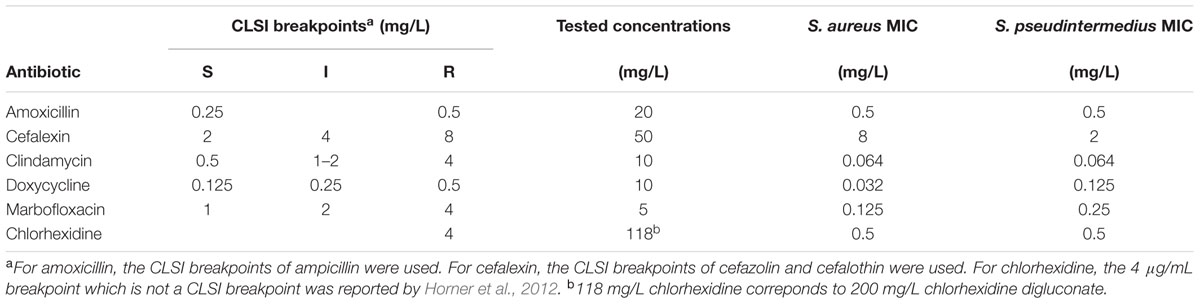
TABLE 1. CLSI and chlorhexidine breakpoints (Horner et al., 2012; CLSI, 2016), tested antibiotic or biocide concentrations on in vitro biofilms and MIC of the antibiotic drugs for the selected strains of Staphylococcus aureus and Staphylococcus pseudintermedius.
Assessment of the Effects of Antibiotics on Biofilms In vitro
After incubation of S. pseudintermedius for 7 h, the suspension contained 8.11 ± 0.33 log10 CFU/mL and the biofilm 7.89 ± 0.20 log10 CFU. For S. aureus at the same time point, the suspension contained 7.32 ± 0.04 log10 CFU/mL and the biofilm contained 8.17 ± 0.14 log10 CFU.
The antimicrobial drugs were added at that time and the bacteria were again counted after 15 h. The numbers of bacteria in the control wells (without any drug) increased slightly overnight. For S. pseudintermedius, the populations increased by 0.71 log10 CFU /mL in the suspension and by 0.85 log10 CFU in the biofilm. For S aureus, the increases were 1.15 log10 CFU/mL and 0.30 log10 CFU, respectively.
The reductions in bacterial counts, after 15 h of drug exposure, in the suspension and in the biofilm for all the tested antibiotics, compared to the control, are given in Tables 2 and 3 and represented in Figures 1 and 2 for S. aureus and S. pseudintermedius, respectively. All tested antibiotics reduced the bacterial counts in the suspension and biofilm of both strains even if some antibiotics showed extremely low activity. For most of the antibiotics, the obtained reduction of the bacterial population was less than 2 log10 CFU. Amoxicillin, cefalexin and doxycycline reduced S. pseudintermedius bacteria by only 0.67 to 0.85 log10 CFU/mL in suspension and by 0.55–0.61 log10 CFU in biofilm. For S. aureus exposed to the same three antibiotics, the reduction ranged from 0.73 to 1.10 log10 CFU/mL in suspension and from 0.50 to 0.57 log10 CFU in biofilm. Clindamycin and marbofloxacin exhibited higher activities than the other drugs, especially against S. aureus. Clindamycin reduced S. pseudintermedius biofilm by 0.75 log10 CFU and S aureus biofilm by 1.84 log10 CFU. For marbofloxacin, the reduction attained 2.9 and 5.4 log10 CFU for S. pseudintermedius and S. aureus biofilms respectively. Marbofloxacin was the only antibiotic which eradicated S. aureus in suspension and gave a final bacterial load of only 3.09 log10 CFU in the biofilm.
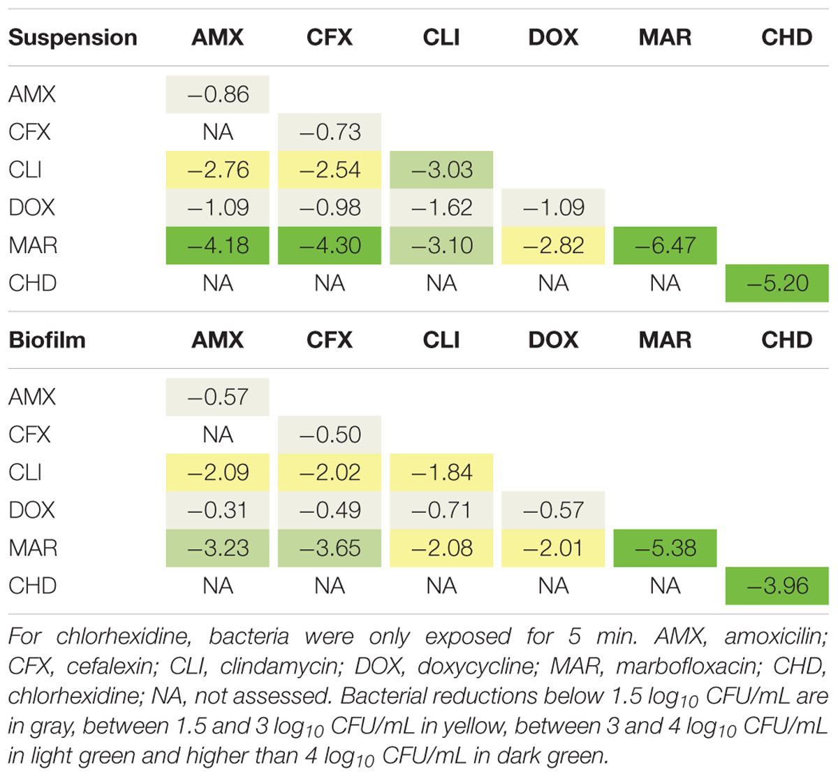
TABLE 2. Bacterial reductions of S. aureus suspension (in log10 CFU/mL) and biofilm (in log10 CFU) after 15-h exposure to one drug or to a two-drug combination.

TABLE 3. Bacterial reductions of S. pseudintermedius suspension (in log10 CFU/mL) and biofilm (in log10 CFU) after 15-h exposure to one drug or to a two-drug combination.
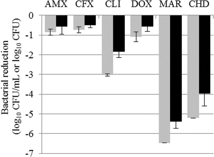
FIGURE 1. Bacterial reductions (mean ± SD) of Staphylococcus aureus suspension (gray bars, in log10 CFU/mL) and biofilm (black bars, in log10 CFU) after 15-h exposure to amoxicillin (AMX), cefalexin (CFX), clindamycin (CLI), doxycycline (DOX), or marbofloxacin (MAR) or after 5- min exposure to chlorhexidine (CHD).
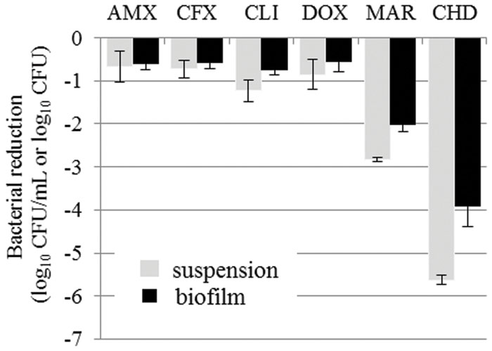
FIGURE 2. Bacterial reductions (mean ± SD) of Staphylococcus pseudintermedius suspension (gray bars, in log10 CFU/mL) and biofilm (black bars, in log10 CFU) after 15-h exposure to amoxicillin (AMX), cefalexin (CFX), clindamycin (CLI), doxycycline (DOX), or marbofloxacin (MAR) or after 5- min exposure to chlorhexidine (CHD).
As none of the tested antibiotics showed bactericidal activity against S. pseudintermedius, we then tested all possible two-drug combinations, except for amoxicillin and cefalexin which share the same bacterial target. The reductions in bacterial counts with the different combinations are shown in Tables 2 and 3. The results obtained by combining the most efficacious drugs, clindamycin and marbofloxacin, are also given in Figure 3. All combinations resulted in less bacterial eradication than the most efficacious of the two drugs tested alone, except for three specific cases where the combination was slightly better. Bacterial reduction in the S. aureus biofilm was 0.25 log10 CFU higher with both the clindamycin–amoxicillin and clindamycin–cefalexin combinations than with clindamycin alone. For S. pseudintermedius, bacterial reduction in the suspension and biofilm was higher, by 0.1 log10 CFU/mL and 0.5 log10 CFU respectively, with the marbofloxacin–amoxicillin combination than with marbofloxacin alone.
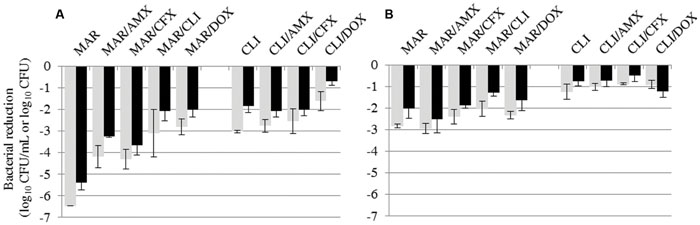
FIGURE 3. Bacterial reductions (mean ± SD) of S. aureus (A) or S. pseudintermedius (B) suspension (gray bars, in log10 CFU/mL) and biofilm (black bars, in log10 CFU) after 15-h exposure to marbofloxacin (MAR) and clindamycin (CLI) alone or in combination with amoxicillin (AMX), cefalexin (CFX), or doxycycline (DOX).
Assessment of the Effects of Chlorhexidine on Biofilms In vitro
Separate experiments were conducted to assess the activity of chlorhexidine on biofilms. We first checked that the use of water to dilute chlorhexidine did not affect the biofilms formed after 7 h incubation. The bacterial counts in the biofilms of control wells incubated for 5 min in water and rinsed twice with water were 7.24 ± 0.25 log10 CFU for S. pseudintermedius and 7.44 ± 0.15 log10 CFU for S. aureus, corresponding to differences of less than 1 log10 CFU (0.65 and 0.73 log10 CFU), as compared to control biofilms in the experiments with antibiotics. After 5 min exposure to 2% chlorhexidine, the mean reduction of S. aureus and S. pseudintermedius biofilms was 4 log10 CFU and eradication was attained in half of the experiments. All bacteria in the suspension were eradicated but the time of exposure to chlorhexidine greatly exceeded 5 min due to the time required for centrifugation before the first rinse.
Discussion
In the light of increasing multidrug resistance in zoonotic staphylococci, optimizing the therapeutic strategies utilized for canine pyoderma has become a human health issue. Although, most cases treated for S. pseudintermedius infection ultimately respond to empirical treatments, the duration of such treatments often exceeds 1 month implying a sustained selective pressure which favors the emergence of resistance in staphylococci and in other commensal bacteria (Hillier et al., 2014). As the bacteria become organized in a biofilm containing many persister cells tolerant to antibiotics (Yang et al., 2015), the predictive value of AST in determining the clinical outcome is very limited and direct assessment of the antibiotic’s activity on biofilms is required. In this study, we showed that most first and second-line antibiotics recommended for pyoderma display very low antibacterial activity on in vitro S. pseudintermedius biofilms.
In this study, S. pseudintermedius and S. aureus biofilms were tested in vitro and no account was taken of the complexity of an in vivo infection, that involves multiple factors such as the immune system, the presence of necrotic or scar tissues and possible hidden intracellular bacteria. Nevertheless, measuring antimicrobial activity on a bacterial population embedded in a biofilm and including a high proportion of persisters is likely to be more efficient in predicting the ultimate clinical outcome than standard AST performed on planktonic bacterial populations in exponential growth phase. We believe that our experiments, even if conducted with one strain per species, could be considered as a first step in the process of drug selection and drug regimen optimization for biofilm-associated infections in veterinary medicine.
After incubation without drugs for 7 or 24 h, more than 7.8 log10 CFU of the bacteria (S. pseudintermedius and S. aureus) were found in the biofilm. This rapid development was also reported in a study showing that S. pseudintermedius could form biofilm within 24 h in vitro with no difference between methicillin-susceptible and methicillin-resistant strains (Singh et al., 2013). Another study by Pompilio et al. (2015) revealed that S. pseudintermedius was able to form a well-structured biofilm consisting of multilayered, mushroom-shaped microcolonies embedded in an abundant EPS matrix. We decided to qualify the activities of our test drugs by examining their efficacies on a “young” biofilm produced after incubation for 7 h. No reduction of biofilm had been observed when the effects of the same antimicrobials had been tested on a 24 h-old biofilm (data not shown). This suggests that an environmental or phenotypic change occurred between 7 and 24 h, which had no effect on the numbers of bacteria but was able to modify their susceptibility to drugs.
We tested some of the first- and second-line drugs recommended for the systemic treatment of pyoderma in veterinary medicine (Hillier et al., 2014). We exposed S. aureus and S. pseudintermedius biofilms for 15 h to concentrations equal or slightly above the peak concentrations observed in vivo in dogs during a recommended dosage regimen. Taking plasma protein binding into account, the actual test concentrations in our system were higher than the active free in vivo concentrations in dogs, especially for doxycycline (90% bound), and we likely assessed the highest possible antibacterial activity of each drug in dogs. Even so, all of the drugs tested exhibited very low killing activity on S. pseudintermedius and S. aureus biofilms, except for the fluoroquinolone, marbofloxacin on S. aureus. Also, by comparing these two staphylococci species, we found that antibacterial activity against S. pseudintermedius tended to be lower than against S. aureus. For example, although the MIC determination classified both species as susceptible to clindamycin and marbofloxacin, bacterial killing was lower for S. pseudintermedius than for S. aureus. Then, for cefalexin, even though S. pseudintermedius was classified as susceptible and S. aureus as resistant, the efficacy against both strains was similarly low. This lower activity of antibiotic drugs on S. pseudintermedius than on S. aureus, which is not predictable by susceptibility testing, is consistent with the difficulty of eradicating pyoderma in dogs. It also implies that a human infection with this zoonotic bacterial species would be hard to eradicate. One study with a S. pseudintermedius strain isolated from a human infection demonstrated that, at concentrations 128-fold higher than the MIC, none of the antibiotics tested (which included vancomycin and linezolid) was able to eradicate a 48-h old biofilm, except for rifampicin (Pompilio et al., 2015). Interestingly, similar observations were reported with Staphylococcus epidermidis, an opportunistic human pathogen responsible for the vast majority of nosocomial catheter-related blood stream infections (Claessens et al., 2015). Even though the strain of S. epidermidis is classified as susceptible to vancomycin, based on the MIC determination, killing activity against a biofilm of the same bacteria was poor (Claessens et al., 2015).
By systematically comparing the bacterial counts in suspension and biofilm, we found that the difference between the two populations was never more than 1 log10 CFU/mL even after exposure to antimicrobial drugs, whereas planktonic bacteria are supposed to be much more readily killed by drugs (Bjarnsholt et al., 2013). This suggests the possible existence of an equilibrium between planktonic and biofilm bacteria, and that bacteria can be released from a biofilm into suspension during antibiotic exposure or that planktonic bacteria associated with a biofilm are not phenotypically the same as the planktonic bacteria exposed to drugs during a MIC determination.
Although amoxicillin, cefalexin and marbofloxacin are all classified as bactericidal in our experiments, marbofloxacin exhibited far greater activity than amoxicillin or cefalexin. This difference in bacterial killing may be due to the fact that fluoroquinolones can kill non-dividing bacteria whereas beta-lactam drugs cannot (Drlica et al., 2008). We showed in our system that the bacterial population at the time of drug challenge exceeded 7.8 log10 CFU in biofilm and 7.3 log10 CFU/mL in suspension and that very little growth occurred in the control wells between 7 and 24 h. This indicates that the bacterial population was in a stationary phase which would favor the activity of marbofloxacin over that of other drugs. The greater but limited in vitro activity of marbofloxacin on S. pseudintermedius, as compared to other drugs in our model, is difficult to connect with clinical outcomes due to the fact that blinded randomized controlled investigations of systemic antimicrobial efficacies in the treatment of canine pyoderma are very rare (Frank and Loeffler, 2012; Summers et al., 2012). However, the generally low antimicrobial activity of the tested drugs would probably be a main factor explaining the need for treatments of long duration to obtain a clinical cure of canine pyoderma and to prevent relapse.
In view of the low antibacterial effects of the first and second-line drugs used in monotherapy, we then assessed the efficacy of two-drug combinations, as is currently being applied in human medicine (Romling and Balsalobre, 2012; Claessens et al., 2015). The addition of a second drug had very slight beneficial or even negative effect on the bacterial killing. According to Jawetz laws (Jawetz, 1968, 1975), these results could perhaps have been anticipated for the combination of bactericidal and bacteriostatic drugs as for example the combination of doxycycline and cefalexin. However, the reduced bacterial activity of marbofloxacin after adding another bactericidal drug, such as amoxicillin or cefalexin, was quite unexpected even though similar results have recently been reported by Yang et al. (2015). Indeed, these authors showed that the efficacy of a combination of ciprofloxacin and vancomycin on S. epidermidis biofilm was less than that of ciprofloxacin alone (Yang et al., 2015). It can be hypothesized that the increased bacterial stress conferred by two drugs resulted in the formation of persister cells (Lewis, 2007). Until sufficient data for each possible combination becomes available, this therapeutic strategy should probably be not recommended.
The poor activity of drugs used singly in our study and the absence of additive activity when used in combination suggest that systemic treatments alone are not the best way to target Staphylococcus spp., especially S. pseudintermedius. For this reason, we also explored an alternative treatment, i.e., external use of chlorhexidine. In our system, a 2% concentration of chlorhexidine was found to kill biofilm bacteria after 5 min exposure. Biofilm bacteria of both strains were eradicated in 1 or 2 wells out of 3. These in vitro results may suggest that a topical shampoo might be more effective than most systemic treatments, the limiting condition being that the application of a chlorhexidine shampoo should come in contact with all the bacteria (treatment of entire surface with no restriction to diffusion (hairs, crusts) in order to attain the bacteria). This excellent efficacy of chlorhexidine is in agreement with another in vitro study which showed that a 4% solution of chlorhexidine killed S. pseudintermedius in less than 1 min (Lloyd and Lamport, 1999). Several clinical studies have also provided evidence of the efficacy of chlorhexidine at least for superficial pyoderma (Bryan et al., 2012; Mueller et al., 2012) and similar efficacy was observed when a topical chlorhexidine digluconate shampoo treatment applied twice weekly for 4 weeks was compared with a systemic administration of amoxicillin–clavulanic acid 25 mg/kg twice daily for 4 weeks (Borio et al., 2015). In addition to the killing activity of chlorhexidine on pathogenic bacteria, a topical treatment has the advantage of not impacting the digestive commensal flora and therefore reducing the selection pressure for resistance in this microbiota.
Although, we did not investigate the development of resistance in the targeted staphylococci all of the tested antibiotic or biocide treatments can potentially induce resistance. The proportion of Methicillin Resistant S. pseudintermedius (MRSP) among S. pseudintermedius isolates from clinical infections in the USA and Europe has risen since 2000 (Frank and Loeffler, 2012) and a link between antimicrobial treatments within 30 days and MRSP infections has been identified in dogs (Weese et al., 2012). Biocide resistances can also develop during chlorhexidine treatment (Johnson et al., 2015). Resistance to chlorhexidine can be conferred by carriage of qac A/B or smr genes conding for efflux pumps. Interestingly, one study reported that, among 247 strains, the qac A/B and smr positive bacteria were more often resistant to some antimicrobial drugs including methicillin, ciprofloxacin, and vancomycin than negative ones (McNeil et al., 2015). This situation confirms the urgent need to implement the stewardship for antibiotic use in veterinary medicine.
Conclusion
This study provides further evidence that S. pseudintermedius and S. aureus biofilms could be highly tolerant to veterinary drugs. Taking in mind that further in vivo investigations on efficacy and resistance development are required, the topical administration of chlorhexidine could provide a promising alternative strategy avoiding the long-term systemic use of inefficacious antimicrobial drugs in animals.
Author Contributions
Substantial contributions to the conception or design of the work and the acquisition, analysis, or interpretation of data for the work; AF, JL, P-LT, AB-M. Drafting the work and revising it critically for important intellectual content; AF, JL, P-LT, AB-M. Final approval of the version to be published; AF, JL, P-LT, AB-M; Agreement to be accountable for all aspects of the work in ensuring that questions related to the accuracy and integrity of any part of the work are appropriately investigated and resolved; AF, JL, P-LT, AB-M.
Conflict of Interest Statement
The authors declare that the research was conducted in the absence of any commercial or financial relationships that could be construed as a potential conflict of interest.
References
Batzias, G. C., Delis, G. A., and Athanasiou, L. V. (2005). Clindamycin bioavailability and pharmacokinetics following oral administration of clindamycin hydrochloride capsules in dogs. Vet. J. 170, 339–345. doi: 10.1016/j.tvjl.2004.06.007
Beco, L., Guaguere, E., Lorente Mendez, C., Noli, C., Nuttall, T., and Vroom, M. (2013). Suggested guidelines for using systemic antimicrobials in bacterial skin infections: part 2- - antimicrobial choice, treatment regimens and compliance. Vet. Rec. 172, 156–160. doi: 10.1136/vr.101070
Bjarnsholt, T., Ciofu, O., Molin, S., Givskov, M., and Hoiby, N. (2013). Applying insights from biofilm biology to drug development - can a new approach be developed? Nat. Rev. Drug Discov. 12, 791–808. doi: 10.1038/nrd4000
Borio, S., Colombo, S., La Rosa, G., De Lucia, M., Damborg, P., and Guardabassi, L. (2015). Effectiveness of a combined (4% chlorhexidine digluconate shampoo and solution) protocol in MRS and non-MRS canine superficial pyoderma: a randomized, blinded, antibiotic-controlled study. Vet. Dermatol. 26:e372. doi: 10.1111/vde.12233
Bryan, J., Frank, L. A., Rohrbach, B. W., Burgette, L. J., Cain, C. L., and Bemis, D. A. (2012). Treatment outcome of dogs with meticillin-resistant and meticillin-susceptible Staphylococcus pseudintermedius pyoderma. Vet. Dermatol. 23:e365. doi: 10.1111/j.1365-3164.2012.01034.x
Claessens, J., Roriz, M., Merckx, R., Baatsen, P., Van Mellaert, L., and Van Eldere, J. (2015). Inefficacy of vancomycin and teicoplanin in eradicating and killing Staphylococcus epidermidis biofilms in vitro. Int. J. Antimicrob. Agents 45, 368–375. doi: 10.1016/j.ijantimicag.2014.11.011
CLSI (2006). Methods for Dilution Antimicrobial Susceptibility Tests for Bacteria That Grow Aerobically, 7th Edn: Approved Standard M7-A7. Wayne, PA: CLSI.
CLSI (2016). VET01-S Performance Standards for Antimicrobial Disk and Dilution Susceptibility Tests for Bacteria Isolated From Animals; 3rd Edn. Wayne, PA: CLSI.
Costerton, J. W., Stewart, P. S., and Greenberg, E. P. (1999). Bacterial biofilms: a common cause of persistent infections. Science 284, 1318–1322. doi: 10.1126/science.284.5418.1318
Davies, D. (2003). Understanding biofilm resistance to antibacterial agents. Nat. Rev. Drug Discov. 2, 114–122. doi: 10.1038/nrd1008
Drlica, K., Malik, M., Kerns, R. J., and Zhao, X. (2008). Quinolone-mediated bacterial death. Antimicrob. Agents Chemother. 52, 385–392. doi: 10.1128/AAC.01617-06
Ferran, A. A., Toutain, P. L., and Bousquet-Melou, A. (2014). Comparison of the reduction in the antibacterial potency of a fluoroquinolone conferred by a single mutation in the quinolone resistance-determining region or by the inoculum size effect. Int. J. Antimicrob. Agents 44, 472–474. doi: 10.1016/j.ijantimicag.2014.07.007
Frank, L. A., and Loeffler, A. (2012). Meticillin-resistant Staphylococcus pseudintermedius: clinical challenge and treatment options. Vet. Dermatol. 23:e256. doi: 10.1111/j.1365-3164.2012.01047.x
Guardabassi, L., Loeber, M. E., and Jacobson, A. (2004). Transmission of multiple antimicrobial-resistant Staphylococcus intermedius between dogs affected by deep pyoderma and their owners. Vet. Microbiol. 98, 23–27. doi: 10.1016/j.vetmic.2003.09.021
Hillier, A., Lloyd, D. H., Weese, J. S., Blondeau, J. M., Boothe, D., Breitschwerdt, E., et al. (2014). Guidelines for the diagnosis and antimicrobial therapy of canine superficial bacterial folliculitis (Antimicrobial Guidelines Working Group of the International Society for Companion Animal Infectious Diseases). Vet. Dermatol. 25, e142–e163. doi: 10.1111/vde.12118
Horner, C., Mawer, D., and Wilcox, M. (2012). Reduced susceptibility to chlorhexidine in staphylococci: is it increasing and does it matter? J. Antimicrob. Chemother. 67, 2547–2559. doi: 10.1093/jac/dks284
Jawetz, E. (1968). The use of combinations of antimicrobial drugs. Annu. Rev. Pharmacol. 8, 151–170. doi: 10.1146/annurev.pa.08.040168.001055
Jawetz, E. (1975). Actions of antimicrobial drugs in combination. Vet. Clin. North Am. 5, 35–50. doi: 10.1016/S0091-0279(75)50002-X
Johnson, N. R., Kazoullis, A., Bobinskas, A. M., Jones, L., Hutmacher, D. W., and Lynham, A. (2015). Bacterial comparison of preoperative rinsing and swabbing for oral surgery using 0.2% chlorhexidine. J. Investig. Clin. Dent. 6, 193–196. doi: 10.1111/jicd.12099
KuKanich, K., and KuKanich, B. (2015). The effect of sucralfate tablets vs. suspension on oral doxycycline absorption in dogs. J. Vet. Pharmacol. Ther. 38, 169–173. doi: 10.1111/jvp.12165
Kung, K., and Wanner, M. (1994). Bioavailability of different forms of amoxycillin administered orally to dogs. Vet. Rec. 135, 552–554.
Lewis, K. (2007). Persister cells, dormancy and infectious disease. Nat. Rev. Microbiol. 5, 48–56. doi: 10.1038/nrmicro1557
Lloyd, D. H., and Lamport, A. I. (1999). Activity of chlorhexidine shampoos in vitro against Staphylococcus intermedius, Pseudomonas aeruginosa and Malassezia pachydermatis. Vet. Rec. 144, 536–537. doi: 10.1136/vr.144.19.536
Loeffler, A., Linek, M., Moodley, A., Guardabassi, L., Sung, J. M., Winkler, M., et al. (2007). First report of multiresistant, mecA-positive Staphylococcus intermedius in Europe: 12 cases from a veterinary dermatology referral clinic in Germany. Vet. Dermatol. 18, 412–421. doi: 10.1111/j.1365-3164.2007.00635.x
McNeil, J. C., Kok, E. Y., Vallejo, J. G., Campbell, J. R., Hulten, K. G., Mason, E. O., et al. (2015). Clinical and molecular features of decreased chlorhexidine susceptibility among nosocomial Staphylococcus aureus isolates at Texas Children’s Hospital. Antimicrob. Agents Chemother. 60, 1121–1128. doi: 10.1128/AAC.02011-15
Mueller, R. S., Bergvall, K., Bensignor, E., and Bond, R. (2012). A review of topical therapy for skin infections with bacteria and yeast. Vet. Dermatol. 23:e362. doi: 10.1111/j.1365-3164.2012.01057.x
Pompilio, A., De Nicola, S., Crocetta, V., Guarnieri, S., Savini, V., Carretto, E., et al. (2015). New insights in Staphylococcus pseudintermedius pathogenicity: antibiotic-resistant biofilm formation by a human wound-associated strain. BMC Microbiol. 15:109. doi: 10.1186/s12866-015-0449-x
Rantala, M., Holso, K., Lillas, A., Huovinen, P., and Kaartinen, L. (2004). Survey of condition-based prescribing of antimicrobial drugs for dogs at a veterinary teaching hospital. Vet. Rec. 155, 259–262. doi: 10.1136/vr.155.9.259
Romling, U., and Balsalobre, C. (2012). Biofilm infections, their resilience to therapy and innovative treatment strategies. J. Intern. Med. 272, 541–561. doi: 10.1111/joim.12004
Rubinstein, E., and Keynan, Y. (2013). Short-course therapy for severe infections. Int. J. Antimicrob. Agents 42(Suppl. ), S22–S24. doi: 10.1016/j.ijantimicag.2013.04.013
Schneider, M., Thomas, V., Boisrame, B., and Deleforge, J. (1996). Pharmacokinetics of marbofloxacin in dogs after oral and parenteral administration. J. Vet. Pharmacol. Ther. 19, 56–61. doi: 10.1111/j.1365-2885.1996.tb00009.x
Silley, P., Rudd, A. P., Symington, W. M., and Tait, A. J. (1988). Pharmacokinetics of cephalexin in dogs and cats after oral, subcutaneous and intramuscular administration. Vet. Rec. 122, 15–17. doi: 10.1136/vr.122.1.15
Singh, A., Walker, M., Rousseau, J., and Weese, J. S. (2013). Characterization of the biofilm forming ability of Staphylococcus pseudintermedius from dogs. BMC Vet. Res. 9:93. doi: 10.1186/1746-6148-9-93
Summers, J. F., Brodbelt, D. C., Forsythe, P. J., Loeffler, A., and Hendricks, A. (2012). The effectiveness of systemic antimicrobial treatment in canine superficial and deep pyoderma: a systematic review. Vet. Dermatol. 23, 305–329, e61. doi: 10.1111/j.1365-3164.2012.01050.x
Van Hoovels, L., Vankeerberghen, A., Boel, A., Van Vaerenbergh, K., and De Beenhouwer, H. (2006). First case of Staphylococcus pseudintermedius infection in a human. J. Clin. Microbiol. 44, 4609–4612. doi: 10.1128/JCM.01308-06
Weese, J. S., Faires, M. C., Frank, L. A., Reynolds, L. M., and Battisti, A. (2012). Factors associated with methicillin-resistant versus methicillin-susceptible Staphylococcus pseudintermedius infection in dogs. J. Am. Vet. Med. Assoc. 240, 1450–1455. doi: 10.2460/javma.240.12.1450
Keywords: biofilm, antimicrobial activity, Staphylococcus aureus, Staphylococcus pseudintermedius, veterinary antimicrobials
Citation: Ferran AA, Liu JJ, Toutain P-L and Bousquet-Mélou A (2016) Comparison of the In vitro Activity of Five Antimicrobial Drugs against Staphylococcus pseudintermedius and Staphylococcus aureus Biofilms. Front. Microbiol. 7:1187. doi: 10.3389/fmicb.2016.01187
Received: 20 April 2016; Accepted: 18 July 2016;
Published: 02 August 2016.
Edited by:
Manuela Caniça, National Institute of Health Dr. Ricardo Jorge, PortugalReviewed by:
Atte Von Wright, University of Eastern Finland, FinlandXian-Zhi Li, Health Canada, Canada
Copyright © 2016 Ferran, Liu, Toutain and Bousquet-Mélou. This is an open-access article distributed under the terms of the Creative Commons Attribution License (CC BY). The use, distribution or reproduction in other forums is permitted, provided the original author(s) or licensor are credited and that the original publication in this journal is cited, in accordance with accepted academic practice. No use, distribution or reproduction is permitted which does not comply with these terms.
*Correspondence: Aude A. Ferran, a.ferran@envt.fr