 Ying Qi1*
- 1 Center for Anti-Aging Research, Shanghai R&D, Nu Skin Enterprises, Shanghai, China
- 2 Center for Anti-Aging Research, Nu Skin Enterprises, Provo, UT, USA
To examine the composition of lanostanoids in Ganoderma lucidum, we have developed a liquid chromatography–mass spectrometry (LC–MS) method by using the ganoderic acids isolated in our laboratory as reference standards. The identity of 14 peaks in the high performance liquid chromatogram (HPLC) of G. lucidum has been confirmed. By using the HPLC retention times of these ganoderic acids and their mass fragmentation patterns established in this paper, one can use LC–MS to analyze G. lucidum without requiring the reference standards of these 14 ganoderic acids. Subsequently, only the HPLC–UV method would be needed to analyze routine samples of G. lucidum.
Introduction
In our ongoing investigations of Traditional Chinese Medicine (TCM) for dietary supplements we are particularly interested in the medicinal mushroom Ganoderma lucidum (W. Curt.:Fr.) Karst because it is a rich producer of lanostanoids. This class of triterpenes has been reported to have distinct pharmacological activities. These include angiotensin-converting-enzyme inhibition, anti-HIV action, antinociceptic action, histamine release inhibition, HMG-CoA reductase inhibition, and liver function stimulation (Mekenna, 1998). The prevalent usage of G. lucidum by cancer patients in Asia also persuaded us to further investigate the active components of G. lucidum. Indeed, we found that the extract of G. lucidum prepared in our labs induced the modulation of secretion from normal human peripheral blood mononuclear leukocytes of cytokines IL-2, IL-4, and IFN-γ (Ma et al., 2002).
Solvent partitioning and repeated chromatography, followed by crystallization resulted in the isolation of 32 lanostanoids from the lipophilic extract of the fruiting body of G. lucidum. These include six previously unknown oxygenated lanostanoids: 8β, 9α-dihydroganoderic acid J (1), methyl 8β, 9α-dihydroganoderate J (2), 20-hydroxylganoderic acid G (3) (Ma et al., 2002), ganoderic acid Sz (4) (Li et al., 2005), 7-oxo-ganoderic acid Z (5), and 15-hydroxy-ganoderic acid S (6) (Figure 1). Compounds 5 and 6 both exhibited inhibitory activities against HMG-CoA reductase and acyl CoA-acyltransferase (Li et al., 2006). We have also isolated 26 known lanostanoids, including ganoderic acids I (7) and C2 (8), 12-deacetylganoderic acid H (9), ganoderic acid G (10), ganoderenic acid B (11), ganoderic acids B (12), AM1 (13), A (14), and H (15), ganoderenic acid D (16), ganoderic acids C (17), F (18), J (19), DM (20), S and Y, methyl ganoderate I, ganoderiols B and F, ganoderol B, ganodermic acid Q, ganodermenonol, ganodermanondiol, ganodermanontriol, lucidone A, and lucidenic A (Ma et al., 2002; Li et al., 2005, 2006; and references cited therein). Their structures were determined by using 1D and 2D NMR and MS spectroscopic methods.
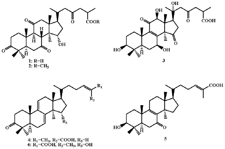
Figure 1. Structures of oxygenated lanostanoids.
Ganoderma lucidum has usually been taken orally as mushroom powder or in its extract form, whether as prescribed by TCM doctors or in modern pill form. There is a need to understand the composition of the natural material ingested. Liquid chromatography–mass spectrometry (LC–MS) has been used wildly to characterize and standardize triterpenes in mushrooms. However, the availability of only limited number of commercial reference standards of lanostanoids has diminished the number of triterpenes to be analyzed (Adamec et al., 2009; Liu et al., 2011). It is not practical to expect every single lab to obtain reference standards by going through laborious and time-consuming steps of isolating and characterizing each individual lanostanoid. The aim of this work is to develop a rapid and conclusive LC–MS method for identification of major triterpenes in Ganoderma mushrooms by using the ganoderic acid samples we isolated.
Materials and Methods
General Experimental Procedures
Melting points were recorded on a Fisher-Johns melting point apparatus. Optical rotations were recorded on a WZZ-1S automatic polarimeter. UV spectra were recorded on a HP-8453 spectrophotometer. IR spectra were recorded on a Nicolet Magna FTIR-750 spectrometer. One- and two-dimensional NMR spectra were recorded on a Bruker AMX 400 spectrometer. Acetonitrile used in LC–MS analysis was supplied by Sigma-Aldrich Co. LLC. Other reagents were all supplied by Sinopharm Chemical Reagent Co. Ltd (Shanghai).
Fungal Material
The mushroom of G. lucidum was identified by Professor Guanyun Gu, School of Pharmacy, Fudan University, Shanghai, People’s Republic of China. A voucher specimen (GL-9807) has been deposited at the Department of Pharmacognosy, Fudan University, Shanghai, People’s Republic of China.
IL-2, IL-4, and IFN-γ Assays
The sample was dissolved in a 0.4% DMSO solution, then incubated with normal human peripheral blood mononuclear leukocytes in a RPMI 1640 buffer containing 10% FBS, 50 unit/mL penicillin, and 50 μg/mL streptomycin at 37°C for 16 h. Levels of IL-2, IL-4, and IFN-γ were measured using ELISA (Welker et al., 1996; Ma et al., 2002).
HMG-CoA Reductase Assay
A phosphate buffer solution contains 100 mM potassium phosphate (pH 7.5), 8 mM G-6-P, 1 mM NADP, 4 mM EDTA, 2 mM DTT, and 0.6 U G-6-P-DH was prepared. The compound was dissolved in a 1% DMSO solution and then pre-incubated with microsomal protein in the phosphate buffer solution at 37°C for 15 min. The reaction was started by the addition of 2.5 μM [14C]HMG-CoA reductase and the reaction was run at 37°C for 15 min. [14C]mevalonate was then quantified. It was determined that compounds 1 and 2 inhibited HMG-CoA reductase with IC50 = 22.3 and 21.7 μM, respectively (Heller and Gould, 1973; Kubo and Strott, 1987; Li et al., 2006).
Acyl CoA-Cholesterol Acyltransferase Assay
The compound was dissolved in a 1% DMSO solution and pre-incubated with Wistar rat hepatic microsomes in the 0.2-M phosphate buffer at 37°C for 15 min. The reaction was started by the addition of 18 μM [14C]palmitoyl CoA-acyltransferase and the reaction was run at 37°C for 15 min. [14C]cholesterol ester was then quantified. The inhibitory activities, IC50, of compounds 1 and 2 against CoA-acyltransferase were determined to be 5.5 and 47.3 μM, respectively (Largis et al., 1989; Li et al., 2006).
Ganoderic Acids
Ganoderma lucidum mushroom fruiting bodies (10 kg) were chipped and extracted with 20 L of 95% ethanol at 80°C three times. After filtering and removing of ethanol, the resulting extract was applied onto a chromatographic silica gel column and eluted with a chloroform/acetone gradient system, followed by a reversed-phase C-18 column chromatographic run with water/methanol. The final purification of the ganoderic acids were accomplished by further high performance liquid chromatogram (HPLC) separation and re-crystallization. Their structure were identified and confirmed by NMR and MS analyses (Ma et al., 2002; Li et al., 2005, 2006). The reference standard solution of each ganoderic acid was prepared by dissolving 1 mg of ganoderic acid in 1 mL of methanol.
Ganoderma lucidum Extract
A G. lucidum extract (ReishiMax GLp™ 500 mg) was introduced into 25 mL of ethyl acetate, sonicated for 30 min, and filtered. The remaining insoluble material was extracted two more times as above. Three filtrates were combined and the solvent was removed under vacuum. The resulting residue was dissolved into 10 mL of methanol for LC–MS analysis.
LC–MS Analysis
The HPLC system was controlled by an Agilent MassChrom data system. Chromatographic separation was performed on a Phenomenex luna C-18 column (5 μm, 250 mm × 4.6 mm. I.D.) from Phenomenex Co. Ltd. A step gradient solvent system was used: 0 min CH3CN/0.1% aqueous CH3COOH (25/75) → 35 min CH3CN/0.1% aqueous CH3COOH (35/65) → 45 min CH3CN/0.1% aqueous CH3COOH (45/55) → 90 min CH3CN with a constant flow rate at 1.0 mL/min. Column temperature was set at 30°C controlled by a column heater–cooler HP 1100. The elution was measured at the wavelength range of 200–500 nm by using a photodiode array detector. Agilent triple-quadrupole ion-trap mass spectrometer was equipped with an atmospheric pressure chemical ionization interface (APCI). The mass spectrometer was mass-calibrated with a polyglycol solution. In order to achieve maximum sensitivity the mass spectrometer conditions were set at 450°C of APCI temperature, 60 psi of nebulizing gas flow rate, 3500 V of capillary voltage, and 325°C of capillary temperature. Full scam spectra from m/z 100 to m/z 1000 Da were obtained in the positive ion mode.
Results and Discussion
An HPLC method was developed to analyze the lipophilic part of the commercial product ReishiMax GLp™. Ganoderic acids were separated by a C-18 column with a step gradient solvent system of CH3CN/0.1% aqueous CH3COOH. Figure 2 showed that the chromatogram of the column eluants measured at the wavelength of 252 nm. Subsequent LC–MS analysis with an ion-trap using APCI ionization technique was followed.
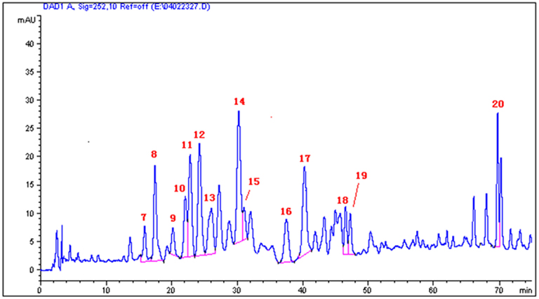
Figure 2. High performance liquid chromatogram of the extract of G. lucidum.
The MS spectra of ganoderic acids 7–20 were established by LC–MS. The APCI mass spectra of these compounds exhibited peaks at m/z [M-nH2O + H]+ and [M-nH2O + H-130]+. The loss of 130 Da in the mass spectra represents the characteristic cleavage of the αβ bond (C20–C22) from the C = O in the side chain (Figure 3). The absence of these prominent fragments of [M-nH2O + H-130]+ in ganoderenic acids B (11) and D (16), and ganoderic acid DM (20) were expected due to the absence of C = O at C23. HPLC retention times (tR) of ganoderic acids 7–20, along with their mass fragments, were established and listed in the Table 1.
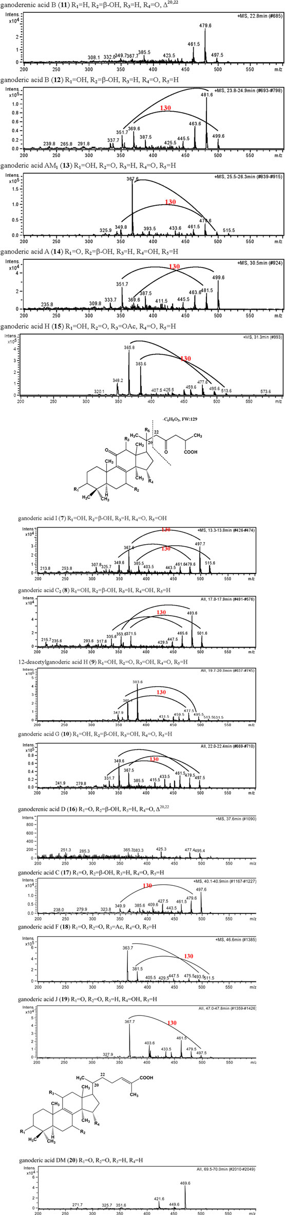
Figure 3. Mass spectra of the major ganoderic acids (7–20) in G. lucidum.
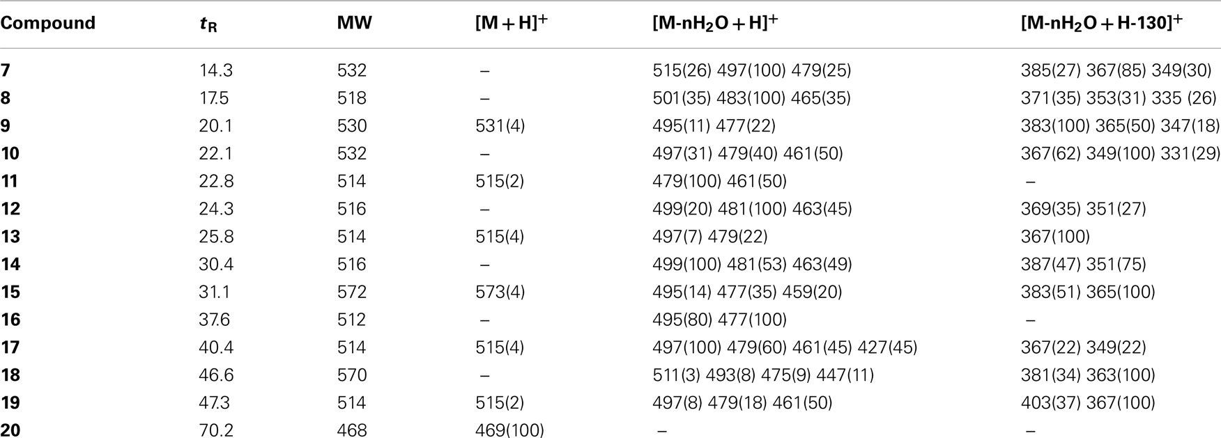
Table 1. Mass fragments and HPLC retention times of ganoderic acids (7–20).
By comparison of the HPLC retention times and mass spectra of the test sample and reference standards of ganoderic acids (Table 1; Figure 3), the identities of 14 major peaks in the chromatogram of ReishiMax GLP™ were confirmed. Thus, one can easily use this approach by performing LC–MS analysis on its samples and then comparing with the data in the Table 1 and mass spectra in Figure 3 in this paper to establish the identity of each chromatographic peak. Subsequently, only HPLC will be needed for routine analysis of G. lucidum. This method has been used in our laboratories and extraction factory to examine the quality and quantity of the triterpene composition in incoming mushrooms, crude extracts, finished goods, and competitor products. Our QC lab has used the HPLC chromatograms as sample fingerprints for the cGMP compliant purpose. We believe this method could also be an effective tool for researchers in future studies of other related Ganoderma species.
Conflict of Interest Statement
The authors declare that the research was conducted in the absence of any commercial or financial relationships that could be construed as a potential conflict of interest.
Acknowledgments
We are grateful to Dr. Jiyuan Ma, Mr. Canjun Li, Ms. Qing Ye, Mr. Jianhua Yin, and Mr. Yanjun Hua for their work in isolation and characterization of ganoderic acids described in this paper.
References
Adamec, J., Jannasch, A., Sudhgaonkar, S., Jedinak, A., Sedlak, M., and Sliva, D. (2009). Development of a new method for improved identification and relative quantification of unknown metabolites in complex samples: determination of a triterpenoid metabolic fingerprint for the in situ characterization of Ganoderma bioactive compounds. J. Sep. Sci. 32, 4052–4058.
Heller, R. A., and Gould, R. G. (1973). Solubilization and partial purification of hepatic 3-hydroxy-3-methylglutaryl coenzyme A reductase. Biochem. Biophys. Res. Commun. 50, 859–865.
Kubo, M., and Strott, C. A. (1987). Differential activity of 3-hydroxy-3-methylglutaryl coenzyme A reductase in zones of the adrenal cortex. Endocrinology 120, 214–221.
Largis, E. E., Wang, C. H., DeVries, V. G., and Schaffer, S. A. (1989). CL 277,082: a novel inhibitor of ACAT-catalyzed cholesterol esterification and cholesterol absorption. J. Lipid Res. 30, 681–690.
Li, C., Li, Y., and Sun, H. H. (2006). New ganoderic acids, bioactive triterpenoid metabolites from the mushroom Ganoderma lucidum. Nat. Prod. Res. 20, 985–991.
Li, C., Yin, J., Guo, G., Zhang, D., and Sun, H. H. (2005). Ganoderic acid Sz, a new lanostanoid from the mushroom Ganoderma lucidum. Nat. Prod. Res. 19, 461–465.
Liu, Y., Liu, Y., Qiu, F., and Di, X. (2011) Sensitive and selective liquid chromatography-tandem mass spectrometry method for the determination of five ganoderic acids in Ganoderma lucidum and its related species. J. Pharm. Biomed. Anal. 54, 717–721.
Ma, J., Ye, Q., Hua, Y., Zhang, D., Cooper, R., Chang, M. N., Chang, J. Y., and Sun, H. H. (2002). New lanostanoids from the mushroom Ganoderma lucidum. J. Nat. Prod. 65, 72–75.
Mekenna, D. J. (ed.). (1998). Natural Dietary Supplements: A Desktop Reference. St. Croix: Institute for Natural Products Research.
Keywords: mushroom, Ganoderma lucidum, lanostanoids, triterpenes, ganoderic acids, LC–MS
Citation: Qi Y, Zhao L and Sun HH (2012) Development of a rapid and confirmatory method to identify ganoderic acids in Ganoderma mushrooms. Front. Pharmacol. 3:85. doi: 10.3389/fphar.2012.00085
Received: 30 November 2011; Paper pending published: 25 January 2012;
Accepted: 16 April 2012; Published online: 07 May 2012.
Edited by:
Xiao-Ling Zhu, Peking University Health Science Center, ChinaReviewed by:
Shu-Hui Zhang, Shanghai University of Traditional Chinese Medicine, ChinaXiao-Ling Zhu, Peking University Health Science Center, China
Copyright: © 2012 Qi, Zhao and Sun. This is an open-access article distributed under the terms of the Creative Commons Attribution Non Commercial License, which permits non-commercial use, distribution, and reproduction in other forums, provided the original authors and source are credited.
*Correspondence: Ying Qi, Center for Anti-Aging Research, Shanghai R&D, Nu Skin Enterprises, No. 116, Building 11, Lane 572, Bi Bo Road, Shanghai 201203, China. e-mail: yingqi@cn.nuskin.com; Hao H. Sun, Center for Anti-Aging Research, Nu Skin Enterprises, 75 West Center Street, Provo, UT 84601, USA. e-mail: hsun@nuskin.com