 Susana Merino
 Eleonora Aquilini
 Kelly M. Fulton2
 Susan M. Twine
 Juan M. Tomás
- 1Departamento de Microbiología, Facultad de Biología, Universidad de Barcelona, Barcelona, Spain
- 2National Research Council, Ottawa, ON, Canada
Plesiomonas shigelloides is the unique member of the Enterobacteriaceae family able to produce polar flagella when grow in liquid medium and lateral flagella when grown in solid or semisolid media. In this study on P. shigelloides 302-73 strain, we found two different gene clusters, one exclusively for the lateral flagella biosynthesis and the other one containing the biosynthetic polar flagella genes with additional putative glycosylation genes. P. shigelloides is the first Enterobacteriaceae were a complete lateral flagella cluster leading to a lateral flagella production is described. We also show that both flagella in P. shigelloides 302-73 strain are glycosylated by a derivative of legionaminic acid (Leg), which explains the presence of Leg pathway genes between the two polar flagella regions in their biosynthetic gene cluster. It is the first bacterium reported with O-glycosylated Leg in both polar and lateral flagella. The flagella O-glycosylation is essential for bacterial flagella formation, either polar or lateral, because gene mutants on the biosynthesis of Leg are non-flagellated. Furthermore, the presence of the lateral flagella cluster and Leg O-flagella glycosylation genes are widely spread features among the P. shigelloides strains tested.
Introduction
Plesiomonas shigelloides is a Gram-negative bacilli flagellated bacterium. This facultative anaerobic bacterium is ubiquitous, has been isolated from different water sources (freshwater or surface water), and animals (wild and domestic; Farmer et al., 1992). In humans, P. shigelloides is associated with diarrheal disease in humans (Brenden et al., 1988). Sometimes could also be the cause of gastroenteritis, including acute secretory gastroenteritis (Mandal et al., 1982), an invasive shigellosis-like disease (McNeeley et al., 1984), and a cholera-like illness (Tsukamoto et al., 1978). Extra intestinal infections, such as meningitis, bacteremia (Billiet et al., 1989), and pseudoappendicitis (Fischer et al., 1988), are also associated with P. shigelloides infection. Of particular concern are the severe cases of meningitis and bacteremia (Fujita et al., 1994) caused by P. shigelloides.
Plesiomonas shigelloides was initially classified in the Vibrionaceae family; however, molecular studies by Martinez-Murcia et al. (1992) indicated that is related to the enterobacterial genus Proteus phylogenetically. Huys and Sings (1999) during studies of Aeromonas spp. genotyping using by the amplified fragment length polymorphism found that P. shigelloides clearly falls out of the major Aeromonas cluster. According to these features the genus Plesiomonas was reclassified to the family Enterobacteriaceae, being the only oxidase-positive member of this family (Garrity et al., 2001). In order to distinguishing different strains of P. shigelloides, two major serotyping schemes, one based on O-antigen lipopolysaccharide (O) and the other one on flagellar (H) antigens. With a total of 102 somatic antigens and 51 flagellar antigens recognized (Aldova and Shimada, 2000).
The flagella biosynthesis, in terms of resources and energy, is a costly commitment for the bacterium (Macnab, 1996). The flagella number is variable, and the distribution most frequently found on pathogenic bacteria are monotrichous (single flagellum) or pertitrichous (multiple flagella around the cell; Macnab, 1996). The flagella expression is dependable of the growth conditions. When grown in plates, several bacterial species produced more flagella than when they grow in liquid medium. Some species, like Proteus mirabilis, have been observed to show an increase in the numbers of flagella. Vibrio parahaemolyticus, have a single polar flagellum in liquid medium, instead when grown on solid medium, produces the polar flagellum (Fla) and peritrichous (or lateral) flagella (Laf; Allison and Hughes, 1991; Allison et al., 1992; Merino et al., 2014). Lateral flagella, were shown in about seven other Vibrio species (some of which evokes a disease spectrum similar to V. parahaemolyticus; Shinoda et al., 1992), while only a reduced number of bacterial species, including Rhodospirillum centenum (a purple photosynthetic bacterium; McClain et al., 2002), Azospirillum spp. (nitrogen-fixing rhizobacteria that colonize plants; Moens et al., 1996), Helicobacter mustelae (the causative agent of chronic gastritis and ulcer disease in ferrets; O’Rourke et al., 1992), P. shigelloides (Inoue et al., 1991), and Aeromonas spp. (opportunistic and gastroenteric pathogens of man; Gavín et al., 2002). Other species that show lateral flagella include Bradyrhizobium japonicum (Kanbe et al., 2007), Photobacterium profundum (Eloe et al., 2008), and Rhodobacter sphaeroides (Poggio et al., 2007). Furthermore, Selenomonas ruminantium subsp. lactilytica is a solely laterally flagellate bacterium (Haya et al., 2011).
Protein glycosylation is one of the most common protein post-translational modifications and consists in the covalent attachment of carbohydrates to amino acids. This mechanism was thought to occur exclusively in eukaryotes. However, protein glycosylation systems have been identified in all forms of life including prokaryotes. N-glycosylation is the covalent linkage to asparagine residues of carbohydrates, while O-glycosylation to serine or threonine residues. O-glycosylation in bacteria has been largely reviewed recently (Iwashkiw et al., 2013). As more bacterial genomes are now available together with bioinformatic analysis coupled with functional analysis, the elucidation of glycosylation pathways achieved increasing, including the identification of many genes that participate in flagellin glycosylation (Iwashkiw et al., 2013). The number of O-glycosylation genes involved is diverse in each bacterial species (Goon et al., 2003; Schirm et al., 2003; Faridmoayer et al., 2007; Iwashkiw et al., 2012). In spite of these advances, the knowledge of glycans structure and composition of which modify from Gram-negative bacteria flagellins is restricted to certain species and has been observed to be strain-dependent [as reviewed by Merino and Tomás (2014)].
In this work we study the genetics of P. shigelloides flagella (polar and lateral), and their flagella post-translational modifications, the first report of flagellar glycosylation in enteric bacteria.
Materials and Methods
Bacterial Strains, their Growth Conditions, and Plasmids Used
The bacterial strains, as well as the plasmids used, are listed on Table 1. Bacteria were grown in TSB broth and TSA medium supplemented if necessary with kanamycin (25 μg/ml), tetracycline (20 μg/ml), and rifampicin (100 μg/ml) when needed.
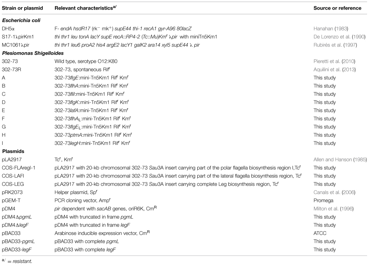
TABLE 1. Bacterial strains and plasmids used.
MiniTn5Km-1 Mutagenesis
Conjugal transfer of miniTn5Km-1 transposition element from Escherichia coli S17-1λpirKm-1 to P. shigelloides 302-73R (wild type strain rifampicin-resistant) was carried out in a conjugal drop as previously described (Aquilini et al., 2013).
Construction of a P. shigelloides Genomic Library
Plesiomonas shigelloides strain 302-73 (serotype O1) genomic DNA was isolated and partially digested with Sau3A as described by Sambrook et al. (1989). The P. shigelloides strain 302-73 genomic library, using cosmid pLA2917 (Allen and Hanson, 1985), was performed as described (Guasch et al., 1996).
General DNA Methods
General DNA manipulations were done essentially as previously described described (Sambrook et al., 1989; Aquilini et al., 2014).
Southern Blot Hybridizations
Southern blotting was performed by capillary transfer (Sambrook et al., 1989) from the gel to a nylon membrane (Hybond N1, Amersham). Probe labeling, hybridization, and detection were carried out as previously described (Aquilini et al., 2014) using the enhanced chemiluminescence labeling and detection system (Amersham) according to the manufacturer’s instructions.
DNA Sequencing and In Silico Analysis of Sequence Data
These studies were previously described (Wilhelms et al., 2013). The dideoxy-chain termination method (Sanger et al., 1977), BLAST (Altschul et al., 1997; Bateman et al., 2002), and Clustal W were used.
Complementation Studies
Complementation of the different mutants carrying the miniTn5 was done as previously described (Aquilini et al., 2013) by conjugal transfer of positive recombinant clones from the genomic library.
Antisera
Anti-P. shigelloides polar flagellum and lateral flagella serum were independently obtained using purified polar flagellum or lateral flagella obtained after cesium chloride, and assayed as previously described for other surface molecules (Tomás et al., 1991; Merino et al., 1992).
Motility Assays (Swarming and Swimming)
The studies were performed as previously described (Wilhelms et al., 2012). Bacterial colonies were picked with a sterile toothpick and deposited into the center of swarm agar or swim agar plate. The plates were incubated up for 16–24 h at 25°C and motility was examined by the migration of bacteria through the agar from the center toward the plate periphery. Swimming motility in liquid medium was observed by phase-contrast microscopy at a magnification of x 400 as previously (Wilhelms et al., 2012).
Transmission Electron Microscopy (TEM)
Transmission electron microscopy (TEM) studies were performed as previously described (Wilhelms et al., 2012).
Flagella Purification
Plesiomonas shigelloides strain 302-73 was grown in TSB for the polar flagellum purification. For the isolation of lateral flagella the strains were grown on TSA and recovered with 100 mM Tris (pH = 7.8). Purified flagella were isolated as previously described (Merino et al., 2014).
Cytoplasmic Fraction
Plesiomonas shigelloides cytoplasmic fraction from strain 302-73 cells grown in TSB at 37°C was obtained as previously described (Wilhelms et al., 2012).
Immunological Methods
Western blot of cytoplasmic fractions or purified flagella was performed as previously described (Wilhelms et al., 2012). Immunoblotting was carried out as described (Towbin and Gordon, 1984) using specific anti-polar or lateral flagellins polyclonal serum (Canals et al., 2006; 1:2000).
Electrospray Liquid Chromatography Mass Spectrometry
Mass spectrometry studies of intact flagellin proteins were carried out using 1 μg or less of protein, as described in our previous work (Wilhelms et al., 2012). Briefly, purified flagellin samples were injected onto a protein microtrap (Michrom Bioresources Inc., Auburn, CA, USA) connected to a gradient HPLC pump (Agilent 1100 HPLC). To resolve the proteins, a gradient of 5–60% solvent B (1 mL/min) over 60 min was used, where Solvent A was 0.1% formic acid in HPLC grade water and solvent B was 0.1% formic acid in acetonitrile. A precolumn splitter was used to direct ∼35 μl/min of the HPLC mobile phase through the trap or column and into the electrospray interface of the QTOF2 (Waters, Milford, MA, USA) or Orbitrap XL Mass Spectrometer (Thermal, CA, USA) to allow real-time monitoring of ion elution profiles. Intact masses of proteins were calculated using MaxEnt (Waters, Beverly, MA, USA) software by spectral deconvolution.
To identify potential glycopeptides, flagellin (50–200 μg) was digested and analyzed as previously described (Wilhelms et al., 2012). Unmodified peptides were identified using MASCOT (Matrix Science, London, UK) as described (Wilhelms et al., 2012). Glycopeptide MS/MS spectra were de novo sequenced as previously described (Wilhelms et al., 2012).
Construction of Defined in Frame Legionaminic Acid Mutants and their Complementation
The chromosomal in-frame pgmL and legF deletion mutants, 302ΔpgmL and 302ΔlegF, respectively, were constructed by allelic exchange as described (Milton et al., 1996), and used by us (Merino et al., 2014). The primers used to obtain the mutants are listed in Table 2. Two DNA fragments (A–B and C–D) were obtained after asymmetric polymerase chain reactions (PCRs), then were annealed at their overlapping region, and a single DNA fragment obtained after PCR using primers A and D. pDM4ΔpgmL and pDM4ΔlegF plasmids were obtained as previously described (Merino et al., 2014). These plasmids were transferred by triparental matings using the E. coli MC1061 (λpir), the mobilizing strain E. coli HB101/pRK2073 and P. shigelloides mutant 302-73R as recipient strain. Colonies grown on plates with chloramphenicol and rifampicin, were confirmed for genome integration of vector by PCR analysis. Colonies grown rifampicin resistant (RifR) and chloramphenicol sensitive (CmS) after sucrose treatment, PCR confirmed for mutation were chosen.
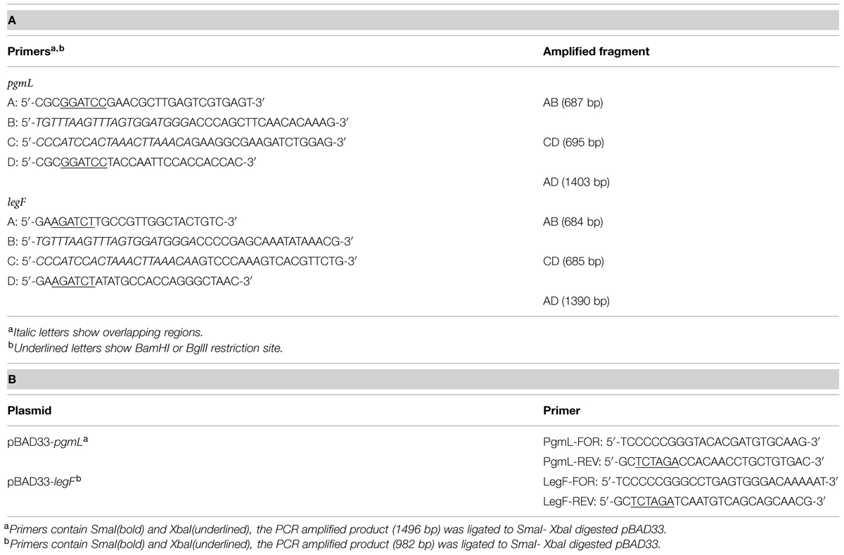
TABLE 2. (A) Primers used in the construction of chromosomal in-frame deletion mutants. (B) Primers used for mutant complementation using vector pBAD33.
Plasmids pBAD33-pgmL and pBAD33-legF were constructed carrying the wild type genes pgmL and legF by PCR amplification of genomic DNA by using specific primer pairs and ligated to the plasmid pBAD33 from ATCC (American Type Culture Collection; see the list of primers in Table 2). Plasmids pBAD33-pgmL and pBAD33-legF were introduced in E. coli DH5α by electroporation, and then by triparental matings were introduced in the corresponding mutants. Induction or repression of genes in pBAD33 was achieved as described in ATCC.
Results
Plesiomonas shigelloides 302-73 [serogroup O1 (Pieretti et al., 2010)] grown in liquid medium or semisolid medium (swimming agar plates) showed the typical three-four flagella located in single point of one cell pole (lophotricus; Figure 1). However, when the agar concentration was increased, the flagellar distribution shifted from single pole to more disperse. The agar concentration seems to be involved in this change in flagella distribution. When the bacteria were grown in solid or semisolid media (swarming agar plates), a complete different flagella distribution was observed. As can be seen in Figure 1 the flagella showed a typical peritrichous distribution over the entire cell surface.
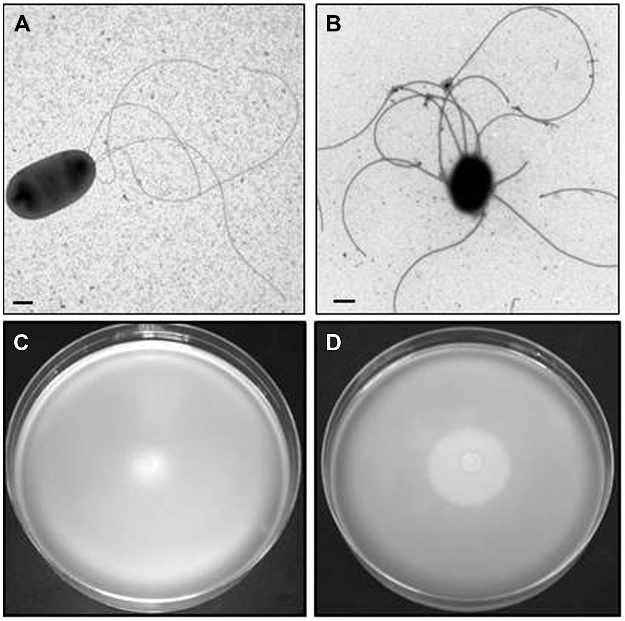
FIGURE 1. Plesiomonas shigelloides 302-73 wild type strain serotype O1. TEM from cells grown in liquid medium (A) and swarming agar plates (B). Motility in swimming (C) and swarming (D) agar plates.
A similar pattern of flagellar distribution with changes in growth medium was observed with 12 P. shigelloides strains. Among these strains eight represented five different serotypes (O1, O2, O3, O17, and O54) while four were non-serotyped strains. The source of the strains was from clinical stools (7) and fish (5), from Japan four of them, four from Spain, three from Brazil, and one from Poland.
MiniTn5Km-1 Mutagenesis
A spontaneous rifampicin-resistant P. shigelloides mutant (named 302-73R) derived from the wild type strain 302-73 was isolated by our group. P. shigelloides 302-73R showed identical pattern of flagella production as described previously for wild type strain. We selected insertional mutants, as described in Materials and Methods, and grouped by their inability to swim, to swarm, or both negative characteristics.
Among an initial screening of 2500 colonies four mutants were selected (initially named A, B, C, and D), based upon inability to swim but retaining the ability to swarm. A further, three mutants (initially named E, F, and G) were selected based upon inability to swarm but retaining ability to swim. Lastly, two mutants (initially named H and I) were selected that were unable to swim or swarm. Mutants A, B, C, and D, when observed by EM in appropriate conditions showed lateral flagella but not polar (Figure 2), while mutants E, F, and G (Figure 3), showed polar but not lateral flagella by EM when grown in appropriate conditions. Mutants H and I were unable to produce polar or lateral flagella observed by EM in any growth conditions (Figure 4). The presence of a single copy of the minitransposon in their genome was determined by Southern blot analysis. We were unable to clone the minitransposon-containing DNA fragment from the mutants using methodologies that were successful in other bacteria (Aquilini et al., 2013).
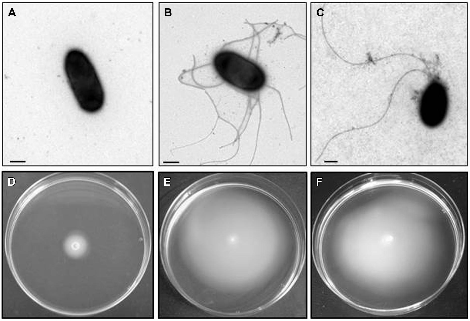
FIGURE 2. Plesiomonas shigelloides A mutant (as an example for the insertional polar flagella mutants). TEM of the A mutant grown in liquid medium (A) and swarming agar plates (B) and complemented mutant with COS-FLAregI-1harboring the corresponding wild type gene grown in liquid medium (C). Bar, correspond to 0.5 μm. Motility of the A mutant in swimming (D) and swarming (E) agar plates. The complemented mutant with COS-FLAregI-1harbouring the corresponding wild type gene in swimming agar plate (F).
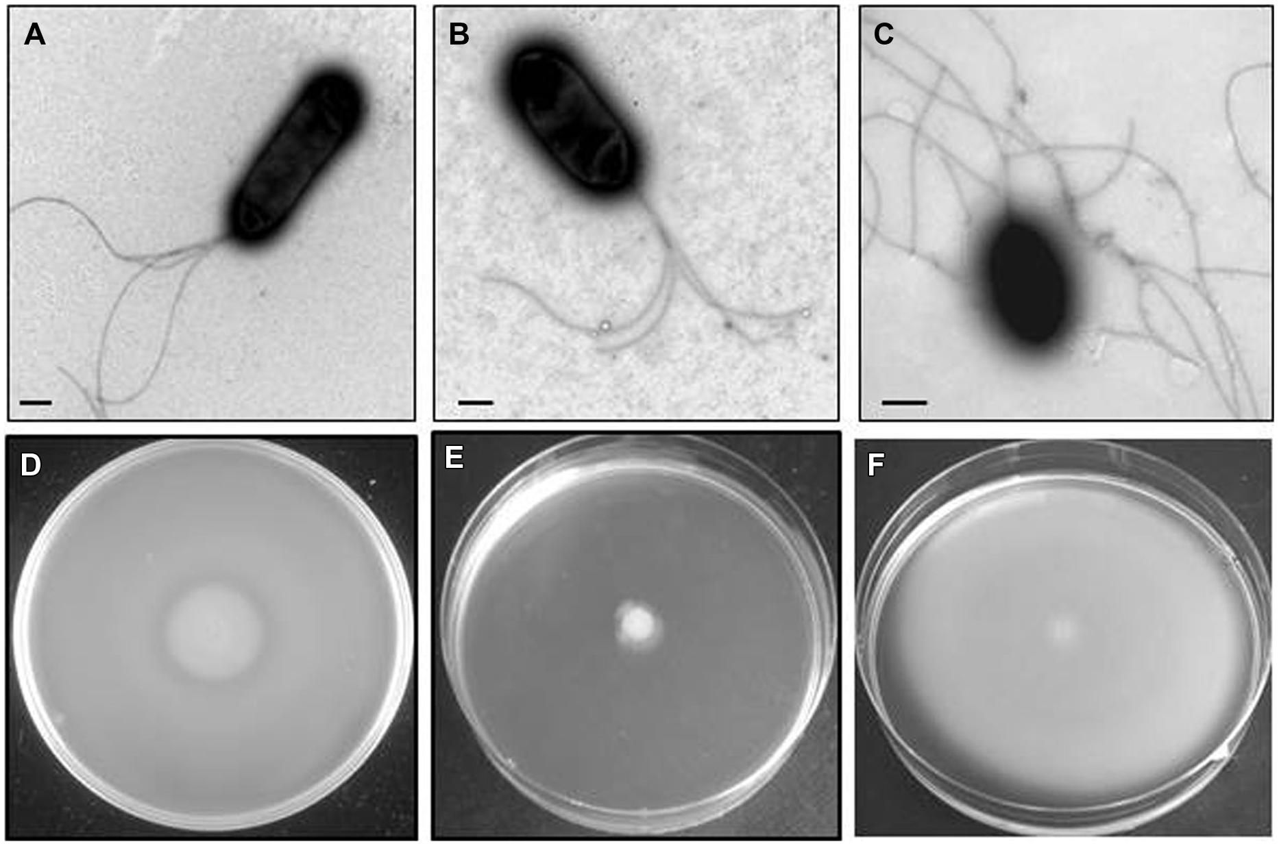
FIGURE 3. Plesiomonas shigelloides E mutant (as an example for the insertional lateral flagella mutants). TEM of the E mutant grown in liquid medium (A) and swarming agar plates (B) and complemented mutant with COS-LAFI harboring the corresponding wild type gene grown in semisolid medium (C). As could be observed in B the polar flagella are constitutively expressed in semisolid medium. Bar, correspond to 0.5 μm. Motility of the E mutant in swimming (D) and swarming (E) agar plates. The complemented mutant with COS-FLAregI-1harbouring the corresponding wild type gene in swarming agar plate (F).
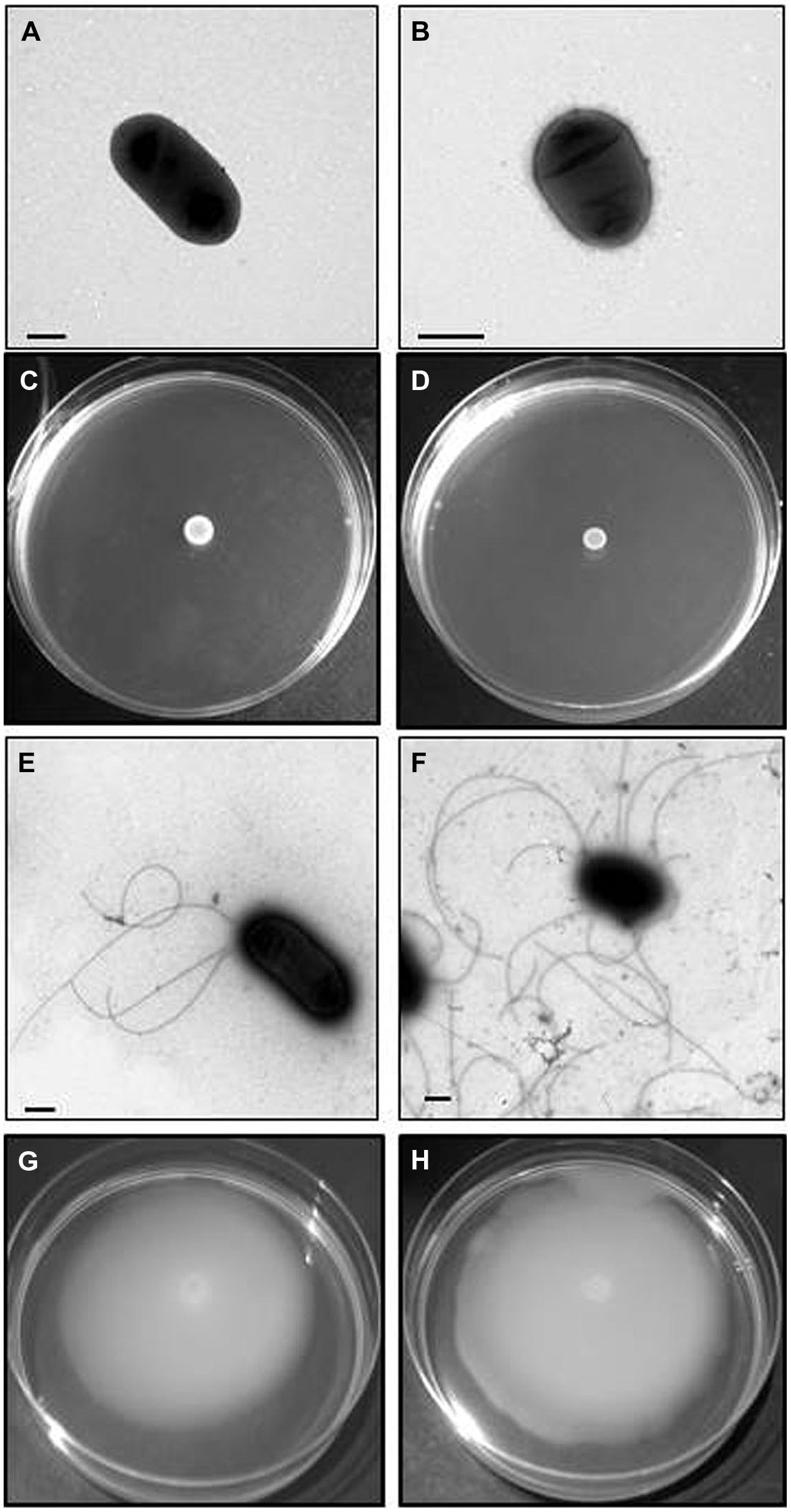
FIGURE 4. Plesiomonas shigelloides H mutant (as an example for the insertional Leg biosynthetic cluster mutants). TEM of the H mutant grown in liquid medium (A) and swarming agar plates (B) and complemented mutant with COS-LAFI harboring the corresponding wild type gene grown in liquid (E) and semisolid medium (F). Bar, correspond to 0.5 μm. Motility of the H mutant in swimming (C) and swarming (D) agar plates. The complemented mutant with COS-LEG harboring the corresponding wild type gene in swimming (G) and swarming (H) agar plates.
Complementation of the mutants, using a cosmid based genomic library of P. shigelloides 302-73 (see Materials and Methods) reversed the phenotype observed, either to swim or swarm in motility plates.
Polar Flagella Mutants
We found several recombinant positive clones able to complement A, B, C, and D mutants. The complementation was studied by the recovery of swimming behavior under appropriate conditions. All complemented mutants were able to produce polar flagella when observed by EM growing in liquid conditions (Figure 2). Sequencing the recombinant positive clones complete inserts revealed the complete region to correspond to PLESHI_03205 to PLESHI_03505 in the complete P. shigelloides 302-73 genome (Piqué et al., 2013).
Polar flagella gene cluster, as shown in Figure 5A, are based in two gene regions (I and II) adjacent to a group of putative biosynthetic Leg genes. In region I there are several genes encoding chemotaxis proteins, including the σ28 factor fliA, cluster from flhB to G, fliK to R, fliE to J, flrA and C, and flaC to J (transcribed in the same direction). This region I, similar to V. parahaemolyticus region two by gene distribution and transcription sense, also lacks the motor genes (McCarter, 2001). Region II, downstream of the putative biosynthetic Leg genes group, contains cluster flgP,O,T, or flgA,M,N with the typical transcription sense in the different Gram-negative bacteria described, two genes encoding chemotaxis proteins, and cluster flgB to L. By gene distribution and transcription sense this region II is similar to region 1 of V. parahaemolyticus and Aeromonas hydrophila (McCarter, 2001; Canals et al., 2006).
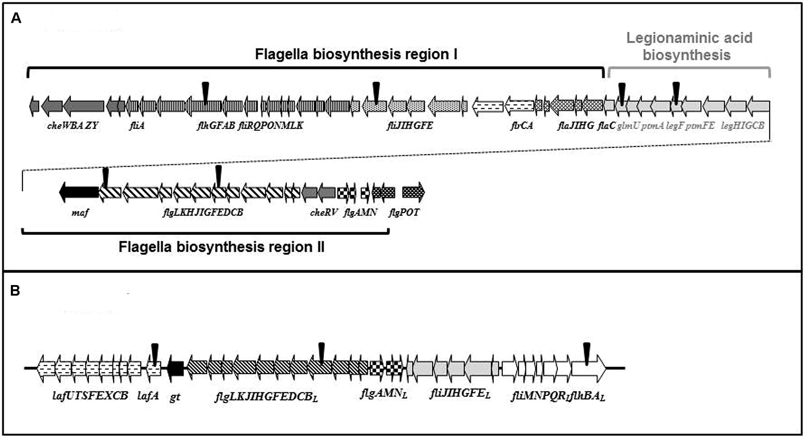
FIGURE 5. Plesiomonas shigelloides 302-73 wild type strain serotype O1 polar (A) and lateral (B) flagella clusters. The polar flagella cluster shows two different regions adjacent to Leg biosynthetic genes. The inverted triangles (▼) indicate the position of the different insertional mutants obtained by miniTn5 mutagenesis.
Table 3 shows the ORFs with their predicted function based on their homology to proteins of known function. Proteins of unknown function were not included. The last gene in this region encoded an ORF (named Gt), which showed homology to domains of a glycosyltransferase. This was provisionally assigned to the polar flagella cluster and not to the putative biosynthetic Leg genes. Once the DNA fragment was completely sequenced, several primers were used to derive the DNA sequence to locate the miniTn5 [A = flgE, B = flhA, C = fliI, and D = flgK (Figure 5A)].
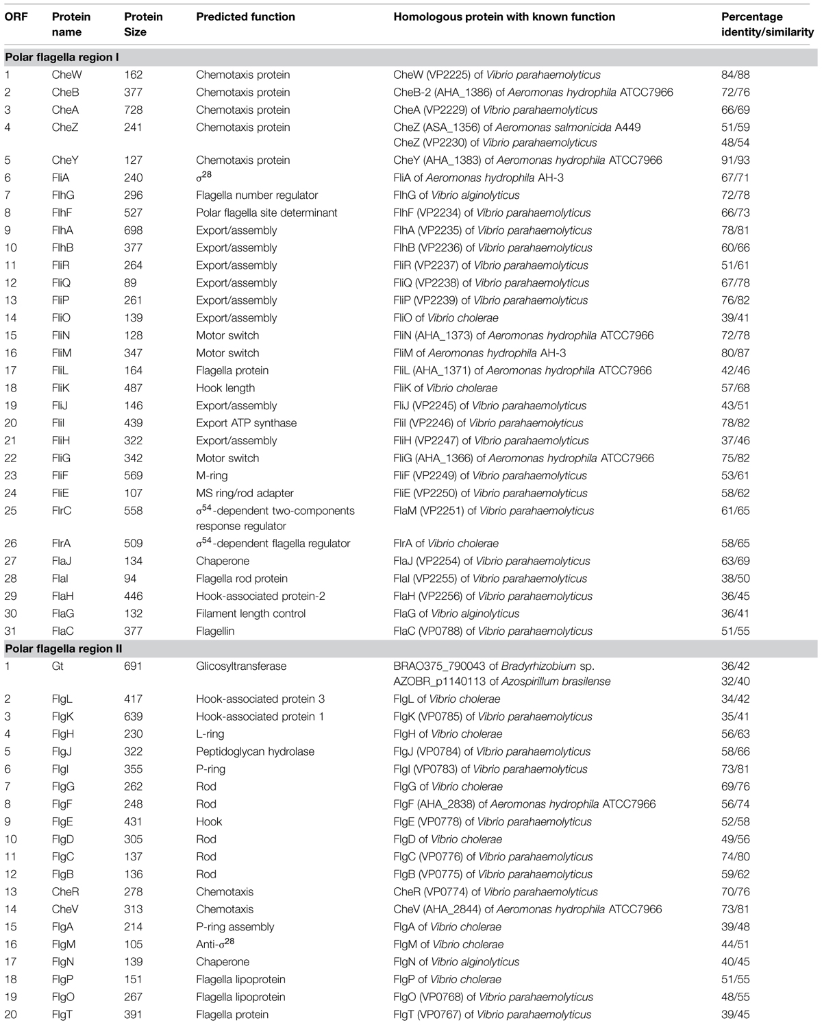
TABLE 3. Characteristics of the P. shigelloides 302-73 strain polar flagella gene regions I and II.
Lateral Flagella Mutants
Several recombinant positive clones complemented E, F, and G mutants separately. Some clones were observed to complement two mutants. The complementation was studied on the basis of recovery of swarming behavior on appropriate growth plates. All complemented mutants were able to produce lateral flagella when observed by EM growing in semisolid conditions (Figure 3). We used the same strategy previously indicated to sequence the entire DNA region contained in the recombinant positive clones. This complete region correspond to PLESHI_07125 to PLESHI_07305 in the complete P. shigelloides 302-73 genome (Piqué et al., 2013).
Lateral flagella gene cluster shows 37 genes grouped in a single region (Figure 5B). Five typical group of genes (lafA to U; flgBL to LL; flgAL,ML,NL; fliEL to JL; and fliML to RL plus flhB-AL) when compared to the most similar A. hydrophila AH-3 lateral flagella region were found. All the genes were found in a unique region similar to A. hydrophila or enteric bacteria. In contrast, in the equivalent region in V. parahaemolyticus is found in two separate regions (Canals et al., 2006; Merino et al., 2006). The group of genes fliEL to JL and fliML to RL plus flhB-AL are adjacent in all the lateral flagella clusters described. The groups of genes have been shown to be transcribed in the same direction in A. hydrophila and divergently in Vibrio, enteric bacteria and P. shigelloides (Merino and Tomás, 2009). Table 4 shows the ORFs with their predicted function based on their homology to proteins of known function. All the protein analogies that were from unknown or not well-established homology were discarded. Between the group of genes flgB-LL and lafA-U, there is a gene encoding for a hypothetical protein without the classical motility accessory factors domains found in A. hydrophyla Maf-5. However, this encoded protein showed a minimal similarity with this Maf-5, and the gene was denoted maf-5 (Parker et al., 2014). Once the DNA fragment was completely sequenced, we used several primers derived from the DNA sequence to locate the miniTn5 in lafA (E), flhAL (F), and flgEL (G; Figure 5B).
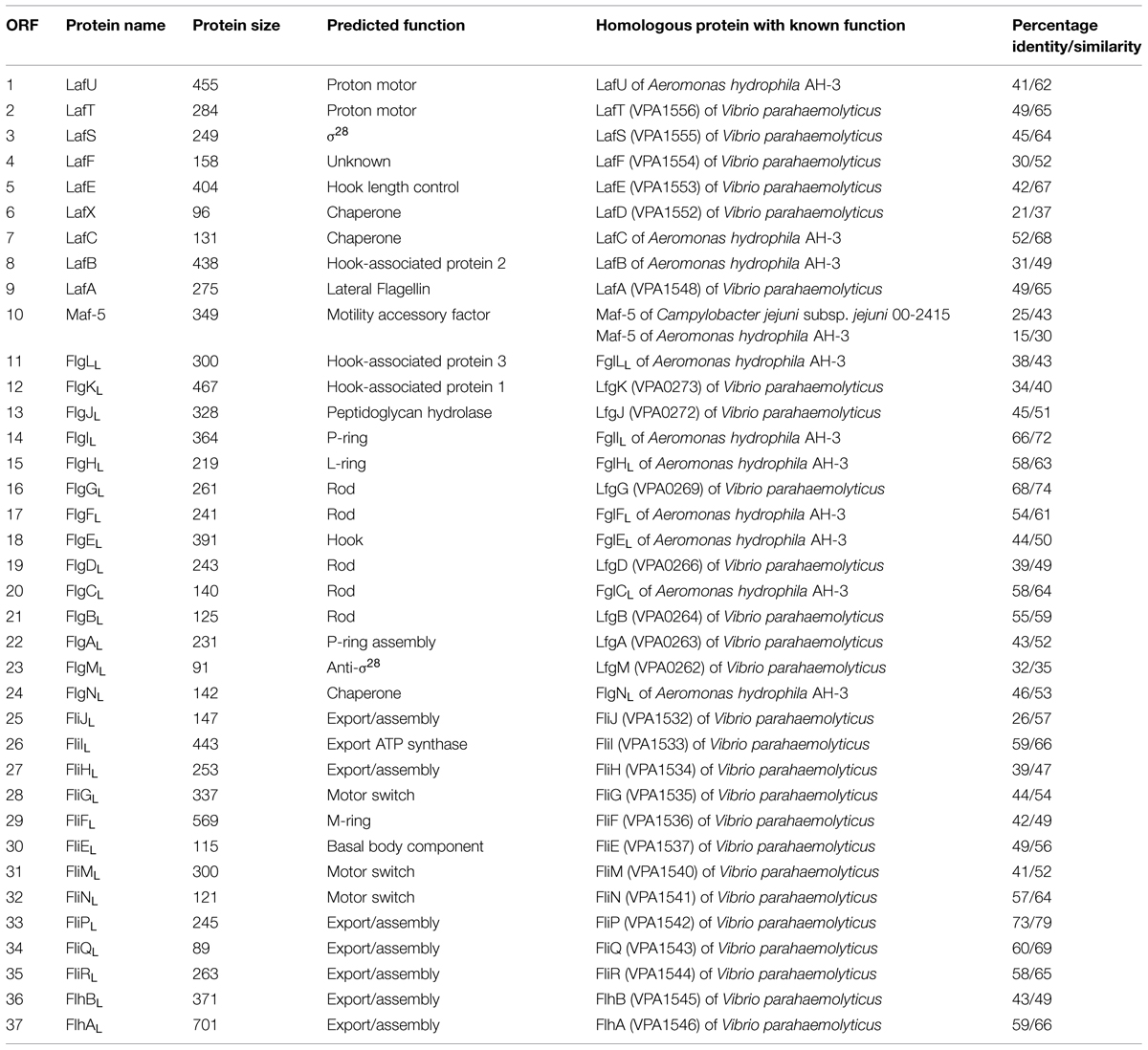
TABLE 4. Characteristics of the P. shigelloides 302-73 strain lateral flagella cluster.
Mutants Unable to Produce Flagella
A single recombinant positive clone was observed to complement both mutants H and I as they recover swimming and swarming in plates. The complemented mutants were able to produce polar and lateral flagella when observed by EM growing in appropriate conditions (Figure 4). Sequencing the entire DNA region in the recombinant positive clone showed this region to contain the group of putative biosynthetic Leg genes (Figure 5A) between region I and II codifying for the polar flagella. This complete region corresponds to PLESHI_03365 to PLESHI_03405 in the complete P. shigelloides 302-73 genome (Piqué et al., 2013).Table 5 shows the ORFs with their predicted function based on their homology to proteins of known function.
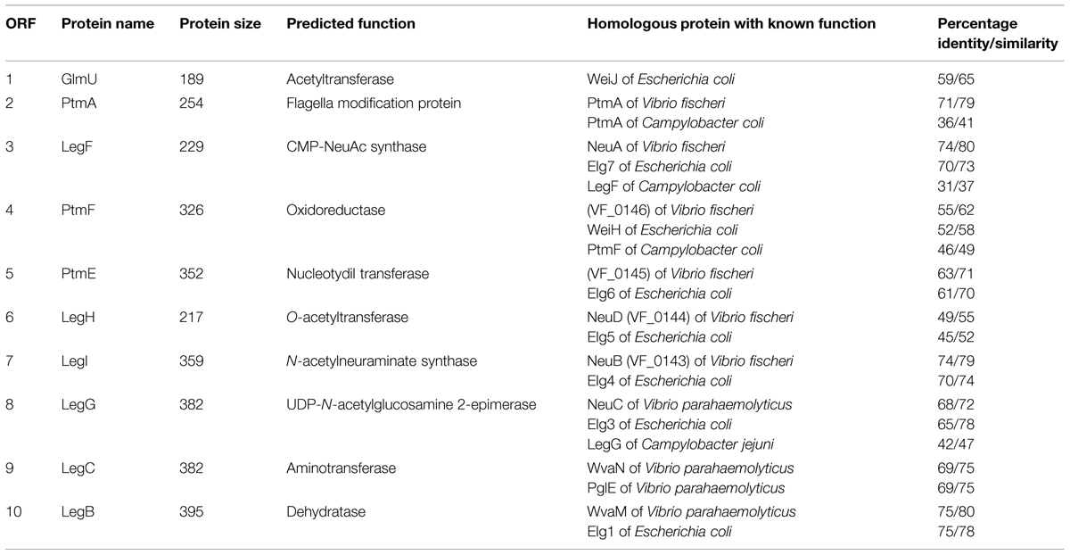
TABLE 5. Characteristics of the P. shigelloides 302-73 strain gene region for legionaminic acid biosynthesis between polar flagella regions I and II.
The Campylobacter jejuni CMP-Leg biosynthetic pathway described involves two segments: synthesis of a GDP-sugar building block and synthesis of the final CMP-nonulosonate which are linked by the N-acetyl transferase GlmU (Schoenhofen et al., 2009). We found all the genes encoding for the necessary two segments of the CMP-Leg biosynthetic pathway in this region besides the one encoding phosphoglucosamine mutase (PgmL) included in the first segment of the biosynthesis. Once the DNA fragment was completely sequenced, we used several primers derived from the DNA sequence to establish that the miniTn5 was located in ptmA (H) and legH (I; Figure 5A).
Flagella Purification
Polar flagellins were purified from the wild type strain after grown in liquid medium and a mixture of polar and lateral flagellins after grown in swarm agar plates (Figure 6A). Lateral flagellin was also isolated from insertion mutant A (unable to produce constitutive polar flagella with unaltered lateral flagella).
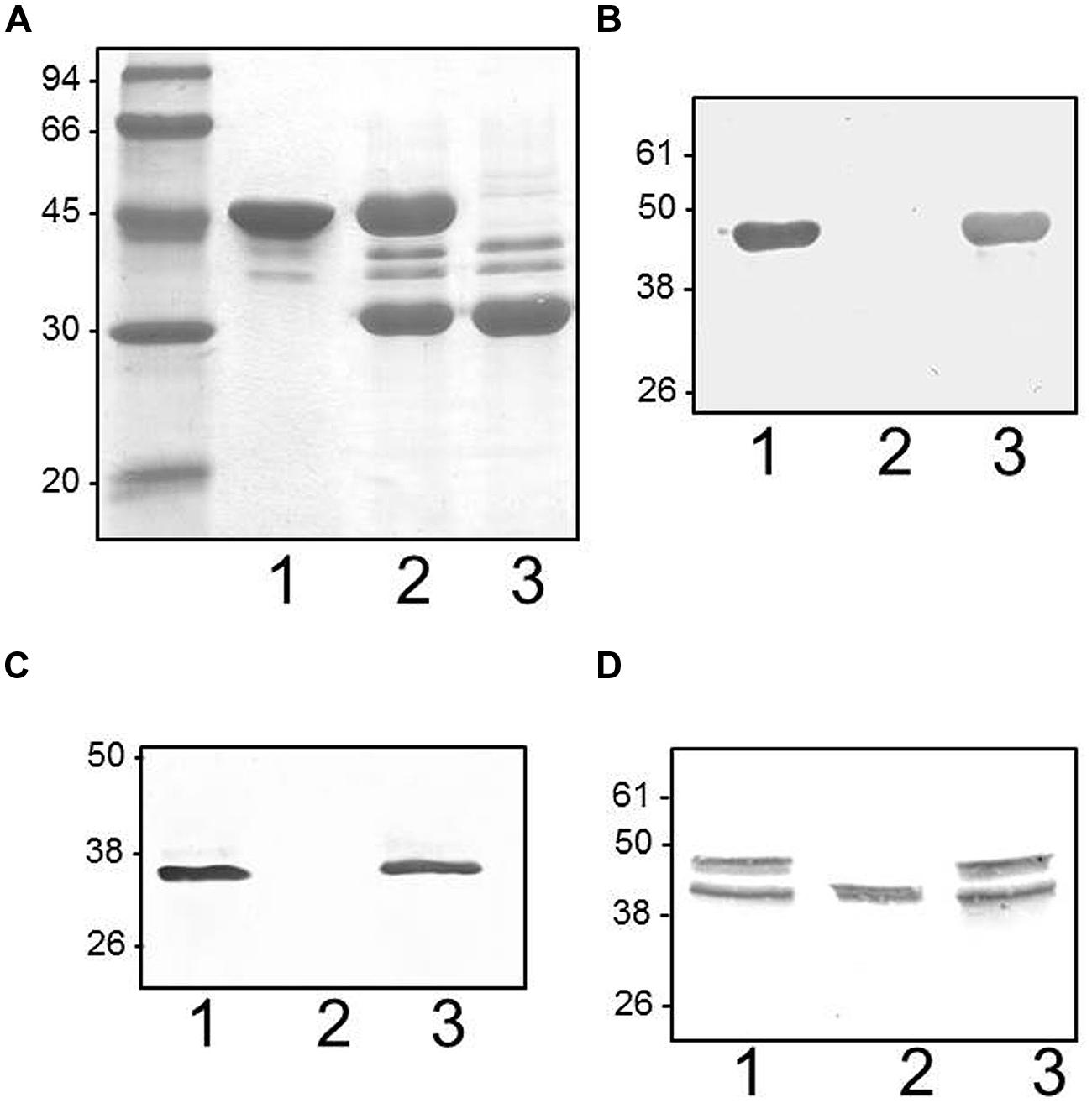
FIGURE 6. (A) Plesiomonas shigelloides 302-73 wild type strain serotype O1 purified flagella according to Section “Materials and Methods” when grown in liquid medium (1) and swarming agar plates (2). As could be observed in 2, and previously indicated in Figure 3, polar flagella are constitutively expressed in semisolid medium. Purified flagella from P. shigelloides insertional polar A mutant grown in swarming agar plates (3). (B). Western blot with specific polar flagella antiserum of purified flagella from wild type (1), P. shigelloides insertional polar H mutant (2), and complemented mutant with COS-LEG harboring the corresponding wild type gene (3) obtained in liquid medium growth. (C) Western blot with specific lateral flagella antiserum of purified flagella from wild type (1), P. shigelloides insertional polar H mutant (2), and complemented mutant with COS-LEG harboring the corresponding wild type gene (3) obtained in swarming agar plates. (D) Western blot with specific polar flagella antiserum of cytoplasmic fractions obtained as described in Section “Materials and Methods” of wild type (1), P. shigelloides insertional polar H mutant (2), and complemented mutant with COS-LEG harboring the corresponding wild type gene (3) obtained in liquid medium growth. The low molecular weight band could correspond to the non-glycosylated form, and the upper band (not present in the mutant) to the glycosylated form.
Intact Mass Analysis of Purified Flagellins
Purified polar flagellin preparations showed a well-resolved ion envelop of multiple charged protein ions, which deconvoluted into three distinct masses at 40201, 40652, and 40931 Da. The mass of the translated gene sequence for polar flagellin was 38710 Da, giving mass excesses of 1491, 1942, and 2221 Da, respectively (data not shown). During front end CID experiments of the purified polar flagellin preparation, labile glycan related ions were observed at m/z 359 and 317. Using increasing cone voltages, fragmentation of this ion at m/z 359 was observed, as shown in Figure 7. The fragment ions observed at m/z 317, 299, 281, 222, and 181 were characteristic fragment ions of nonulosonic acids, such as pseudaminic or legionaminic acid.
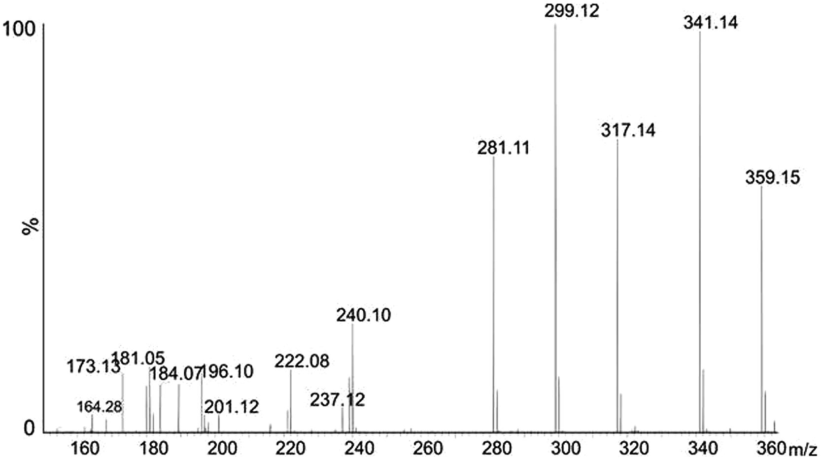
FIGURE 7. Glycan fragmentation pattern. Front end collision induced dissociation of polar flagellin protein, showing low m/z region. Fragment ions characteristic of an acetylated nonulosonic acid sugar are present.
From the observed mass of 316.124, the top ranked plausible elemental formula was C13H21N2O6, suggestive that this moiety is a carbohydrate. The additional glycan ion observed at m/z 359, gave a top ranked plausible elemental formula C15H23N2O8, suggesting this species to be a nonulosonic acid with an additional of an acetyl group. An intense fragment ion was observed at m/z 341, most likely a loss of water from the glycan ion observed at m/z 359.
The preparation containing purified polar and lateral flagellins showed a more complex elution profile when HPLC separated, with two sequentially eluting protein peaks. The area under each peak was combined separately and each showed a complex ion envelope. The ion envelope of the first eluted protein deconvoluted into two distinct masses at 39325, 40678 Da. The second eluting protein ion envelope deconvoluted to give a single protein mass at 30940. It is possible that the larger MW proteins correspond to the polar flagellin and the 30 kDa protein the lateral flagellin. The A mutant that is unable to produce polar flagella showed only this second eluting peak when grown in swarming conditions (Figure 6A). In each case, the measured molecular mass is greater than that of the translated gene sequence for each protein. This suggests that both polar and lateral flagellins are post-translational modified. Front end CID experiments showed almost identical profiles when compared with the polar flagellin preparation, with intense ions observed at m/z 359, 317. These data suggest that both polar and lateral flagellins are modified with the same nonulosonic acid sugar, with or without acetylation.
Bottom Up Mass Spectrometry Studies of Flagellins
Tandem mass spectrometry studies of tryptic digests of purified polar flagellins identified a number of unmodified peptides. De novo sequencing of the MS/MS data showed a number of spectra that were identified as flagellin peptides and harboring mass excess of 316 Da. Also observed was an intense ion at m/z 317, suggestive of a glycan oxonium ion. Figure 8A shows the MS/MS spectrum of the polar flagellin glycopeptide AIASLSTATINK, modified with a putative 316 Da glycan. Peptide type y and b fragment ions are annotated and confirm the peptide sequence. In addition, low m/z fragment ions that did not correspond to peptide type y or b ions were also observed at m/z 317, 299, 281, 240, 221, 196, and 181. Combined with the mass excess, glycan oxonium ion and putative glycan fragment ions, the data suggest the flagellin peptides to be modified with a legionaminic acid like glycan.
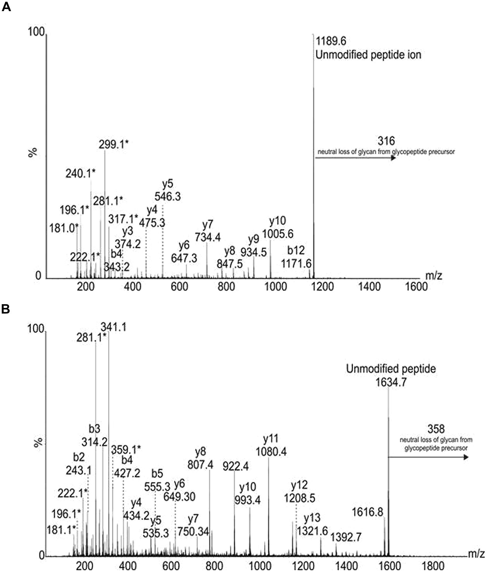
FIGURE 8. Tandem mass spectra of flagellin glycopeptides (A) from polar flagellin, peptide AIASLSTAINK, modified with a 316 Da glycan. Peptide type y and b ions are indicated, confirming the peptide sequence. In addition, glycan related fragment ions are indicated with an asterisk (∗), and include a glycan oxonium ion at m/z 317, and related ions at m/z 299, 281, 240, 221, and 181. (B) From lateral flagellin, the peptide type y and b fragment ions indicate a sequence of ELALQSANGTNTTADK. In this case the peptide is modified with a 358 Da glycan, as observed with a neutral loss of 358 from the glycopeptide precursor and the glycan oxonium ion at m/z 359. A loss of water is observed from this oxonium ion, in addition glycan related fragment ions are indicated with an asterisk (∗).
The purified polar and lateral flagellins were also digested with trypsin and analyzed by tandem mass spectrometry, identifying a number of unmodified flagellin peptides. Once again, de novo sequencing showed several flagellin peptides from both polar and lateral flagellins to be modified with putative glycan moieties. The lateral flagellin (LafA) harbored peptides modified with glycans of 316 and 358 Da (Figure 8B). In some cases peptides were showed to harbor both glycans. It was not clear from the data whether two monosaccharides were modifying two separate amino acids, or whether a single disaccharide was modifying at one site.
The polar flagellin was also observed to be modified with 316 and 358 Da glycan moieties. In some cases, glycan chains comprised of multiple 358 Da glycans were observed; in other cases a single modification of 316 or 358 Da was noted. Very low levels of peptides harboring distinct glycan masses were observed, such as the peptide AIASLSTATINK, was observed to be modified with either 316 Da glycan, or a 523 or 481 Da glycan. Glycan related ions were observed in each case, with intense ions observed at m/z 524 and 184 or m/z 424 and 184. The ion at m/z 184 was also observed in front end CID experiments with the intact polar and lateral flagellin preparations, and gave a top ranked plausible elemental formula of C9H12O4, suggesting that it is a related nonulosonic acid type sugar. The low abundance of these glycopeptides made any further analyses challenging.
Legionaminic Acid Biosynthetic Mutants
The insertional mutants in ptmA (H) and legH (I) were unable to produce polar or lateral flagella under induced conditions, as shown by TEM or by immunodetection (Figure 6B) or lateral flagellins (Figure 6C) in purified flagella. The introduction of the P. shigelloides wild type genes was observed to recover the production of polar and lateral flagella in the mutants. This was demonstrated using immunodections, as shown in Figures 6B,C. These data prompted us to examine the production of the polar flagellin in the mutants by immunodetection. Western blot analysis shows presence of polar flagellin the cytoplasmic subcellular fraction. Interestingly, only a single protein band was observed, with a lower than expected molecular weight (Figure 6D). Wild type flagellin typically migrates as two distainct bands, both detectable by Western blot. We speculate that the single, lower molecular weight species is a non-glycosylated form of flagellin. The complemented mutants showed the same cytoplasmic polar flagellin molecular weight bands as observed with wild type strain. Similarly, where lateral flagellin was detected in the cytoplasmic fraction, it was observed at a lower molecular weight, likely the non-modified form of the protein. Then, the lack of polar and lateral flagella formation observed in the mutants is not by the lack of flagellin protein or the master regulator transcription.
In order to prove at the genomic level that mutations in the CMP-Leg biosynthetic pathway were responsible for the phenotypic traits shown by insertional mutants H and I, two in-frame pgmL and legF deletion mutants were generated, 302ΔpgmL and 302ΔlegF, respectively. Our genomic studies indicates that all the genes of the Leg pathway are included in the cluster between polar region I and II, with the exception of the PgmL ortholog which is found in another region of the chromosome [703.5 peg 1785 (Piqué et al., 2013)]. PgmL or GlmM, phosphoglucosamine mutase, is involved in the first step to produce GDP-GlcNAc. LegF, CMP-legionaminic acid synthase is the final enzyme of the second step to produce CMP-Leg. Using TEM, neither mutant was observed to produce polar or lateral flagella under induced conditions. Both show the same phenotypic traits as insertional mutants H and I. When mutants 302ΔpgmL and 302ΔlegF were complemented with their single corresponding wild type gene (pBAD33-pgmL and pBAD33-legF, respectively) under inducing conditions (plus arabinose) all the wild type phenotypic traits (production of polar and lateral flagella or swimming and swarming motilities) were fully recovered. Control plasmid pBAD33 alone under inducing conditions (plus arabinose) was unable to do it.
Lateral Flagella and Leg O-Flagella Glycosylation Gene Distribution on P. shigelloides
In order to test if the presence of lateral flagella and Leg O-flagella glycosylation genes is a specific feature for the strain studied, the 12 previously mentioned P. shigelloides strains used for PCR studies were eight strains representing five different serotypes (O1, O2, O3, O17, and O54) plus 4 non-serotyped strains described in Material and Methods. Initially, genomic DNA from 302-73 strain was used as template for PCR amplification with two sets of oligonucleotides: 5′-ATCGCGTCTGAAAGGCTAC-3′ and 5′-CTGCGCCATAGAACTACCC-3′ which amplified a 2160 bp DNA fragment from lateral flagella cluster (partial lafA and complete maf-5); and another oligonucleotide set (5′-CGGGTTAAAGCTATCCCATC-3′ and 5′-CCAATGACAGCTGAATCTCC-3′) amplified a 1985 bp DNA fragment from Leg biosynthesis genes (partial legH and complete legI). DNA fragments of the same size (2160 and 1985 bp, respectively) were PCR amplified for all the genomic DNAs from the strains studied, as shown by the results shown in Figure 9. DNA sequence of the amplified fragments confirmed the presence of the lateral and Leg biosynthetic genes. In addition, in all the amplified maf-5 and legI fragments the presence of a sequence coding for the N-terminal amino acid residues of lafA and legH genes, respectively, were found adjacent to maf-5 or legI, suggesting that in the analyzed strains the genomic location is the same as that found in P. shigelloides wild type strain 302-73 (Figure 5).
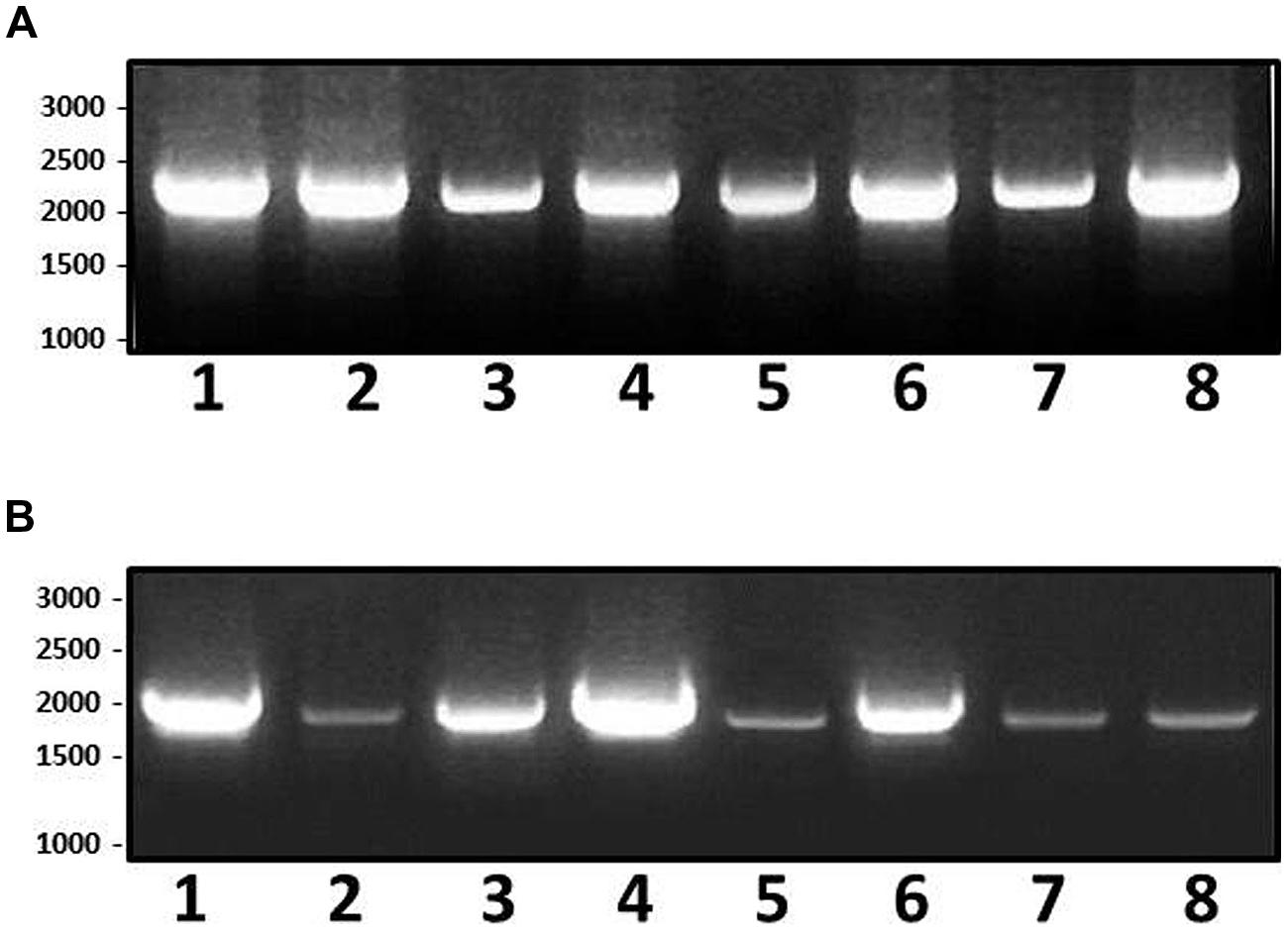
FIGURE 9. Polymerase chain reaction (PCR) amplified bands from genomic P. shigelloides DNAs of strains: 302-73, serotype O1 (1); 306-73, serotype O2 (2); 307-73, serotype O3 (3); 343-73, serotype O17 (4), PCM2647, serotype O54 (5), C1, non-serotyped from Spain (6); P12, non-serotyped from Spain (7); and lt645, non-serotyped from Brazil (8); using the set of oligonucleotides for lateral flagella cluster (A, 2160 bp DNA fragment) and Leg biosynthesis genes (B, 1985 bp DNA fragment).
Discussion
Motility is an essential mechanism in adaptation to different environments for free living bacteria. Bacteria showed three flagella types classified according to their location on a cell: peritrichous, polar, and lateral. It has been reported dual flagella systems in some polar flagellated bacteria when grow in viscous environments or surfaces. This fact allows bacteria to swarm on solid or semisolid media by a mixed flagellation (polar and lateral flagella). P. shigelloides has been observed to express mixed flagellation (Inoue et al., 1991).
Two P. shigelloides 302-73 different gene clusters were described, one exclusively involved in lateral flagella biosynthesis, and a second containing the polar flagella genes distributed in two regions spaced by putative glycosylation genes. It is characteristic of the bacteria with dual flagella systems to separate both in different gene clusters (McCarter, 2001; Canals et al., 2006; Merino et al., 2006; Merino and Tomás, 2009). Of note, P. shigelloides is the first Enterobacteriaceae with lateral flagella production as shown herein.
Plesiomonas shigelloides lateral gene cluster is nearly identical to the lateral gene cluster of A. hydrophila according to the gene grouping and transcription direction, with the exception of the group of genes fliML to RL plus flhB-AL which are transcribed in opposite direction (Canals et al., 2006). However, no lafK ortholog could be detected in P. shigelloides lateral gene cluster. This gene has been reported in all the lateral gene clusters, including the non-functional in the Enterobacteriaceae (Canals et al., 2006; Merino and Tomás, 2009). A non-functional Flag-2 flagella cluster with large similarity to V. parahaemolyticus lateral flagella system, was found in different E. coli enteroaggregative or Yersinia pestis or pseudotuberculosis strains (Ren et al., 2004). However, as we proved, P. shigelloides lateral gene cluster is fully functional.
The transcriptional hierarchy of V. parahaemolyticus lateral flagella is one of the Gammaproteobacteria model. LafK (σ54-associated transcriptional activator) is the master regulon in this model, controlling Class II lateral flagella genes transcription. Class II genes contains the σ28 factor (fliAL) which is involved in transcription of Class III lateral flagella genes (Stewart and McCarter, 2003). In V. parahaemolyticus the absence of polar flagellum induces the expression of lateral flagella in liquid medium, and LafK is able to compensate the lack of FlaK (σ54-associated polar transcriptional activator) and activate polar flagellum class promoters. A. hydrophila lateral flagella transcriptional hierarchy represents the second Gammaproteobacteria model. Class I gene transcription in A. hydrophila lateral flagella is σ70-dependent as LafK in contrast to describe in V. parahaemolyticus (Stewart and McCarter, 2003). It is important to point out that A. hydrophila lateral flagella genes are transcribed in liquid and solid or semisolid media, and unlike V. parahaemolitycus the genes are not induced by mutation of polar flagellum genes. The transcription hierarchy of A. hydrophila lateral flagella is complex because LafK is not strictly their master lateral flagella regulator, and many clusters of genes are LafK independently transcribed (Wilhelms et al., 2013). A. hydrophila LafK protein is unable to not compensate the lack of FlrA, which is the polar-flagellum regulator (σ54-associated transcriptional activator for polar flagellum), a situation that happens in V. parahaemolyticus (Wilhelms et al., 2013). This point is in agreement with A. hydrophila FlrA mutation not affect lateral flagella besides that abolishes polar flagellum formation in liquid and on solid surfaces (Wilhelms et al., 2013).
The P. shigelloides polar flagella gene regions show greater similarity to those reported in Vibrio or Aeromonas than the regions in Enterobacteriaceae [e.g., E. coli or S. typhimurium (Chilcott and Hughes, 2000)]. Bacteria with peritrichous flagella, such as E. coli and Salmonella, showed three hierarchy levels. The σ70 is required for transcription of class I and II genes, and class I promoter responds to different regulatory factors and transcribes the FlhDC master activator, which allowed the class II σ70-dependent promoter expression. At the top of the Vibrio sp. or A. hydrophyla polar flagella hierarchy is σ54-associated transcriptional activator (FlrA, named FleQ in Pseudomonas aeruginosa) which activates class II genes σ54-dependent promoters. Class II promoters encode a two component signal-transducing system (Vibrio sp. or A. hydrophyla FlrBC and FleSR in P. aeruginosa) whose regulator (FlrC/FleR) activates class III genes σ54-dependent promoters.
In the P. shigelloides polar flagella region I only flrA and C orthologs were observed. P. shigelloides FlrA shows the characteristic three domains (FleO, σ54 -interaction domain and family regulatory protein Fis) like in Vibrio sp. or A. hydrophila (Kim and McCarter, 2004; Wilhelms et al., 2011). Class II promoters encode a two component signal-transducing system (FlrBC of Vibrio sp. or A. hydrophila and FleSR in P. aeruginosa) whose regulator (FlrC/FleR) activates class III σ54-dependent promoters. However, when analysis of P. shigelloides FlrC encoded protein, revealed the corresponding domains for FlrB and C. Thus, P. shigelloides FlrC contains two domains of Vibrio sp. or A. hydrophila FlrB (PAS domain and His Kinase A) as well as two domains of Vibrio sp. or A. hydrophila FrlC (σ54-interaction domain and family regulatory protein Fis). We suggest that P. shigelloides FlrC could be able to activate class III genes σ54-dependent promoters as observed in Vibrio sp. or A. hydrophila. No FlrB ortholog was observed in the P. shigelloides 302-73 genome (Piqué et al., 2013). It could be suggested that in P. shigelloides, FlrB and C functions are developed by a single bifunctional protein encoded by the single flrC as it happens for some LPS-core biosynthetic genes (Jiménez et al., 2009). Taken together, the data presented hererin, no lafK or separate flrB in P. shigelloides, indicate that their lateral and polar flagella transcriptional hierarchy represents a different Gammaproteobacteria model that requires further study.
Among this large P. shigelloides polar flagella gene cluster, genes were identified between the two polar flagella regions, the presence of genes putatively linked to glycosylation. These genes were not found in other Enterobacteriaceae studied. O-glycosylation could be performed by a mechanism dependent or not of an oligosaccharyltransferase (OTase; Kim and McCarter, 2004; Iwashkiw et al., 2013). The O-glycosylation frequently affects protein stability, flagella filament assembly, bacterial adhesion, biofilm formation, and virulence in general as has been described in several bacteria (Lindenthal and Elsinghorst, 1999; Logan, 2006; Faridmoayer et al., 2008; Egge-Jacobsen et al., 2011; Iwashkiw et al., 2013; Lithgow et al., 2014). The predominant O-glycans linked to flagellins are mainly derivatives of pseudaminic acid (PseAc, where Ac represents an acetamido group) and in a minor extent an acetamidino form of legionaminic acid (LegAm, where Am represents acetamidino; Merino et al., 2014). Both are nine-carbon sugars related to sialic acid. The flagellin glycosylation pathways in both cases have been elucidated, including the Pse pathway of Helicobacter pylori and C. jejuni (Fox, 2002), the Leg pathway of C. jejuni (Schoenhofen et al., 2009). Until today the Leg flagella glycosylation has been restricted to C. jejuni or coli. The CMP-legionaminic acid biosynthetic pathway in C. jejuni involves two steps: synthesis of a GDP-GlcNAc and synthesis of the final CMP-Leg (Schoenhofen et al., 2009). The insertional mutants obtained ptmA (H) and legH (I), represent key eznymes in the first and second steps of the CMP-Leg biosynthesis, confirming the observation data that both mutants are unable to produce polar or lateral flagella. Furthermore, the in frame mutants obtained in pgmL and legF, one enzyme of the first step and the last enzyme of the second step of the CMP-Leg biosynthesis, respectively, clearly confirmed the legionaminic acid polar and lateral glycosylation as both mutants are unable to produce polar or lateral flagella as it happens with the insertional mutants.
Mass spectrometry studies show that both flagella in P. shigelloides strain 302-73 are glycosylated by a derivative of Leg, and is also indicated by the presence of Leg biosynthetic pathway genes nearby the polar flagella gene regions. It is the first Enterobacteriaceae reported to harbor O-glycosylation modification on both polar and lateral flagella. Moreover, it is also the first bacteria reported to express a lateral flagella glysosylated by Leg. We also demonstrated that flagella O-glycosylation is essential for bacterial flagella formation, either polar or lateral. However, the flagella O-glycosylation is not determinant for cytoplasmic flagellin production as can be observed by immunodetection studies.
The P. shigelloides homologous recombination rates are extremely high (Salerno et al., 2007), like naturally transformable species as Streptococcus pneumoniae. In the rest of Enterobacteriaceae the recombination rate is much lower. The high recombination observed in this bacterium could offer a reason for P. shigelloides variety of LPS-core structures (Salerno et al., 2007). The PCR experiments using several P. shigelloides strains and lateral flagella or Leg pathway genes, with the motility and EM studies, demonstrated that presence of lateral flagella and Leg O-flagella glycosylation is a widely spread feature, not a strain specific observation. Furthermore, the maintenance of these genes among the different strains besides the recombination rate observed for P. shigelloides, indicates the importance of glycosylated polar and lateral flagella production for this bacterium.
Conflict of Interest Statement
The authors declare that the research was conducted in the absence of any commercial or financial relationships that could be construed as a potential conflict of interest.
Acknowledgments
This work was supported by Plan Nacional de I + D + i (Ministerio de Educación, Ciencia y Deporte and Ministerio de Sanidad, Spain) and from Generalitat de Catalunya (Centre de Referència en Biotecnologia). We thank Maite Polo for her technical assistance and the Servicios Científico-Técnicos from University of Barcelona.
References
Aldova, E., and Shimada, T. (2000). New O and H antigens of the international antigenic scheme for Plesiomonas shigelloides. Folia Microbiol. 45, 301–304. doi: 10.1007/BF02817550
Allen, L. N., and Hanson, R. S. (1985). Construction of broad host-range cosmid cloning vector: identification of genes necessary for growth of Methylobacterium organophilum on methanol. J. Bacteriol. 161, 955–962.
Allison, C., Coleman, N., Jones, P. L., and Hughes, C. (1992). The ability of Proteus mirabilis to invade human urothelial cells is coupled to motility and swarming differentiation. Infect. Immun. 60, 4740–4746.
Allison, C., and Hughes, C. (1991). Bacterial swarming: an example of prokaryotic differentiation and multicellular behaviour. Sci. Prog. 75, 403–422.
Altschul, S. F., Madden, T. L., Schäffer, A. A., Zhang, J., Zhang, Z., Miller, W., et al. (1997). Gapped BLAST and PSI-BLAST: a new generation of protein database search programs. Nucleic Acids Res. 25, 3389–3402. doi: 10.1093/nar/25.17.3389
Aquilini, E., Merino, S., Regué, M., and Tomás, J. M. (2014). Genomic and proteomic studies on Plesiomonas shigelloides lipopolysaccharide core biosynthesis. J. Bacteriol. 196, 556–567. doi: 10.1128/JB.01100-13
Aquilini, E., Merino, S., and Tomás, J. M. (2013). The Plesiomonas shigelloides wbO1 gene cluster and the role of O1-antigen LPS in pathogenicity. Microb. Pathog. 63, 1–7. doi: 10.1016/j.micpath.2013.05.010
Bateman, A., Birney, E., Cerruti, L., Durbin, R., Etwiller, L., Eddy, S. R., et al. (2002). The Pfam protein families database. Nucleic Acids Res. 30, 276–280. doi: 10.1093/nar/30.1.276
Billiet, J., Kuypers, S., Van Lierde, S., and Verhaegen, J. (1989). Plesiomonas shigelloides meningitis and septicaemia in a neonate: report of a case and review of the literature. J. Infect. 19, 267–271. doi: 10.1016/S0163-4453(89)90809-8
Brenden, R. A., Miller, M. A., and Janda, J. M. (1988). Clinical disease spectrum and pathogenic factors associated with Plesiomonas shigelloides infections in humans. Rev. Infect. Dis. 10, 303–316. doi: 10.1093/clinids/10.2.303
Canals, R., Altarriba, M., Vilches, S., Horsburgh, G., Shaw, J. G., Tomás, J. M., et al. (2006). Analysis of the lateral flagellar gene system of Aeromonas hydrophila AH-3. J. Bacteriol. 188, 852–862. doi: 10.1128/JB.188.3.852-862.2006
Chilcott, G. S., and Hughes, K. T. (2000). Coupling of flagellar gene expression to flagellar assembly in Salmonella enterica serovar Typhimurium and Escherichia coli. Microbiol. Mol. Biol. Rev. 64, 694–708. doi: 10.1128/MMBR.64.4.694-708.2000
De Lorenzo, V., Herrero, M., Jakubzik, U., and Timmis, K. N. (1990). Mini-Tn5 transposon derivatives for insertion mutagenesis, promoter probing, and chromosomal insertion of cloned DNA in gram-negative eubacteria. J. Bacteriol. 172, 6568–6572.
Egge-Jacobsen, W., Salomonsson, E. N., Aas, F. E., Forslund, A. L., Winther-Larsen, H., Maier, J., et al. (2011). O-linked glycosylation of the PilA pilin protein of Francisella tularensis: identification of the endogenous protein-targeting oligosaccharyltransferase and characterization of the native oligosaccharide. J. Bacteriol. 193, 5487–5497. doi: 10.1128/JB.00383-11
Eloe, E. A., Lauro, F. M., Vogel, R. F., and Bartlett, D. H. (2008). The deep-sea bacterium Photobacterium profundum SS9 utilizes separate flagellar systems for swimming and swarming under high-pressure conditions. Appl. Environ. Microbiol. 74, 6298–6305. doi: 10.1128/AEM.01316-08
Faridmoayer, A., Fentabil, M. A., Haurat, M. F., Yi, W., Woodward, R., Wang, P. G., et al. (2008). Extreme substrate promiscuity of the Neisseria oligosaccharyl transferase involved in protein O-glycosylation. J. Biol. Chem. 283, 34596–34604. doi: 10.1074/jbc.M807113200
Faridmoayer, A., Fentabil, M. A., Mills, D. C., Klassen, J. S., and Feldman, M. F. (2007). Functional characterization of bacterial oligosaccharyltransferases involved in O-linked protein glycosylation. J. Bacteriol. 189, 8088–8098. doi: 10.1128/JB.01318-07
Farmer, J. J. III, Arduino, M. J., and Hickman-Brenner, F. W. (1992). “The genera Aeromonas and Plesiomonas,” in The Prokaryotes, Vol. 3, eds A. Balows, H. G. Trupper, M. Dworkin, H. Wim, and K. H. Schleifer (New York, NY: Springer-Verlag), 3012–3043.
Fischer, K., Chakraborty, T., Hof, H., Kirchner, T., and Wamsler, O. (1988). Pseudoappendicitis caused by Plesiomonas shigelloides. J. Clin. Microbiol. 26, 2675–2677.
Fox, J. G. (2002). The non-H pylori helicobacters: their expanding role in gastrointestinal and systemic diseases. Gut 50, 273–283. doi: 10.1136/gut.50.2.273
Fujita, K., Shirai, M., Ishioka, T., and Kakuya, F. (1994). Neonatal Plesiomonas shigelloides septicemia and meningitis: a case and review. Acta Paediatr. Jpn. 36, 450–452. doi: 10.1111/j.1442-200X.1994.tb03222.x
Garrity, G. M., Winters, M., and Searles, D. B. (2001). “Taxonomic outline of the procaryotic genera,” in Bergey’s Manual of Systematic Bacteriology, 2nd Edn, eds G. M. Garrity, D. R. Boone, and R. W. Castenholz (New York, NY: Springer-Verlag), 13.
Gavín, R., Rabaan, A. A., Merino, S., Tomás, J. M., Gryllos, I., and Shaw, J. G. (2002). Lateral flagella of Aeromonas species are essential for epithelial cell adherence and biofilm formation. Mol. Microbiol. 43, 383–397. doi: 10.1046/j.1365-2958.2002.02750.x
Goon, S., Kelly, J. F., Logan, S. M., Ewing, C. P., and Guerry, P. (2003). Pseudaminic acid, the major modification on Campylobacter flagellin, is synthesized via the Cj1293 gene. Mol. Microbiol. 50, 659–671. doi: 10.1046/j.1365-2958.2003.03725.x
Guasch, J. F., Piqué, N., Climent, N., Ferrer, S., Merino, S., Rubires, X., et al. (1996). Cloning and characterization of two Serratia marcescens genes involved in core lipopolysaccharide biosynthesis. J. Bacteriol. 178, 5741–5747.
Hanahan, D. (1983). Studies on transformation of Escherichia coli with plasmids. J. Mol. Biol. 166, 557–580. doi: 10.1016/S0022-2836(83)80284-8
Haya, S., Tokumaru, Y., Abe, N., Kaneko, J., and Aizawa, S. (2011). Characterization of lateral flagella of Selenomonas ruminantium. Appl. Environ. Microbiol. 77, 2799–2882. doi: 10.1128/AEM.00286-11
Huys, G., and Sings, J. (1999). Evaluation of a fluorescent amplified fragment length polymorphism (FAFLP) methodology for the genotypic discrimination of Aeromonas taxa. FEMS Microbiol. Lett. 177, 83–92. doi: 10.1111/j.1574-6968.1999.tb13717.x
Inoue, K., Kosako, Y., Suzuki, K., and Shimada, T. (1991). Peritrichous flagellation in Plesiomonas shigelloides strains. Jpn. J. Med. Sci. Biol. 44, 141–146. doi: 10.7883/yoken1952.44.141
Iwashkiw, J. A., Seper, A., Weber, B. S., Scott, N. E., Vinogradov, E., Stratilo, C. H., et al. (2012). Identification of a general O-linked protein glycosylation system in Acinetobacter baumannii and its role in virulence and biofilm formation. PLoS Pathog. 8:e1002758. doi: 10.1371/journal.ppat.1002758
Iwashkiw, J. A., Vozza, N. F., Kinsella, R. L., and Feldman, M. F. (2013). Pour some sugar on it: the expanding world of bacterial protein O-linked glycosylation. Mol. Microbiol. 89, 14–28. doi: 10.1111/mmi.12265
Jiménez, N., Vilches, S., Lacasta, A., Regué, M., and Merino, S. (2009). A bifunctional enzyme in a single gene catalyzes the incorporation of GlcN into the Aeromonas core LPS. J. Biol. Chem. 284, 32995–33005. doi: 10.1074/jbc.M109.038828
Kanbe, M., Yakasagi, J., Zehner, S., Göttfert, M., and Aizawa, S. I. (2007). Characterization of two sets of sub-polar flagella in Bradyrhizobium japonicum. J. Bacteriol. 189, 1083–1089. doi: 10.1128/JB.01405-06
Kim, Y. K., and McCarter, L. L. (2004). Cross-regulation in Vibrio parahaemolyticus: compensatory activation of polar flagellar genes by the lateral flagellar regulator LafK. J. Bacteriol. 186, 4014–4018. doi: 10.1128/JB.186.12.4014-4018.2004
Lindenthal, C., and Elsinghorst, E. A. (1999). Identification of a glycoprotein produced by enterotoxigenic Escherichia coli. Infect. Immun. 67, 4084–4091.
Lithgow, K. V., Scott, N. E., Iwashkiw, J. A., Thomson, E. L. S., Foster, L. J., Feldman, M. F., et al. (2014). A general protein O-glycosylation system within the Burkholderia cepacia complex is involved in motility and virulence. Mol. Microbiol. 92, 116–137. doi: 10.1111/mmi.12540
Logan, S. M. (2006). Flagellar glycosylation—a new component of the motility repertoire? Microbiology 152, 1249–1262. doi: 10.1099/mic.0.28735-0
Macnab, R. M. (1996). “Flagella and motility,” in Escherichia coli and Salmonella typhimurium: Celular and Molecular Biology, eds F. C. Neidhardt, R. Curtiss III, J. L. Ingraham, E. C. C. Lin, K. B. Low, B. Magasanik et al. (Washington, DC: ASM Press), 123–145.
Mandal, B. K., Whale, K., and Morrison, B. C. (1982). Acute colitis due to Plesiomonas shigelloides. Br. Med. J. 285, 1539–1540. doi: 10.1136/bmj.285.6354.1539-a
Martinez-Murcia, A. J., Benlloch, S., and Collins, M. D. (1992). Phylogenetic interrelationships of members of the genera Aeromonas and Plesiomonas as determined by 16S ribosomal DNA sequencing: lack of congruence with results of DNA-DNA hybridizations. Int. J. Syst. Bacteriol. 42, 412–421. doi: 10.1099/00207713-42-3-412
McCarter, L. L. (2001). Polar flagellar motility of the Vibrionaceae. Microbiol. Mol. Biol. Rev. 65, 445–462. doi: 10.1128/MMBR.65.3.445-462.2001
McClain, J., Rollo, D. R., Rushing, B. G., and Bauer, C. E. (2002). Rhodospirillum centenum utilizes separate motor switch components to control lateral and polar flagellum rotation. J. Bacteriol. 184, 2429–2438. doi: 10.1128/JB.184.9.2429-2438.2002
McNeeley, D., Ivy, P., Craft, J. C., and Cohen, I. (1984). Plesiomonas: biology of the organism and diseases in children. Pediatr. Infect. Dis. J. 3, 176–181. doi: 10.1097/00006454-198403000-00023
Merino, S., Camprubí, S., Albertí, S., Benedí, V. J., and Tomás, J. M. (1992). Klebsiella pneumoniae resistance mechanisms to complement mediated killing. Infect. Immun. 60, 2529–2535.
Merino, S., Fulton, K. M., Twine, S. M., Wilhelms, M., Molero, R., and Tomás, J. M. (2014). Aeromonas hydrophila flagella glycosylation: involvement of a lipid carrier. PLoS ONE 9:e89630. doi: 10.1371/journal.pone.0089630
Merino, S., Shaw, J. G., and Tomás, J. M. (2006). Bacterial lateral flagella: an inducible flagella system. FEMS Microbiol. Lett. 263, 127–135. doi: 10.1111/j.1574-6968.2006.00403.x
Merino, S., and Tomás, J. M. (2009). “Lateral flagella systems,” in Pili and Flagella: Current Research and Future Trends, ed. K. Jarrell (Norfolk, VA: Caister Academic Press), 171–188.
Merino, S., and Tomás, J. M. (2014). Gram-negative flagella glycosylation. Int. J. Mol. Sci. 15, 2840–2857. doi: 10.3390/ijms15022840
Milton, D. L., O’Toole, R., Horstedt, P., and Wolf-Watz, H. (1996). Flagellin a is essential for the virulence of Vibrio anguillarum. J. Bacteriol. 178, 1310–1319.
Moens, S., Schloter, M., and Vanderleyden, J. (1996). Expression of the structural gene, laf1, encoding the flagellin of the lateral flagella in Azospirillum brasilense Sp7. J. Bacteriol. 178, 5017–5019.
O’Rourke, J., Lee, A., and Fox, J. G. (1992). An ultrastructural study of Helicobacter mustelae and evidence of specific association with gastric mucosa. J. Med. Microbiol. 36, 420–427. doi: 10.1099/00222615-36-6-420
Parker, J. L., Lowry, R. C., Couto, N. A. S., Wright, P. C., Stafford, G. P., and Shaw, J. G. (2014). Maf dependent bacterial flagellin glycosylation occurs before chaperone binding and flagelar T3SS export. Mol. Microbiol. 92, 258–272. doi: 10.1111/mmi.12549
Pieretti, G., Carillo, S., Lindner, B., Lanzetta, R., Parrilli, M., Jimenez, N., et al. (2010). The complete structure of the core of the LPS from Plesiomonas shigelloides 302-73 and the identification of its O-antigen biological repeating unit. Carbohydr. Res. 345, 2523–2528. doi: 10.1016/j.carres.2010.09.007
Piqué, N., Aquilini, E., Alioto, T., Miñana-Galbis, D., and Tomás, J. M. (2013). Genome sequence of Plesiomonas shigelloides strain 302-73 (serotype O1). Genome Announc. 1, e00404–e00413. doi: 10.1128/genomeA.00404-13
Poggio, S., Abreu-Goodger, C., Fabela, S., Osorio, A., Dreyfus, G., Vinuesa, P., et al. (2007). A complete set of flagellar genes acquired by horizontal transfer coexists with the endogenous flagellar system in Rhodobacter sphaeroides. J. Bacteriol. 189, 3208–3216. doi: 10.1128/JB.01681-06
Ren, C. P., Beatson, S., Parkhill, J., and Pallen, M. J. (2004). The Flag-2 locus, an ancestral gene cluster, is potentially associated with a novel flagellar system from Escherichia coli. J. Bacteriol. 187, 1207–1209.
Rubirés, X., Saigí, F., Piqué, N., Climent, N., Merino, S., Rubires, X., et al. (1997). A gene (wbbL) from Serratia marcescens N28b (O4) complements the rfb-50 mutation of Escherichia coli K-12 derivatives. J. Bacteriol. 179, 7581–7586.
Salerno, A., Delétoile, M., Lefevre, I., Ciznar, I., Krovacek, K., Grimont, P., et al. (2007). Recombining population structure of Plesiomonas shigelloides (Enterobacteriaceae) revealed by multilocus sequence typing. J. Bacteriol. 189, 7808–7818. doi: 10.1128/JB.00796-07
Sambrook, J., Fritsch, E. F., and Maniatis, T. (1989). Molecular Cloning: A Laboratory Manual, 2nd Edn. Cold Spring Harbor, NY: Cold Spring Harbor Laboratory Press.
Sanger, F., Nicklen, S., and Coulson, A. R. (1977). DNA sequencing with chain-terminating inhibitors. Proc. Natl. Acad. Sci. U.S.A. 74, 5463–5467. doi: 10.1073/pnas.74.12.5463
Schirm, M., Soo, E. C., Aubry, A. J., Austin, J., Thibault, P., and Logan, S. M. (2003). Structural, genetic and functional characterization of the flagellin glycosylation process in Helicobacter pylori. Mol. Microbiol. 48, 1579–1592. doi: 10.1046/j.1365-2958.2003.03527.x
Schoenhofen, I. C., Vinogradov, E., Whitfield, D. M., Brisson, J. R., and Logan, S. M. (2009). The CMP-legionaminic acid pathway in Campylobacter: biosynthesis involving novel GDP-linked precursors. Glycobiology 19, 715–725. doi: 10.1093/glycob/cwp039
Shinoda, S., Yakiyama, I., Yasui, S., Kim, Y., Ono, B., and Nakagami, S. (1992). Lateral flagella of vibrios: serological classification and genetical similarity. Microbiol. Immunol. 36, 303–309. doi: 10.1111/j.1348-0421.1992.tb01667.x
Stewart, B. J., and McCarter, L. L. (2003). Lateral flagellar gene system of Vibrio parahaemolyticus. J. Bacteriol. 185, 4508–4518. doi: 10.1128/JB.185.15.4508-4518.2003
Tomás, J. M., Camprubí, S., Merino, S., Davey, M. R., and Williams, P. (1991). Surface exposure of O1 serotype lipopolysaccharide in Klebsiella pneumoniae strains expressing different K antigens. Infect. Immun. 59, 2006–2011.
Towbin, H., and Gordon, J. (1984). Immunoblotting and dot immunobinding-current status and outlook. J. Immunol. Methods 72, 313–340. doi: 10.1016/0022-1759(84)90001-2
Tsukamoto, T., Kinoshita, Y., Shimada, T., and Sakazaki, R. (1978). Two epidemics of diarrhoeal disease possibly caused by Plesiomonas shigelloides. J. Hyg. 80, 275–280. doi: 10.1017/S0022172400053638
Wilhelms, M., Fulton, K. M., Twine, S. M., Tomás, J. M., and Merino, S. (2012). Differential glycosylation of polar and lateral flagellins in Aeromonas hydrophila AH-3. J. Biol. Chem. 287, 27851–27862. doi: 10.1074/jbc.M112.376525
Wilhelms, M., Gonzalez, V., Tomás, J. M., and Merino, S. (2013). Aeromonas hydrophila lateral flagella gene transcriptional hierarchy. J. Bacteriol. 195, 1436–1445. doi: 10.1128/JB.01994-12
Keywords: Plesiomonas shigelloides, polar flagella, lateral flagella, O-glycosylation, legionaminic acid
Citation: Merino S, Aquilini E, Fulton KM, Twine SM and Tomás JM (2015) The polar and lateral flagella from Plesiomonas shigelloides are glycosylated with legionaminic acid. Front. Microbiol. 6:649. doi: 10.3389/fmicb.2015.00649
Received: 30 January 2015; Accepted: 15 June 2015;
Published: 26 June 2015.
Edited by:
Nelson Cruz Soares, University of Cape Town, South AfricaReviewed by:
Jason Warren Cooley, University of Missouri, USAAkos T. Kovacs, Friedrich Schiller University Jena, Germany
Copyright © 2015 Merino, Aquilini, Fulton, Twine and Tomás. This is an open-access article distributed under the terms of the Creative Commons Attribution License (CC BY). The use, distribution or reproduction in other forums is permitted, provided the original author(s) or licensor are credited and that the original publication in this journal is cited, in accordance with accepted academic practice. No use, distribution or reproduction is permitted which does not comply with these terms.
*Correspondence: Juan M. Tomás, Departamento de Microbiología, Facultad de Biología, Universidad de Barcelona, Diagonal 643, Barcelona 08071, Spain, jtomas@ub.edu