 Evelyne Mann1,2*
 Beate Pinior3
 Stefanie U. Wetzels2,4
 Barbara U. Metzler-Zebeli2,5
 Martin Wagner1,2
 Stephan Schmitz-Esser1,2
- 1Institute of Milk Hygiene, Milk Technology and Food Science, Department for Farm Animals and Veterinary Public Health, University of Veterinary Medicine, Vienna, Austria
- 2Research Cluster ‘Animal Gut Health’, Department for Farm Animals and Veterinary Public Health, University of Veterinary Medicine, Vienna, Austria
- 3Institute for Veterinary Public Health, Department for Farm Animals and Veterinary Public Health, University of Veterinary Medicine, Vienna, Austria
- 4Institute of Animal Nutrition and Functional Plant Compounds, Department for Farm Animals and Veterinary Public Health, University of Veterinary Medicine, Vienna, Austria
- 5University Clinic for Swine, Department for Farm Animals and Veterinary Public Health, University of Veterinary Medicine, Vienna, Austria
The exploration of microbiomes in lymphatic organs is relevant for basic and applied research into explaining microbial translocation processes and understanding cross-contamination during slaughter. This study aimed to investigate whether metabolically active bacteria (MAB) could be detected within tonsils and mandibular lymph nodes (MLNs) of pigs. The hypervariable V1-V2 region of the bacterial 16S rRNA genes was amplified from cDNA from tonsils and MLNs of eight clinically healthy slaughter pigs. Pyrosequencing yielded 82,857 quality-controlled sequences, clustering into 576 operational taxonomic units (OTUs), which were assigned to 230 genera and 16 phyla. The actual number of detected OTUs per sample varied highly (23–171 OTUs). Prevotella zoogleoformans and Serratia proteamaculans (best type strain hits) were most abundant (10.6 and 41.8%, respectively) in tonsils and MLNs, respectively. To explore bacterial correlation patterns between samples of each tissue, pairwise Spearman correlations (rs) were calculated. In total, 194 strong positive and negative correlations |rs| ≥ 0.6 were found. We conclude that (i) lymphatic organs harbor a high diversity of MAB, (ii) the occurrence of viable bacteria in lymph nodes is not restricted to pathological processes and (iii) lymphatic tissues may serve as a contamination source in pig slaughterhouses. This study confirms the necessity of the EFSA regulation with regard to a meat inspection based on visual examinations to foster a minimization of microbial contamination.
Introduction
Bacterial cross-contamination during pig slaughter is a hidden, invisible process, and is complex to monitor. The porcine gastrointestinal tract and its appending organs are considered most hazardous for microbial contamination of pork. The hazard deriving from lymphatic organs is comparatively low if tissues are not mobilized or opened up. However, a contamination during the handling of lymph nodes or tonsils while processing carcass halves is easily overlooked, particularly if lymphatic tissues lack any signs of pathological alteration. Pursuant to a former regulation of post-mortem pork inspection, Council Directive (EC) No. 854/2004, mandibular lymph nodes (MLNs) were required to be incised and examined during meat inspection (MI; European Parliament and the Council of the European Union, 2004). This directive has recently been repealed by a commission regulation with regard to an only visual MI. Additional palpation and incision of tissues, for example lymph nodes, is permitted if a possible risk to public health is suspected (European Commission, 2014). The regulation is intended to aim for an MI focusing on visual detection of pathologies and, consequently, minimalizing the probable microbial contamination which may derive from incised organs during MI.
This study was designed to provide valid information about the viable part of the bacterial diversity in lymphatic organs and to support this decision. The key question of this study was whether metabolically active bacteria (MAB) can be detected within healthy tonsils and MLNs of slaughter pigs.
Culture-independent methods have arisen during the last 20 years to assess the full diversity of microorganisms on food and to overcome cultivation biases (Giraffa and Neviani, 2001; Cocolin et al., 2011). High-throughput sequencing has revolutionized the ability to investigate bacterial microbiome compositions in farm animals and recently the diversity of microbes occupying different niches in the gastrointestinal tract, the soft palate and enteric lymph nodes of pigs has been described (Lowe et al., 2012; Mann et al., 2014a,b; Zhao et al., 2015). However, these methods have rarely been used for spoilage analysis of carcasses until now.
Palatine tonsils are often incompletely removed during MI. During the pig’s life, palatine tonsils are constantly involved in immune surveillance, whereby the crypts paradoxically provide a colonization niche for many commensal as well as pathogenic organisms (Horter et al., 2003). These organisms were primarily ingested or inhaled and therefore tonsils are also a routine sampling site for pathogen surveillance (Horter et al., 2003). Two culture-dependent and independent DNA-based community analyses of porcine tonsils (Lowe et al., 2011, 2012) and one study examining the impact of the porcine nasal microbiome on methicillin-resistant Staphylococcus aureus carriage (Weese et al., 2014) have been published recently, describing highly diverse microbial DNA patterns.
The MLN drains several superficial structures of the face, the intermandibular space and the nostrils area. A variety of invasive bacteria which translocate to the MLN in the mammalian body have been described so far, e.g., Salmonella Typhimurium or Streptococcus suis in pigs (Madsen et al., 2002; Vieira-Pinto et al., 2005), Brucella microti in red foxes (Scholz et al., 2009) or Mycobacterium bovis in boars (Naranjo et al., 2006). In an early study it was suggested that autochthonous bacteria are constantly translocating in low numbers from the gastrointestinal tract of mammals to the mesenteric lymph nodes (Berg, 1995). Bacteria were detected in mesenteric lymph nodes of healthy mice, transported by dendritic cells (Macpherson and Uhr, 2004; Macpherson and Smith, 2006; Obata et al., 2010). Recently, three independent in-depth sequencing studies have provided evidence that the occurrence of high loads of microbial DNA in lymph nodes is not restricted to invasive pathogens and that a diverse microbiome exists in the lymph nodes of pigs, rats, and mule deer (Wittekindt et al., 2010; Cuenca et al., 2014; Mann et al., 2014a). Even if cultivation efforts have proven the existence of viable bacteria in lymph nodes; e.g., bacilli, Enterobacteriaceae, Clostridiaceae, Corynebacteriaceae, and Mycobacteriaceae (Dahlinger et al., 1997; Pate et al., 2004; Mann et al., 2015), the diversity of the metabolically active part has not been investigated until now.
Materials and Methods
Sampling Procedure
Porcine tissues were sampled at an IFS (International Food Standard) certified slaughterhouse in Austria. The MI was carried out by veterinarians and included incision of the MLN and removal of the palatine tonsils. Carcass halves (n = 8) were sampled for residual tonsil tissue (1–2 cm3), which remained in the carcass after the initial removal of the tonsils, and the incised MLN. All pigs included in the study originate from different farms. The surface of the incised MLNs was disinfected by dipping the MLN into 70% ethanol. Subsequently, a half of the incised MLN was sampled: Biopsies of 1 g were taken from an uncut part of the MLN. Tonsil residues and MLN biopsies were cut into small pieces using sterile scalpel blades and all lymphatic tissues were stored separately in RNAlater (Life Technologies, Vienna, Austria) for 2–3 h at room temperature until RNA extraction.
Extraction Procedures and Preparation of 16S rRNA Amplicon Libraries
RNA was isolated from 1 g tonsil and lymph node tissue using the Power SoilTM Total RNA Isolation kit (Mo Bio Laboratories, Carlsbad, CA, USA) according to the manufacturer’s instructions. DNA was removed by the DNase Kit ‘Turbo DNA-free’ (Thermo Fisher Scientific, Vienna, Austria) and the integrity of the RNA was checked with an Agilent Bioanalyzer (Agilent Technologies, Palo Alto, CA, USA). RNA was transcribed into cDNA by the RevertAid H Minus First Strand cDNA Synthesis Kit (Thermo Fisher Scientific, Vienna, Austria) and adjusted to 25 ng/μl in DEPC-treated water (Fermentas GmbH, St. Leonia, NJ, USA). The V1–V2 region of 16S rRNA genes was amplified with the primers F27 (5′-AGA GTT TGA TCC TGG CTC AG-3′; Weisburg et al., 1991) and R357 (5′-CTG CTG CCT YCC GTA-3′; Dorsch and Stackebrandt, 1992). The PCR reactions (50 μl) included Fast Start Buffer (1x concentration), 2.5 U High Fidelity Enzyme, 200 μM each of dNTPs, 0.4 μM barcoded primers (Eurofins MWG, Ebersberg, Germany), 2.5 mM MgCl2, PCR-grade water (Roche Diagnostics, Mannheim, Germany) and 125 ng cDNA. After denaturation at 95°C (3 min), amplification was carried out by 38 cycles at 95°C (45 s), annealing at 56°C (45 s), and by extension at 72°C (1 min), with a final extension for 7 min. Amplicons were purified (Transgenomic Inc., Omaha, NE, USA). Subsequently, amplicons were eluted and DNA concentrations were determined with a PicoGreen® dsDNA Assay Kit (Life Technologies, Carlsbad, CA, USA). Amplicons were quality checked using a 2100 Bio Analyzer (Agilent Technologies, Waldbronn, Germany). Emulsion PCRs of pooled samples were performed with the GS Titanium MV emulsion PCR Kit (Roche 454 Life Science).
Pyrosequencing, Read Processing, and Statistics
Sequencing was performed with the GS-FLX Titanium Sequencing Kit “XLR70” (Roche 454 Life Science) at the Medical University of Graz (Center for Medical Research, Core Facility Molecular Biology, Austria). All sequences derived from GS-FLX sequencing were processed together with the software mothur, version 1.34.0 (Schloss et al., 2009). The description of the workflow, published elsewhere (Schloss and Westcott, 2011), served as template for processing. Low-quality sequences, primers and barcodes were trimmed with a minimum average quality score of 35 (window size of 50 bp), a minimum sequence length of 162 bp and an allowed number of differences to primer- and barcode- sequences of 2 and 1, respectively. A filter for maximal homopolymer length of 8 was applied. “pre.cluster” and “chimera.uchime” commands, both implemented in mothur, reduced sequencing errors and excluded chimeric sequences.
In total, 82,857 sequences passed the quality control. Uncorrected pairwise distances (“dist.seqs” command in mothur) were used as input for the assignment to OTUs (operational taxonomic units), which were calculated based on a distance limit of 0.03. In a second control step, OTUs containing less than five sequences were removed. The RDP naïve Bayesian rRNA Classifier (Wang et al., 2007) and the SILVA SSU reference database v102 (Pruesse et al., 2007) were used for taxonomic classification of sequences. The “classify.otu” command in mothur was used to assign a taxonomy to each OTU. For estimation of species diversity, data were bootstrapped (500 replicates) and species richness estimators and diversity indices were calculated with Explicet (Robertson et al., 2013).
Statistically significant differences in relative abundance with regard to sampling sites (MLNs and tonsils) were calculated using ‘metastats’ in mothur, which is based on the homonymous bioinformatics program (White et al., 2009). Means were reported ±standard deviation (SD). Fold-changes describe the abundance change of a phylotype of one sampling site compared to another sampling site. The significance level was set to p < 0.05. To control the false discovery rate at 10%, only significant phylotypes (p < 0.05) with q-values ≤ 0.1 were considered. The 30 most abundant OTUs of both sampling sites were additionally classified against type strains using the Greengenes database1 (DeSantis et al., 2006). The tonsil dataset was additionally compared with a published tonsil dataset, which was based on cloning and Sanger sequencing (Lowe et al., 2011). Sequences were processed together with mothur and a Venn diagram of OTUs (distance limit of 0.03) was generated with the Venn diagram plotter (PNNL, Richland, WA). Discriminant analyses were calculated in JMP Pro (SAS Institute, North Carolina, USA). The heatmap was created using JColorGrid (Joachimiak et al., 2006).
Correlation Networks
In a next step, we focused on the mathematical and topological features of the pairwise Spearman correlations (rs) between the 30 most abundant genera within the eight samples of each tissue. In this context, rs were computed for the MLNs and tonsils group. The resulting correlation matrix was converted into a network. According to graph theory, a graph or network (G = V,E) consists of nodes V (e.g., vi,vj) and edges E (Pinior et al., 2012a, 2015), where the nodes correspond to genera and the edges represent the correlation values between these genera [i.e., ei = cor(vi,vj)]. Thus, an ei can be considered as the pair of nodes it connects (Pinior et al., 2012b), whereby the relation between the nodes can be weighted (e.g., correlation values). In the next step, the graph was visualized as an arc-diagram2. The network calculations and visualizations were implemented using the R statistical computing environment (R Core Team, 20143).
Accession Number of Pyrosequencing Data
Pyrosequencing data are available in the EMBL SRA database under the accession number PRJEB9066.
Results
All reads derived from pyrosequencing (16 samples) were processed together. Between 2,779 and 8,952 pyrotags per sample passed the quality control. A total of 576 OTUs were assigned containing more than five sequences per OTU and these OTUs were used for all downstream analyses. Species richness and diversity indices for both sampling sites are listed in Table 1. Values of richness and diversity indices did not differ significantly among sampling sites, but species richness estimators indicate a slightly higher bacterial diversity in tonsils compared to MLNs.
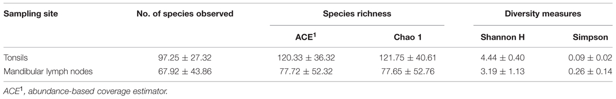
TABLE 1. Species richness and diversity measures of tonsils and mandibular lymph nodes (MLNs). Mean values ± standard deviation are listed.
Rarefaction curves were calculated for all samples and were depicted in Supplementary Figure S1A. High diversity coverage was achieved for MLNs with curves reaching asymptotes. Tonsils samples were sequenced sufficiently, but some tonsil curves did not reach complete saturation.
The actual number of detected OTUs varied highly among samples (23–171 OTUs). MLNs harbored between 23 and 171 OTUs (median = 66 OTUs) and tonsils between 49 and 158 OTUs (median = 105 OTUs). The total microbial diversity detected in MLNs and tonsils consisted of 355 OTUs and 278 OTUs, respectively.
Rank abundance curve dynamics (Supplementary Figure S1B) were similar to dynamics found in complex microbial communities of other biological niches (e.g., gastrointestinal microbiomes). A high proportion of very low abundant OTUs was dominated by some highly abundant OTUs: 10 OTUs of MLNs and 23 OTUs of tonsils reached relative abundances of >1%. The 30 most abundant OTUs over both sampling sites are depicted in Figure 1. Detailed information on relative abundances of OTUs and the associated p-values are listed in Supplementary Table S1. In tonsils, OTU 8, OTU 18, and OTU 1 dominated (best hits: Prevotella zoogleoformans, Treponema pedis, and Serratia proteamaculans) with relative abundances between 5.3 and 10.6%. In MLNs, OTU 1 (best hit: Serratia proteamaculans) was highly dominant (41.8% of all sequences), followed by OTU 15 and OTU 3 (best hits: Pseudomonas marginalis and Herbaspirillum huttiense) with 5.6 and 4.1% relative abundance (Figure 1). All closest reference strains of the OTUs detected in our study have previously been described as occurring in the gastrointestinal tract of mammals, on plants or environmental samples.
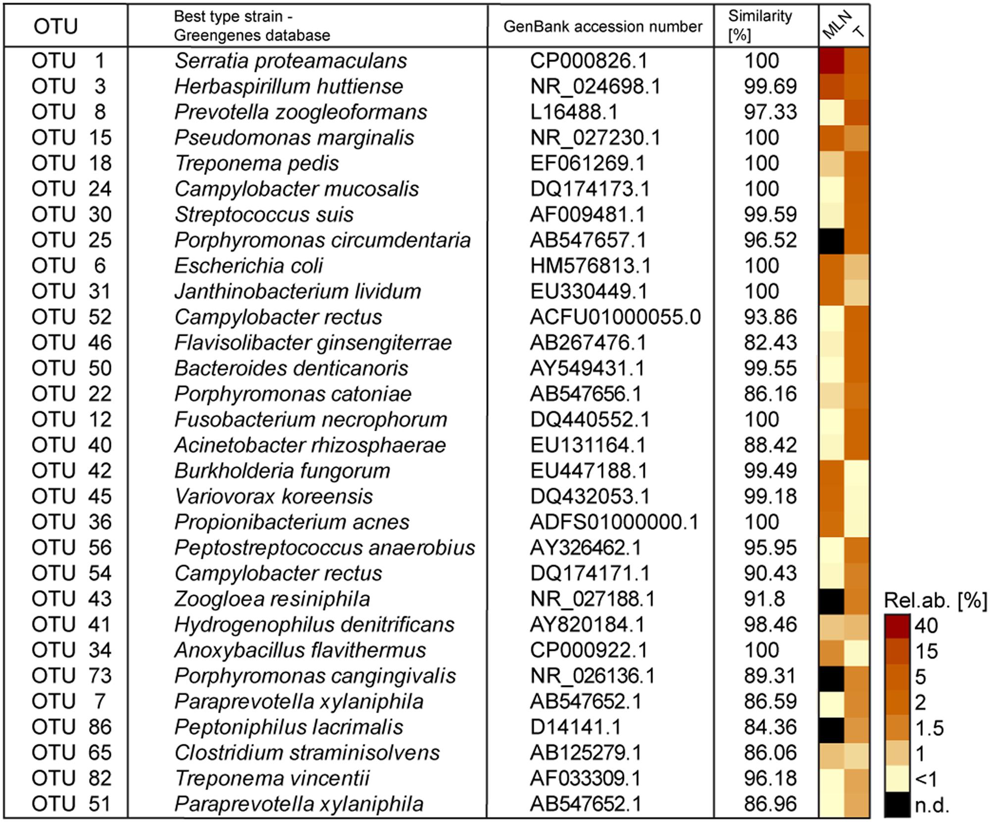
FIGURE 1. The 30 most abundant operational taxonomic units (OTUs) detected in tonsils and mandibular lymph nodes (MLNs) are shown in the heatmap. The best type strain hits (Greengenes database) and its accession numbers as well as similarity percentage is listed. Sampling sites were abbreviated as follows: T, tonsils; MLNs, mandibular lymph nodes. N.d. = not detected.
Considering all 576 OTUs, 32% were shared between tonsil and MLN samples (Figure 2). These shared OTUs include also highly abundant OTUs: From the 30 most abundant OTUs, 87% were detected in both sampling sites. Comparing the relative abundances of OTUs between the two datasets, OTU 8 (best hit: Prevotella zoogleoformans), OTU 24 (best hit: Campylobacter mucosalis), OTU 12 (best hit: Fusobacterium necrophorum), and OTU 56 (best hit: Peptostreptococcus anaerobius) were significantly increased in tonsils compared to MLN tissues (106.0-, 88.6-, 510.0-, and 91.4-fold change, respectively). OTU 45 (best hit: Variovorax koreensis) was significantly enriched in MLNs compared to tonsils (26.8-fold change).
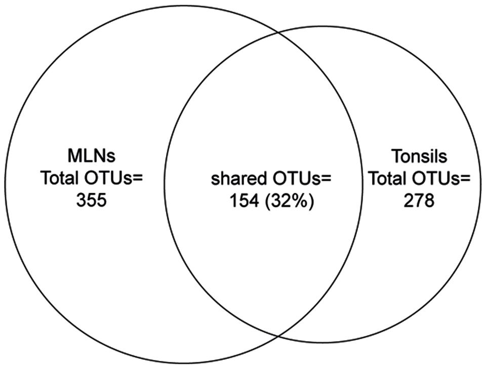
FIGURE 2. Venn diagram showing the number of shared OTUs between the two sampling sites. The size of the circles is in proportion to the number of OTUs detected in each sampling site.
Based on the mothur OTU classification, we assigned all OTUs to their respective genus (n = 230). The complete list of genera detected in each tissue and relative abundances of genera and p-values can be found in Supplementary Tables S2 and S3. In accordance to results on OTU level, Serratia, Herbaspirillum, and Pseudomonas were highly abundant in MLNs, whereas Prevotella, Treponema, Campylobacter, and Porphyromonas were highly abundant in tonsils. To explore bacterial correlation similarities between samples in a tissue group, pairwise Spearman correlations (rs) were calculated for abundant genera. Figure 3 shows that in the tonsil network Pseudomonas, Herbaspirillum, Serratia, and Janthinobacterium highly positively correlated among each other (rs > 0.9). A strong negative correlation (rs = –0.9) was calculated for Treponema and Pasteurella. In the MLN network Treponema, Anaerovirgula, and Proteocatella highly positively correlated with each other (rs > 0.9). Highly positive correlations were also found between Gemella, Porphyromonas, and Fusobacterium (rs > 0.9). Additionally, Comamonas and Paracoccus, Porphyromonas and Fusobacterium, Modestobacter and Roseomonas, and Serratia and Pseudomonas highly positively correlated (rs > 0.9).
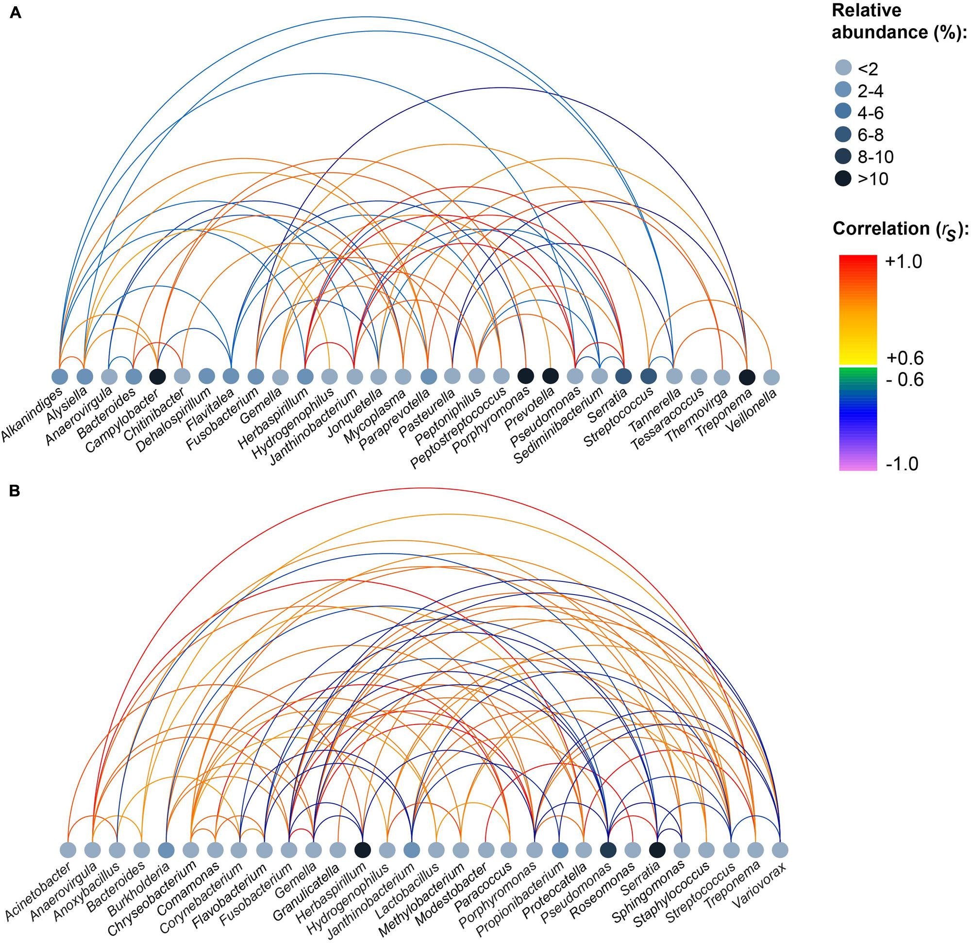
FIGURE 3. Strong positive and negative correlations between genera within all samples of each sampling site. (A) Tonsils, (B) mandibular lymph nodes. Pairwise Spearman correlations (rs) between the most abundant genera were computed for each tissue. Relative abundances of genera are indicated by node color. The color of the edges indicates the degree of correlation. The figure depicts correlations |rs| ≥ 0.6.
Tonsils and MLN samples clustered distinctly in the discriminant analysis (Figure 4). In the MLN dataset, Serratia, Herbaspirillum, Pseudomonas, Janthinobacterium, and Variovorax had the highest component loadings. In the tonsil dataset, Prevotella, Paraprevotella, Porphyromonas, and Peptostreptococcus vectors had highest loadings (loading plot data not shown).
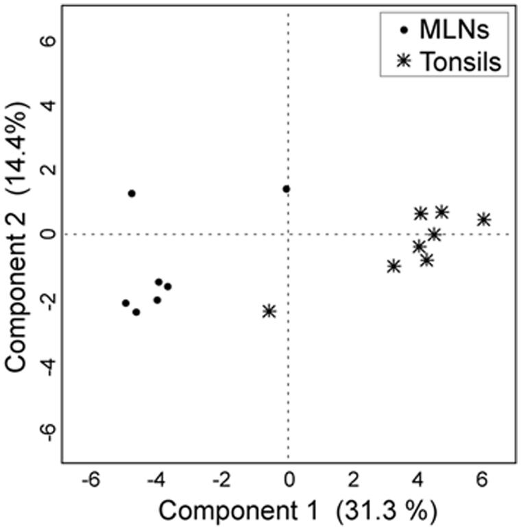
FIGURE 4. Discriminant analysis with the first two principal components. For the discriminant analysis the 30 most abundant genera were used as covariates and the sampling site as the categorial variable. Each point corresponds to one sample.
In total 16 phyla were identified, Proteobacteria, Firmicutes, and Bacteroidetes being most abundant (86.6% of all reads; Figure 5). Proteobacteria was significantly lower in tonsils compared to MLNs (2.5-fold change). Bacteroidetes, Spirochaetes, and Tenericutes were significantly increased in tonsils compared to MLNs (8.1-, 11.7-, and 28.4-fold change, respectively). Exact values of relative abundances at phylum level, SD and p-values are available in Supplementary Table S4.
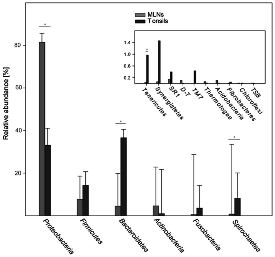
FIGURE 5. Relative abundances of bacterial phyla. Abundant phyla with more than 2% mean abundance are shown for each sampling site (tonsils, MLNs = mandibular lymph nodes). In the inlay, relative abundances of rare phyla (<2% mean abundance) are depicted. The phylum Deinococcus–Thermus is abbreviated with D–T, Thermodesulfobacteria with TSB. Error bars represent standard error of the means. Significance in one sampling site compared to another is marked with an asterisk and is defined as p ≤ 0.05 and q ≤ 0.1.
Discussion
In this study, the metabolically active part of the microbiome of cervical lymphatic tissues of slaughter pigs was examined for the first time.
Being aware of current discussions about artifacts that might be introduced by pyrosequencing (Quince et al., 2009, 2011), we set up stringent quality controls to reduce potential sequencing biases. However, reads from pyrosequencing are short (median value = 182 bp) and therefore the ‘best hits’ provided in this manuscript should be interpreted carefully, especially if sequence similarity is below 99%.
The Metabolically Active Microbiome of Porcine Palatine Tonsils
Pigs have five types of tonsils including lingual, paraepiglottic, pharyngeal, tubal, and palatine tonsils. Tonsils contain tonsillar crypts and lymphoid follicles with M cells located within deep crypts, aiming at providing a first ‘handling site’ for host immune cells (Horter et al., 2003). Although tonsils are routinely used in porcine disease surveillance for many pathogens (Bonardi et al., 2003; Wertheim et al., 2009), the nasal and palatine region of pigs has previously only been investigated to a limited extent concerning the diversity of microorganisms. In this study, we describe a substantial MAB diversity in palate tonsils. In contrast to our RNA-based approach, Lowe et al. used a DNA-based approach for examining the tonsil microbiome (Lowe et al., 2012). The actual richness detected in tonsils in the DNA-based approach amounted to four times the number of OTUs found in our study. This can be explained by a high number of dead bacteria that had already been processed by the host immune cells or bacterial DNA fragments detected. The high number of OTU 8 (best hit: Prevotella zoogleoformans) and OTU 18 (best hit: Treponema pedis) in tonsil samples was not surprising: In a study examining the porcine nasal microbiota based on DNA-based sequencing, Treponema had already been described as being the predominant microorganism (Weese et al., 2014). Prevotella are well-known colonizers of human tonsils as well (Jensen et al., 2013). The genus Prevotella is also known to be an abundant colonizer of the pig’s gastrointestinal tract, especially of the colonic region (46% relative abundance) and Prevotella consists of a huge variety of bacterial species (Mann et al., 2014b). The correlation network of tonsils revealed strong correlations between Pseudomonas, Herbaspirillum, Serratia, and Janthinobacterium indicating a constant co-occurrence pattern of these genera in all tonsil samples. Janthinobacterium and Pseudomonas were low abundant species which might benefit from the presence of Serratia, which can break down many carbon sources.
A previous study based on DNA-sequencing defined a core microbiome (Actinobacillus, Alkanindiges, Fusobacterium, Haemophilus, Lactobacillus, Moraxella, Pasteurella, Peptostreptococcus, Streptococcus, and Veillonella) of pig tonsils based on the examination of two healthy pig herds (Lowe et al., 2012). With the exception of Haemophilus, all genera listed were also detected in our study, confirming the results of the above-mentioned study but also showing that these organisms are viable in tonsils.
In accordance with the results reported here, we suggest that Prevotella, Paraprevotella, Porphyromonas, Treponema, and Campylobacter should be included in the definition of the core microbiome of porcine palatine tonsils, as these four organisms were highly abundant in our MAB tonsil dataset (between 5% and 18% relative abundance) and were present in all tonsils examined. These organisms, together with the ‘core genera’ defined by Lowe et al. (2012) accounted for 51.3% of all sequences. The organisms suggested above were also detected in Lowe’s DNA-based sequencing study (Lowe et al., 2012), but in lower abundance. At phylum level, the DNA-based sequencing study of Lowe et al. (2012) described 73% Proteobacteria in tonsils. Interestingly, Bacteroidetes, which was the most abundant metabolically active phylum in tonsils in our study, were described as low abundant (0.8% of total sequences) by Lowe et al. (2012). One explanation would be a high proportion of dead Proteobacteria that may have overwhelmed metabolically active Bacteroidetes and other phyla in DNA-based datasets. This would be in accordance with cultivation data of microbes from porcine tonsils, where only about 20% could be assigned to genera belonging to Proteobacteria (Lowe et al., 2011). However, as stated by Lowe et al. (2012), a possible amplification bias during PCR reactions in the DNA-based approach would also explain this difference. Interestingly, a DNA-based deep sequencing study examining human tonsils described a similar proportion of phyla in healthy adults as detected in our study, and found a high proportion of Proteobacteria linked with recurrent tonsillitis in children (Jensen et al., 2013). Although we assume that the Bacteroidetes fraction might have been strongly underestimated by Lowe et al. (2011), conclusions about a possible pathological process linked to a high number of Proteobacteria would need further investigations. In another study, Lowe et al. (2011) provided extensive DNA-based 16S rRNA clone libraries from pig tonsils. We processed these sequences together with our tonsil dataset and revealed 30 OTUs shared, which cover 47.3% of all sequences. This indicates a substantial overlap of abundant OTUs (Supplementary Figure S2).
The Metabolically Active Microbiome of Porcine MLN
In this study, we provide the first metabolically active microbiome of porcine MLNs consisting of commensals and pathogens. Thus, translocation of bacteria from the area of distribution to local lymph nodes is not restricted to pathological processes. The samples harbored between 23 and 171 OTUs (median = 66 OTUs), being in accordance with the number of OTUs detected in a DNA-based in-depth sequencing study of ileocaecal lymph nodes (Mann et al., 2014a). Previous studies have shown that the number of OTUs can vary widely in healthy (but also in pathologically altered) ileocaecal lymph nodes (Mann et al., 2014a). We assume that the exact sampling location (T-cell area fully covered or not) could contribute to differences in diversity. The overall diversity of MAB found in MLN was unexpectedly high (390 OTUs) and accounted for 60% of OTUs detected in ileocaecal lymph nodes with a DNA-based survey (Mann et al., 2014a). Interestingly 48.7% of all MLN sequences accounted for Serratia, of which 86% belonged to OTU 1 (best hit: Serratia proteamaculans). All pigs examined in this study harbored a high abundance of Serratia proteamaculans in MLNs. In the early 1950s, this Gram-negative, facultatively anaerobic bacterium was already proven to survive in lymph node tissue (Lepovetsky et al., 1953). Serratia proteamaculans was detected in moderate abundance (0.5%) in ileocaecal lymph nodes with the DNA-based survey (Mann et al., 2014a). Considering the high abundance of metabolically active Serratia which was found in this study, we assume that physiological conditions in lymph nodes foster their survival. Serratia proteamaculans has previously been isolated from human pneumonia samples, animal habitats and other environmental samples, indicating the flexibility of the growth of this species (Bollet et al., 1993; Dworkin and Falkow, 2006; Kajikazawa et al., 2007). It is known to be able to utilize a huge variety of carbon sources (Bollet et al., 1993; Dworkin and Falkow, 2006), and to be an opportunistic food-borne pathogen (Kajikazawa et al., 2007).
Herbaspirillum, also detected in high abundance (19.1%) in all pigs in our study, was not found in ileocaecal lymph nodes (Mann et al., 2014a). In total, 83% of Herbaspirillum sequences belonged to OTU 3 (best hit: Herbaspirillum huttiense), commonly found in soil- and animal- environments but also known to be present in human respiratory tracts (Spilker et al., 2008). It has been suggested that Herbaspirillum huttiense may contribute to lung diseases in humans (Spilker et al., 2008). In the MLN network, the number of strong correlations was more than double than that of the tonsil network. To our knowledge, most of the correlating genera (e.g., Porphyromonas or Treponema) have not previously been described in porcine lymph nodes until now.
Present results indicate that translocation processes to local lymph nodes are not restricted to lymph nodes draining the gastrointestinal tract, and are not restricted to pathological processes. Interestingly, the relatively high number of MAB found in pigs in the present study give us a direct hint to a possible survival scenario of various species in host immune cells. Dendritic cells for example are known to keep living bacteria and are ineffective in eliminating them, in contrast to macrophages (Macpherson and Uhr, 2004; Macpherson and Smith, 2006). However, further research investigating the exact position and function of MAB in lymph nodes is required.
This is the first RNA-based study exploring MAB in porcine lymphatic organs. Further studies using mRNA-based approaches might give insights into gene expression of the MAB and hence contribute to an increased understanding of microbial activity in lymphatic organs. With the methodological toolbox used, the relatedness between the bacterial community in lymphatic organs, the gut bacterial community and the slaughterhouse-specific bacterial community could be characterized and compared to link the occurrence of phylotypes to pork contamination in ongoing studies.
Conclusion
This study confirms the necessity of the EFSA regulation with regard to an MI based on visual MI and assures that this decision can contribute to minimalize microbial contamination.
Conflict of Interest Statement
The authors declare that the research was conducted in the absence of any commercial or financial relationships that could be construed as a potential conflict of interest.
Acknowledgments
We thank Ingeborg Klymiuk (Center for Medical Research, Medical University Graz, Austria) for directing the pyrosequencing approach. The authors are grateful to Clair Firth, Karin Lebl, Karin Frühwirth, and Eugen Mann for their comments and scientific support, which substantially helped to improve the manuscript. The present research was funded by the science funds of the Land Niederoesterreich, Austria (project ID: MIKRORIND). EM was partially funded by the Austrian Science Fund (FWF) project I 1251-B25 to SS-E, SW was funded by the WWTF project D-I.INFLACOW, LS12-010.
Supplementary Material
The Supplementary Material for this article can be found online at: http://journal.frontiersin.org/article/10.3389/fmicb.2015.01362
Footnotes
- ^ http://greengenes.lbl.gov
- ^ http://gastonsanchez.com/software/arcdiagram_introduction.pdf
- ^ http://www.r-project.org
References
Berg, R. D. (1995). Bacterial translocation from the gastrointestinal tract. Trends Microbiol. 3, 149–154. doi: 10.1016/S0966-842X(00)88906-4
Bollet, C., Grimont, P., Gainnier, M., Geissler, A., Sainty, J. M., and Demicco, P. (1993). Fatal pneumonia due to Serratia proteamaculans subsp. quinovora. J. Clin. Microbiol. 31, 444–445.
Bonardi, S., Brindani, F., Pizzin, G., Lucidi, L., D’incau, M., Liebana, E., et al. (2003). Detection of Salmonella spp., Yersinia enterocolitica and verocytotoxin-producing Escherichia coli O157 in pigs at slaughter in Italy. Int. J. Food Microbiol. 85, 101–110. doi: 10.1016/S0168-1605(02)00504-4
Cocolin, L., Dolci, P., and Rantsiou, K. (2011). Biodiversity and dynamics of meat fermentations: the contribution of molecular methods for a better comprehension of a complex ecosystem. Meat Sci. 89, 296–302. doi: 10.1016/j.meatsci.2011.04.011
Cuenca, S., Sanchez, E., Santiago, A., El Khader, I., Panda, S., Vidal, S., et al. (2014). Microbiome composition by pyrosequencing in mesenteric lymph nodes of rats with CCl4-induced cirrhosis. J. Innate Immun. 6, 263–271. doi: 10.1159/000356454
Dahlinger, J., Marks, S. L., and Hirsh, D. C. (1997). Prevalence and identity of translocating bacteria in healthy dogs. J. Vet. Int. Med. 11, 319–322. doi: 10.1111/j.1939-1676.1997.tb00473.x
DeSantis, T. Z., Hugenholtz, P., Larsen, N., Rojas, M., Brodie, E. L., Keller, K., et al. (2006). Greengenes, a chimera-checked 16S rRNA gene database and workbench compatible with ARB. Appl. Environ. Microbiol. 72, 5069–5072. doi: 10.1128/AEM.03006-05
Dorsch, M., and Stackebrandt, E. (1992). Some modifications in the procedure of direct sequencing of PCR amplified 16S rDNA. J. Microbiol. Methods 16, 271–279. doi: 10.1016/0167-7012(92)90017-X
Dworkin, M., and Falkow, S. (2006). The Prokaryotes: Proteobacteria, Gamma Subclass. New York, NY: Springer Science and Business Media.
European Commission (2014). Annexes to regulation (EC) No 853/2004 and (EC) No 854/2004 of the european parliament and of the council and commission regulation (EC) No 2074/2005. OJEUL 69, 95–98.
European Parliament and the Council of the European Union (2004). Regulation (EC) No 854/2004 of the european parliament and of the council of 29 April 2004 laying down specific rules for the organisation of official controls on products of animal origin intended for human consumption. OJEUL 226, 83–127.
Giraffa, G., and Neviani, E. (2001). DNA-based, culture-independent strategies for evaluating microbial communities in food-associated ecosystems. Int. J. Food Microbiol. 67, 19–34. doi: 10.1016/S0168-1605(01)00445-7
Horter, D. C., Yoon, K. J., and Zimmerman, J. J. (2003). A review of porcine tonsils in immunity and disease. Anim. Health. Res. Rev. 4, 143–155. doi: 10.1079/AHRR200358
Jensen, A., Fago-Olsen, H., Sorensen, C. H., and Kilian, M. (2013). Molecular mapping to species level of the tonsillar crypt microbiota associated with health and recurrent tonsillitis. PLoS ONE 8:e56418. doi: 10.1371/journal.pone.0056418
Joachimiak, M. P., Weisman, J. L., and May, B. (2006). ColorGrid: software for the visualization of biological measurements. BMC Bioinformatics 7:225. doi: 10.1186/1471-2105-7-225
Kajikazawa, T., Sugita, T., and Nishikawa, A. (2007). Comprehensive identification of bacteria in processed fresh edible sea urchin using 16S ribosomal DNA sequence analysis: the products contain various food poisoning-related bacteria and opportunistic bacterial pathogens. J. Health Sci. 53, 756–759. doi: 10.1248/Jhs.53.756
Lepovetsky, B. C., Weiser, H. H., and Deatherage, F. E. (1953). A microbiological study of lymph nodes, bone marrow and muscle tissue obtained from slaughtered cattle. Appl. Microbiol. 1, 57–59.
Lowe, B. A., Marsh, T. L., Isaacs-Cosgrove, N., Kirkwood, R. N., Kiupel, M., and Mulks, M. H. (2011). Microbial communities in the tonsils of healthy pigs. Vet. Microbiol. 147, 346–357. doi: 10.1016/j.vetmic.2010.06.025
Lowe, B. A., Marsh, T. L., Isaacs-Cosgrove, N., Kirkwood, R. N., Kiupel, M., and Mulks, M. H. (2012). Defining the “core microbiome” of the microbial communities in the tonsils of healthy pigs. BMC Microbiol. 12:20. doi: 10.1186/1471-2180-12-20
Macpherson, A. J., and Smith, K. (2006). Mesenteric lymph nodes at the center of immune anatomy. J. Exp. Med. 203, 497–500. doi: 10.1084/Jem.20060227
Macpherson, A. J., and Uhr, T. (2004). Induction of protective IgA by intestinal dendritic cells carrying commensal bacteria. Science 303, 1662–1665. doi: 10.1126/science.1091334
Madsen, L. W., Bak, H., Nielsen, B., Jensen, H. E., Aalbaek, B., and Riising, H. J. (2002). Bacterial colonization and invasion in pigs experimentally exposed to Streptococcus suis serotype 2 in aerosol. J. Vet. Med. B Infect. Dis. Vet. Public Health 49, 211–215. doi: 10.1046/j.1439-0450.2002.00491.x
Mann, E., Dzieciol, M., Metzler-Zebeli, B. U., Wagner, M., and Schmitz-Esser, S. (2014a). Microbiomes of unreactive and pathologically altered ileocecal lymph nodes of slaughter pigs. Appl. Environ. Microbiol. 80, 193–203. doi: 10.1128/AEM.03089-13
Mann, E., Schmitz-Esser, S., Zebeli, Q., Wagner, M., Ritzmann, M., and Metzler-Zebeli, B. U. (2014b). Mucosa-associated bacterial microbiome of the gastrointestinal tract of weaned pigs and dynamics linked to dietary calcium-phosphorus. PLoS ONE 9:e86950. doi: 10.1371/journal.pone.0086950
Mann, E., Dzieciol, M., Pinior, B., Neubauer, V., Metzler-Zebeli, B. U., Wagner, M., et al. (2015). High diversity of viable bacteria isolated from lymph nodes of slaughter pigs and its possible impacts for food safety. J. Appl. Microbiol. 119, 1420–1432. doi: 10.1111/jam.12933
Naranjo, V., Hofle, U., Vicente, J., Martin, M. P., Ruiz-Fons, F., Gortazar, C., et al. (2006). Genes differentially expressed in oropharyngeal tonsils and mandibular lymph nodes of tuberculous and nontuberculous European wild boars naturally exposed to Mycobacterium bovis. FEMS Immunol. Med. Microbiol. 46, 298–312. doi: 10.1111/j.1574-695X.2005.00035.x
Obata, T., Goto, Y., Kunisawa, J., Sato, S., Sakamoto, M., Setoyama, H., et al. (2010). Indigenous opportunistic bacteria inhabit mammalian gut-associated lymphoid tissues and share a mucosal antibody-mediated symbiosis. Proc. Natl. Acad. Sci. U.S.A. 107, 7419–7424. doi: 10.1073/pnas.1001061107
Pate, M., Pirs, T., Zdovc, I., Krt, B., and Ocepek, M. (2004). Haemolytic Rhodococcus equi isolated from a swine lymph node with granulomatous lesions. J. Vet. Med. B Infect. Dis. Vet. Public Health 51, 249–250. doi: 10.1111/j.1439-0450.2004.00758.x
Pinior, B., Conraths, F., Petersen, B., and Selhorst, T. (2015). Decision support for risk managers in the case of deliberate food contamination: the dairy industry as an example. Omega 53, 41–48. doi: 10.1016/j.omega.2014.09.011
Pinior, B., Konschake, M., Platz, U., Thiele, H. D., Petersen, B., Conraths, F. J., et al. (2012a). The trade network in the dairy industry and its implication for the spread of contamination. J. Dairy Sci. 95, 6351–6361. doi: 10.3168/jds.2012-5809
Pinior, B., Platz, U., Ahrens, U., Petersen, B., Conraths, F., and Selhorst, T. (2012b). The German milky way: trade structure of the milk industry, and possible consequences of a food crisis. J. Chain. Netw. Sci. 12, 25–39. doi: 10.3920/JCNS2012.x001
Pruesse, E., Quast, C., Knittel, K., Fuchs, B. M., Ludwig, W., Peplies, J., et al. (2007). SILVA: a comprehensive online resource for quality checked and aligned ribosomal RNA sequence data compatible with ARB. Nucleic Acids Res. 35, 7188–7196. doi: 10.1093/nar/gkm864
Quince, C., Lanzen, A., Curtis, T. P., Davenport, R. J., Hall, N., Head, I. M., et al. (2009). Accurate determination of microbial diversity from 454 pyrosequencing data. Nat. Methods 6, 639–641. doi: 10.1038/nmeth.1361
Quince, C., Lanzen, A., Davenport, R. J., and Turnbaugh, P. J. (2011). Removing noise from pyrosequenced amplicons. BMC Bioinformatics 12:38. doi: 10.1186/1471-2105-12-38
R Core Team (2014). R: A Language and Environment for Statistical Computing. Vienna: R Foundation for Statistical Computing.
Robertson, C. E., Harris, J. K., Wagner, B. D., Granger, D., Browne, K., Tatem, B., et al. (2013). Explicet: graphical user interface software for metadata-driven management, analysis and visualization of microbiome data. Bioinformatics 29, 3100–3101. doi: 10.1093/bioinformatics/btt526
Schloss, P. D., and Westcott, S. L. (2011). Assessing and improving methods used in operational taxonomic unit-based approaches for 16S rRNA gene sequence analysis. Appl. Environ. Microbiol. 77, 3219–3226. doi: 10.1128/AEM.02810-10
Schloss, P. D., Westcott, S. L., Ryabin, T., Hall, J. R., Hartmann, M., Hollister, E. B., et al. (2009). Introducing mothur: open-source, platform-independent, community-supported software for describing and comparing microbial communities. Appl. Environ. Microbiol. 75, 7537–7541. doi: 10.1128/AEM.01541-09
Scholz, H. C., Hofer, E., Vergnaud, G., Le Fleche, P., Whatmore, A. M., Al Dahouk, S., et al. (2009). Isolation of Brucella microti from mandibular lymph nodes of red foxes, Vulpes vulpes, in lower Austria. Vector Borne Zoonotic Dis. 9, 153–156. doi: 10.1089/vbz.2008.0036
Spilker, T., Uluer, A. Z., Marty, F. M., Yeh, W. W., Levison, J. H., Vandamme, P., et al. (2008). Recovery of Herbaspirillum species from persons with cystic fibrosis. J. Clin. Microbiol. 46, 2774–2777. doi: 10.1128/JCM.00460-08
Vieira-Pinto, M., Temudo, P., and Martins, C. (2005). Occurrence of Salmonella in the ileum, ileocolic lymph nodes, tonsils, mandibular lymph nodes and carcasses of pigs slaughtered for consumption. J. Vet. Med. B Infect. Dis. Vet. Public Health 52, 476–481. doi: 10.1111/j.1439-0450.2005.00892.x
Wang, Q., Garrity, G. M., Tiedje, J. M., and Cole, J. R. (2007). Naive Bayesian classifier for rapid assignment of rRNA sequences into the new bacterial taxonomy. Appl. Environ. Microbiol. 73, 5261–5267. doi: 10.1128/AEM.00062-07
Weese, J. S., Slifierz, M., Jalali, M., and Friendship, R. (2014). Evaluation of the nasal microbiota in slaughter-age pigs and the impact on nasal methicillin-resistant Staphylococcus aureus (MRSA) carriage. BMC Vet. Res. 10:69. doi: 10.1186/1746-6148-10-69
Weisburg, W. G., Barns, S. M., Pelletier, D. A., and Lane, D. J. (1991). 16S ribosomal DNA amplification for phylogenetic study. J. Bacteriol. 173, 697–703.
Wertheim, H. F., Nghia, H. D., Taylor, W., and Schultsz, C. (2009). Streptococcus suis: an emerging human pathogen. Clin. Infect. Dis. 48, 617–625. doi: 10.1086/596763
White, J. R., Nagarajan, N., and Pop, M. (2009). Statistical methods for detecting differentially abundant features in clinical metagenomic samples. PLoS Comput. Biol. 5:e1000352. doi: 10.1371/journal.pcbi.1000352
Wittekindt, N. E., Padhi, A., Schuster, S. C., Qi, J., Zhao, F., Tomsho, L. P., et al. (2010). Nodeomics: pathogen detection in vertebrate lymph nodes using meta-transcriptomics. PLoS ONE 5:e13432. doi: 10.1371/journal.pone.0013432
Keywords: 16S rRNA sequencing, lymph node, tonsil, contamination, slaughter house, microbiota
Citation: Mann E, Pinior B, Wetzels SU, Metzler-Zebeli BU, Wagner M and Schmitz-Esser S (2015) The Metabolically Active Bacterial Microbiome of Tonsils and Mandibular Lymph Nodes of Slaughter Pigs. Front. Microbiol. 6:1362. doi: 10.3389/fmicb.2015.01362
Received: 08 May 2015; Accepted: 17 November 2015;
Published: 15 December 2015.
Edited by:
Jean-christophe Augustin, Ecole Nationale Vétérinaire d’Alfort, FranceReviewed by:
Kiiyukia Matthews Ciira, Mount Kenya University, KenyaKalliopi Rantsiou, University of Turin, Italy
Copyright © 2015 Mann, Pinior, Wetzels, Metzler-Zebeli, Wagner and Schmitz-Esser. This is an open-access article distributed under the terms of the Creative Commons Attribution License (CC BY). The use, distribution or reproduction in other forums is permitted, provided the original author(s) or licensor are credited and that the original publication in this journal is cited, in accordance with accepted academic practice. No use, distribution or reproduction is permitted which does not comply with these terms.
*Correspondence: Evelyne Mann, evelyne.mann@vetmeduni.ac.at