 Eva J. Scharinger
 Richard Dietrich
 Tobias Wittwer2
 Erwin Märtlbauer
 Kristina Schauer
- 1Department of Veterinary Science, Faculty of Veterinary Medicine, Ludwig-Maximilians-Universität München, Oberschleißheim, Germany
- 2R-Biopharm AG, Darmstadt, Germany
The ubiquitous and opportunistic pathogen Cronobacter sakazakii is responsible for severe meningitis, sepsis, and necrotizing enterocolitis in neonates and infants associated with ingestion of contaminated powdered infant formula (PIF). The current ISO method for isolation and detection of Cronobacter spp. is laborious, time-consuming and expensive. In this study, a multiplexed lateral flow test strip was developed to rapidly detect and simultaneously serotype O1 and O2 C. sakazakii serotypes. The assay is based on two monoclonal antibodies (MAb) that specifically bind to the lipopolysaccharides (LPS) of these pathogens. The test strip provides results very quickly; C. sakazakii could be detected in pure culture within 15 min with a sensitivity of 107 CFU/ml. After non-selective enrichment for 18 h as low as one Cronobacter cell per g PIF could be detected. Moreover, the established lateral flow assay (LFA) offers excellent specificity showing no cross-reactivity with other C. sakazakii serotypes, Cronobacter species or Enterobacteriaceae tested. These characteristics, together with several advantages such as speed, simplicity in performance, low analysis cost, and no requirement of specialized skills or sophisticated equipment make the developed multiplexed LFA suitable for reliable detection and serotyping of C. sakazakii serotypes O1 and O2.
Introduction
Cronobacter spp. are opportunistic foodborne pathogens and can cause severe infections in all age groups, with neonates being the most susceptible and affected group for invasive disease (Lai, 2001). However, the number of neonatal infections with Cronobacter caused by contaminated PIF might be underestimated globally (Tothova et al., 2011). The genus comprises of seven species among which Cronobacter sakazakii are primarily associated with neonatal infections such as necrotizing enterocolitis (NEC), bacteremia and meningitis resulting in a high mortality rate of 40–80% (Bowen and Braden, 2006; Jason, 2012). At present, the species C. sakazakii consists of five serotypes O1–O4 and O7 (Blažková et al., 2015). Predominantly the serotypes O1 and O2 are isolated from clinical as well as from food products, suggesting that some serotypes of C. sakazakii might be more virulent than others (Blažková et al., 2015; Yan et al., 2015a,b; Scharinger et al., 2016). Therefore, it is important for epidemiological studies to not only identify the genus and species but also the serotype of the pathogen.
Epidemiologically, neonatal C. sakazakii infections are linked to the consumption of contaminated powdered infant formula (PIF) (Iversen and Forsythe, 2004; Drudy et al., 2006; Craven et al., 2010). As Cronobacter spp. does not survive the standard pasteurization process, a Cronobacter contamination critical for causing disease can only occur during filling and packaging and/or during reconstitution of PIF (Nazarowec-White and Farber, 1997; Parra-Flores et al., 2015). Furthermore, a hallmark of Cronobacter spp. is its extreme desiccation resistance: this bacterium may be recovered even after 2.5 years from a dairy food, in contrast to other PIF-associated bacteria such Salmonella enterica serovar Enteritidis and Escherichia coli which were already undetectable after 15 months (Barron and Forsythe, 2007). Therefore, a microbial risk assessment for E. sakazakii and other bacteria in PIF was developed by the WHO/FAO.
The contamination of PIF with Cronobacter spp. is being monitored using optimized conventional microbiological methods published by the International Standards Organization (ISO) and the International Dairy Federation (IDF) (ISO, 2006). However, these methods are laborious and definitive identification of Cronobacter species–without serotyping–takes up to 6 days, because diverse enrichments and biochemical confirmation steps are required. Besides culture-based methods, other rapid, highly sensitive and specific approaches for Cronobacter detection such as fluorescence in situ hybridization (FISH) (Almeida et al., 2009), DNA microarray (Wang et al., 2009), polymerase chain reaction (PCR) (Zimmermann et al., 2014; Yan and Fanning, 2015; Zhou et al., 2016), and antibody-based immunoassays (Hochel and Škvor, 2009; Blažková et al., 2011; Park et al., 2012; Xu et al., 2014; Scharinger et al., 2016) have been reported. All these techniques require specialized equipment and trained staff, consequently, they become expensive and laboratory-based.
Ideally, a new and improved diagnostic tool to detect Cronobacter should be more rapid, reliable, efficient, inexpensive and user-friendly. Among the diagnostic tests, the immunochromatographic strip assays satisfy all these prerequisites and some lateral flow tests for different pathogenic bacteria or their agents have already been successfully developed in order to enhance food safety (Kawatsu et al., 2008; Fang et al., 2014; Cox et al., 2015; Wu et al., 2015; Raeisossadati et al., 2016). Therefore, the aim of this study was to develop a multiplexed lateral flow assay (LFA) for rapid detection and simultanous serotyping of C. sakazakii serotypes O1 and O2, based on the recently described highly serotype specific monoclonal antibodies (MAbs) (Scharinger et al., 2016). The ability of the established lateral flow test strip to specifically detect C. sakazakii was confirmed by analyzing artificially contaminated PIF samples after enrichment in buffered peptone water (BPW) in comparison to the standard ISO reference method. This new multiplexed LFA shows great promise to complement or replace the time-consuming and culture-based methods for Cronobacter detection and has a high potential for on-site detection.
Materials and Methods
Bacterial Strains
The bacterial strains used in this study are listed in Table 1. All strains were grown in Luria-Bertani (LB) medium at 37°C with constant shaking. For solid media, 15 g/l agar was added. To measure the number of colony forming units (CFU), bacteria were quantified by plating 10-fold serial dilutions on Chromogenic Cronobacter Isolation Agar (CCI, Oxoid) or LB agar plates. The genus, species and serotype of Cronobacter strains were identified according to the proposed PCR-based schema for Cronobacter spp. (Lehner et al., 2006; Stoop et al., 2009; Jarvis et al., 2011; Sun et al., 2012; Carter et al., 2013; Yan et al., 2015a).
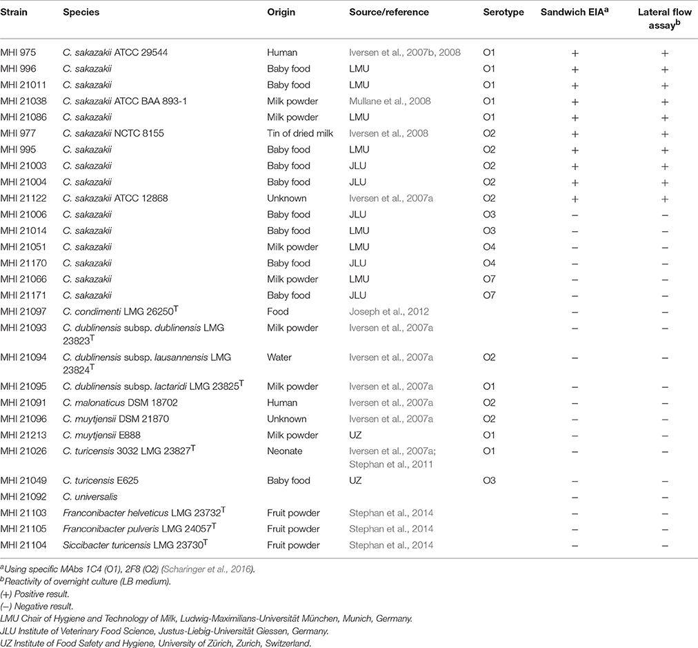
Table 1. Reactivity of bacterial strains used in this study.
Monoclonal Antibodies
MAbs 1C4 and 2F8 specific to C. sakazakii serotypes O1 and O2 respectively were described in a previous study (Scharinger et al., 2016).
Preparation of the Multiplexed Lateral Flow Test Strip
The LFA was carried out in a dipstick format (Figure 1A) which is typically composed of four parts: a sample pad for loading of the sample, a conjugate pad with the deposited detection molecule, a reaction nitrocellulose membrane for formation of the detectable colored lines and finally, an absorbent pad enhancing the capillary driving force and adsorbing non-reacting substances. In our developed lateral flow test strip, the sample pad was omitted and replaced by a longer conjugate pad. All parts are connected to each other and incorporated in the plastic backing card to ensure robustness of the LFA. The strips were prepared by the R-Biopharm AG in Germany. The detection zone of the strip is composed of three test lines, dispensed by an automatic dispenser on the nitrocellulose membrane (FF120 HP, Whatman), and contained (1) immobilized anti-dinitrophenyl (DNP) antibodies and (2) streptavidin as test lines for C. sakazakii serotype O1 and O2, respectively, and (3) goat-anti-mouse antibodies (Sigma-Aldrich, Steinheim, Germany) as a control line. As tracer conjugate, an anti-digoxigenin (DIG) antibody (Sigma-Aldrich, Steinheim, Germany) was conjugated to colloidal gold particles (diameter = 40 nm) and sprayed onto the glass fiber conjugate pad (SureWick, Merck Millipore). To achieve a sandwich format for the LFA, C. sakazakii serotype O1 and O2 specific antibodies were either conjugated to DNP and DIG (MAb 1C4 specific for C. sakazakii serotype O1) or to biotin and DIG (MAb 2F8 specific for C. sakazakii serotype O2). The mixed DNP- and biotin-labeled antibodies MAb 1C4-DNP (3.90 μg/ml) and MAb 2F8-biotin (2.49 μg/ml) were combined in dilution buffer (100 mM NaCl, 0.5% BSA, 0.5% Tween 20, 0.049% sodium azide in 50 mM phosphate buffer; pH 8.0) and designated as solution A, while DIG-labeled antibodies (MAb 1C4 5.31 μg/ml and MAb 2F8 2.85 μg/ml) represent solution B.
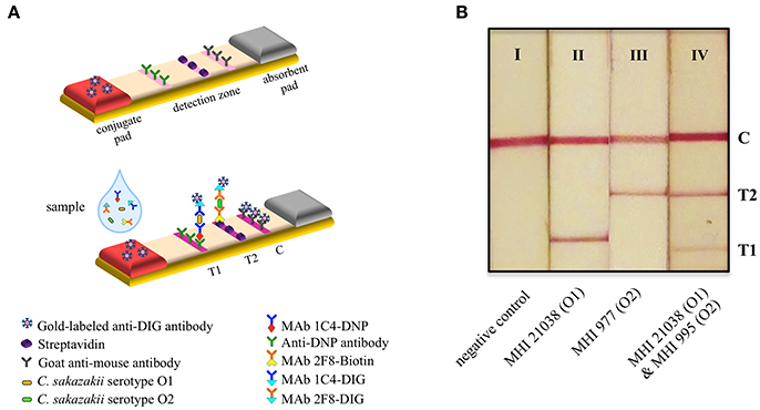
Figure 1. Principle and application of the multiplexed lateral flow test strip. (A) Schematic representation of the developed immunochromatographic assay. (B) Lateral flow test strips used for detection of C. sakazakii in inoculated PIF samples. Interpretation of the LFA results: I, negative (only one red line for the control line), II, and III, positive for C. sakazakii serotypes O1 and O2 (two red lines at the readout are: control line and test line 1 or test line 2, respectively), IV, positive for both C. sakazakii serotypes O1 and O2 (three red lines at the readout are: control line and two test line 1 and 2). C, control line; T1 and T2, test lines 1 and 2.
During the experiment, 100 μl of the bacterial suspension or pre-enriched baby food-sample was mixed with 400 μl dilution buffer. Fifty microliters of this mixture was combined with 50 μl of solution A and 50 μl of solution B. After 5 min of incubation at room temperature, 100 μl of this mixture was applied to the strip. The test results could be observed within 15 min of applying the sample. The signal intensities of the test and control lines were evaluated visually.
Inclusivity and Exclusivity Tests
The reactivity and specificity of the used MAbs 1C4 and 2F8 has been determined in a previous study by testing more than 100 Cronobacter spp. and other Enterobacteriaceae using EIAs (Scharinger et al., 2016). For this reason, only a few representative strains were used in this study to verify the specificity of the established LFA (Table 1). In order to investigate the inclusivity of the developed immunochromatographic strip, the reactivity of five C. sakazakii serotype O1 and five C. sakazakii serotype O2 strains was analyzed with the lateral flow test strip. The exclusivity of the LFA was tested by analyzing two strains each of the C. sakazakii serotypes O3-O4 and O7 as well as other Cronobacter species and foodborne pathogens often associated with C. sakazakii in PIF (Table 1). The tested strains were cultured in LB medium overnight to an average bacterial cell count of 109 CFU/ml. Then, the overnight cultures were mixed with dilution buffer, solutions A and B as described above, before applying 100 μl of this mixture directly to the lateral flow test strips.
Detection Limit
The limit of detection (LOD) of the LFA was determined by analyzing overnight culture of five strains of C. sakazakii serotype O1 and O2 each and with C. sakazakii inoculated follow-on formula milk powder. The pure bacterial cultures were tested in the LFA with and without 10-fold serial dilutions and the Cronobacter cell counts were determined in parallel as described above. The used C. sakazakii strains are listed in Table 1. To determine the LOD of the LFA in PIF, 10 g of follow-on formula milk powder inoculated with C. sakazakii strains MHI 21011 (serotype O1) and MHI 977 (serotype O2) at a level of 1 CFU per g were analyzed by the LFA. These samples were enriched in 90 ml BPW in accordance with the ISO standard method by incubating at 37°C for 20 h without agitation. PIF in BPW without bacteria served as a negative control. After incubation times of 8, 12, 16, 18, and 20 h, sample aliquots (three replicates for each sample and time point) were analyzed by the lateral flow test strip. In parallel, the exact CFU of each sample was determined according to the ISO method on CCI agar.
Evaluation of the Test with Standard Reference Material
nutriCert®-Cronobacter spp. in milk powder (25 g) reference material was obtained from the DRRR (Deutsches Referenzbüro für Ringversuche und Referenzmaterialien; German reference office for food proficiency testing and reference materials, Kempten, Germany). The standard reference material included two positive samples, each contaminated with C. sakazakii ATCC 29544 (serotype O1), and one negative sample without Cronobacter. Each sample was dissolved 1/10 in 225 ml BPW, incubated at 37°C without agitation for 18 h and then analyzed by the LFA. For control purposes, the Cronobacter cell counts were determined in parallel as described above.
Artificially Spiked Baby Food Experiments
Three different kinds of dried baby food, including two initial formulas (from birth on), two follow-on formulas (from 6th month on) and two plant-based baby food (mash) were obtained from a local German drugstore and tested negative for the presence of Cronobacter spp. using the detection method ISO/TS 22964:2006. For spiking experiments, each sample (10 g) was mixed with 90 ml BPW and artificially contaminated with three different strains of C. sakazakii serotype O1 (MHI 996, MHI 21011, MHI 21038) and O2 (MHI 977, MHI 995, MHI 21122). The inoculation of samples was carried out with 1 ml of a dilution of the C. sakazakii overnight culture corresponding to 1 or 10 CFU. In parallel, the real CFU/ml of each inoculum was determined by plating on CCI agar. Artificially contaminated samples were enriched in accordance with the ISO standard. After 18 h of non-selective enrichment as well as after a subsequent selective enrichment in mLST/vancomycin broth (20 h), samples were analyzed by the lateral flow test strip. In parallel, the CFU of each sample was determined by plating 10-fold serial dilutions on CCI agar.
Detection of C. sakazakii in Naturally Contaminated Samples
In total five Cronobacter-positive, in Mossel-Bouillon enriched samples were provided from a local PIF producing factory. Three environmental swabs, consisted of two floor samples before cleaning as well as one after cleaning, and two samples from the produced PIF were analyzed with the Assurance GDS™ Enterobacter sakazakii (BioControl, Bellvue, USA) by the factory laboratory. These enriched samples were directly applied to the lateral flow test strips according to the established protocol.
Results
Detection Principle of the Developed Multiplexed Lateral Flow Test Strip
The developed LFA is based on a sandwich-EIA format as illustrated in Figure 1A. In contrast to the usual LFA design in which gold-labeled antigen-specific antibodies serve as detection reagent, here a universally applicable experimental set-up was realized. This approach is based on the use of hapten-specific antibodies or reagents (streptavidin) for both capturing the immune complexes on the test membrane and visualizing bound immune complexes. This toolbox approach offers high-flexibility and broad versatility to the simple and fast realization of LFAs as the often poorly reproducible gold-labeling of the primary, antigen-specific antibodies is avoided. Prior to being applied to the lateral flow test strip, the sample was pre-incubated with solution A and B containing differently labeled anti-C. sakazakii antibodies (see Material and Methods). During the incubation of 5 min, a sandwich complex consisting of the bacterial cell and the different labeled antibodies forms. In the case of C. sakazakii serotype O1, DIG-, and DNP-labeled antibodies (MAbs 1C4) bind to the LPS structures on the bacterial surface, whereas in the case of C. sakazakii serotype O2 the sandwich complex is composed of the bacteria bound to a DIG- and a biotin-labeled antibodies (MAbs 2F8). After being applied to the lateral flow strip, the complex is bound by either the anti-DNP antibodies (test line 1) or streptavidin (test line 2) (Figure 1A). Gold labeled anti-DIG antibodies from the conjugation pad will bind to the DIG-labeled anti-Cronobacter antibody and lead to the formation of a visible red band. Unbound anti-DIG antibodies will be immobilized on the control line (goat-anti-mouse antibody). Coloration of the control line indicates the correct performance of the LFA. The excess fluid and any unreacted substances are absorbed by the absorbent pad. Observation of both a colored test line 1 or test line 2 and a colored control line on the membrane is a positive result indicating the presence of C. sakazakii serotype O1 or C. sakazakii serotype O2 in the sample. If both Cronobacter serotypes are present in a sample three colored bands would be obtained. A single colored control line represents a negative result (Figure 1B). In addition, the stability and non-bleaching of the colloidal gold-tracer enables the conservation of results for ≥10 years, provided that the fluid flow on the strip was interrupted after 15 min of reaction time.
Specificity and Sensitivity of the Lateral Flow Test Strip
To determine the specificity of the LFA, inclusivity and exclusivity tests were performed with pure bacterial cultures. For this purpose, five strains of C. sakazakii serotype O1 and O2 each were analyzed as well as two strains of each C. sakazakii serotypes O3, O4, O7, and ten Cronobacter spp.-strains, including C. condimenti, C. dublinensis, C. malonaticus, C. muytjensii, C. turicensis, and C. universalis. Three additional strains of the Enterobacteriaceae family, namely Franconibacter helveticus, Franconibacter pulveris, and Siccibacter turicensis were also tested because they often simultaneously occur in the same PIF. The CFU of the applied bacterial cultures ranged between 108 and 109 CFU/ml. For C. sakazakii serotype O1- and O2-strains (n = 10) the corresponding serotype specific colored test line and the colored control line appeared in all cases within 15 min after application of the sample to the strip. Thus, the serotypes of the C. sakazakii strains were identified correctly. For all other tested strains (n = 19) only the colored control line appeared on the test strip. Thus, no indication for false positive or false negative results was obtained (Table 1).
The sensitivity of the multiplexed lateral flow test strip was evaluated using the spiked PIF format. For this purpose, PIF samples were enriched for 8, 12, 16, 18, and 20 h in BPW and then analyzed by the LFA. In parallel, the number of CFU was determined by plating serial dilutions on CCI agar. As shown in Figure 2, after 8 h of enrichment, the bacterial concentration of C. sakazakii strains MHI 21011 (O1) and MHI 977 (O2) did not exceed 7 × 10⁵ CFU/ml and were not detectable with the lateral flow test strip. After 12 h, strain MHI 21011 (O1) reached a bacterial cell count of 1.3 × 10⁷ CFU/ml and was detectable by clearly visible test line 1 on the lateral flow test strip, while at the same time point strain MHI 977 (O2) was not detectable due to the lower cell-count of 6.7 × 10⁶ CFU/ml. After 16 h of enrichment, strain MHI 977 (O2) with a bacterial cell count of 7.5 × 10⁸ CFU/ml was also detectable. Longer enrichment of up to 20 h did not improve the reactivity of the test (Figure 2).
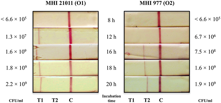
Figure 2. Detectability of C. sakazakii serotypes O1 and O2 after non-selective enrichment. PIF samples (10 g) were inoculated with 10 CFU of either strain MHI 21011 (C. sakazakii serotype O1) or MHI 977 (C. sakazakii serotype O2) and enriched in BPW. The samples were collected at 8, 12, 16, 18, and 20 h and analyzed by the LFA. The bacterial cell counts (CFU/ml) of the samples were determined by plating onto CCI agar.
Detection of C. sakazakii in Reference Material
nutriCert®-Cronobacter spp. in milk powder reference material (three samples at 25 g) was subjected to the cultured-based ISO detection procedure. After a non-selective enrichment step, samples were directly analyzed by the LFA and plated onto CCI agar to determine the CFU/ml. C. sakazakii serotype O1 could be detected in PIF sample 1 and PIF sample 3 at a bacterial cell count of 1.2 × 10⁹ CFU/ml. PIF sample 2 (negative control) was negative in the lateral flow test strip which was confirmed by the absence of bacterial growth on CCI agar (Table 2).
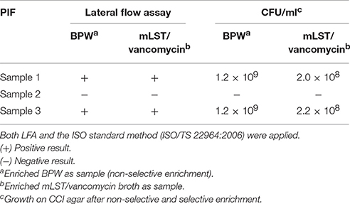
Table 2. Analysis of reference material “nutriCert®-Cronobacter spp. in milk powder” after non-selective and selective enrichment.
Detection of C. sakazakii in Artificially Contaminated Food Samples
Different commercially available infant food formulas, including initial milk formula, follow-on formula and plant-based baby food (mash) from two different manufacturers were chosen to test the applicability of the developed multiplexed LFA for the reproducible and sensitive detection of C. sakazakii. Samples of 10 g were dissolved in 90 ml BPW and each was subsequently inoculated with a strain of three different C. sakazakii serotype O1- (MHI 996, MHI 21011, MHI 21038) and three C. sakazakii serotype O2- (MHI 977, MHI 995, MHI 21122) strains. Except for strain MHI 21122 (origin unknown), all strains were originally isolated from baby food or milk powder/dried milk and thus were used for the artificial contamination of the infant food formulas in order to mimic natural conditions. Considering the zero tolerance limit laid down within the EU for Cronobacter in PIF (Anomymous, 2007), extreme low inoculum concentrations of one and ten bacterial CFU per 10 g milk powder were chosen to spike the samples prior to enrichment according to the ISO standard method. After non-selective pre-enrichment in BPW (18 h) and a subsequent selective enrichment in mLST/vancomycin broth (20 h), samples were applied to the lateral flow test strip and in parallel plated onto CCI agar to determine the bacterial cell count. Infant food formula samples without Cronobacter contamination served as negative controls and did not produce any false positive results in the LFA analyses. During validation study it turned out that 14 intrinsically with Cronobacter spiked PIF samples were tested negative both in LFA and by the ISO standard method and thus were excluded from the further statistical analysis (Figure 3). All these PIF samples has been spiked with low bacterial concentrations ranging from 0.8 to 3.1 CFU/10 g PIF.
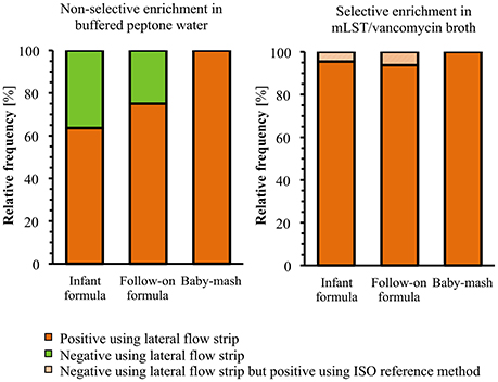
Figure 3. Overview of the LFA results obtained for artificially contaminated baby food samples (n = 58) after non-selective and selective enrichment in comparison to the ISO standard detection method.
In the contaminated initial milk formula, both C. sakazakii serotypes O1 and O2 could be detected in 63.6% (n = 7) of the spiked samples after the non-selective enrichment using the LFA (Figure 3). After a subsequent selective enrichment in mLST/vancomycin broth, 90.9% (n = 10) of all samples inoculated with C. sakazakii serotype O1 were tested positive. One false negative result was obtained for samples inoculated with 1 CFU of C. sakazakii MHI 21038 strain (Table 3). For C. sakazakii serotype O2, 100% (n = 11) of the samples tested positive after selective enrichment in mLST/vancomycin broth. Based on the CFU number of Cronobacter determined from the enriched cultures, most of the LFA-negative samples had not reached a bacterial concentration of >6.7 × 10⁶ CFU/ml. Thus, the lowest bacterial concentration that the LFA was able to detect was as low as 3 × 10⁷ CFU/ml (O1 serotype) and 1.3 × 10⁷ CFU/ml (O2 serotype) (Table 3).
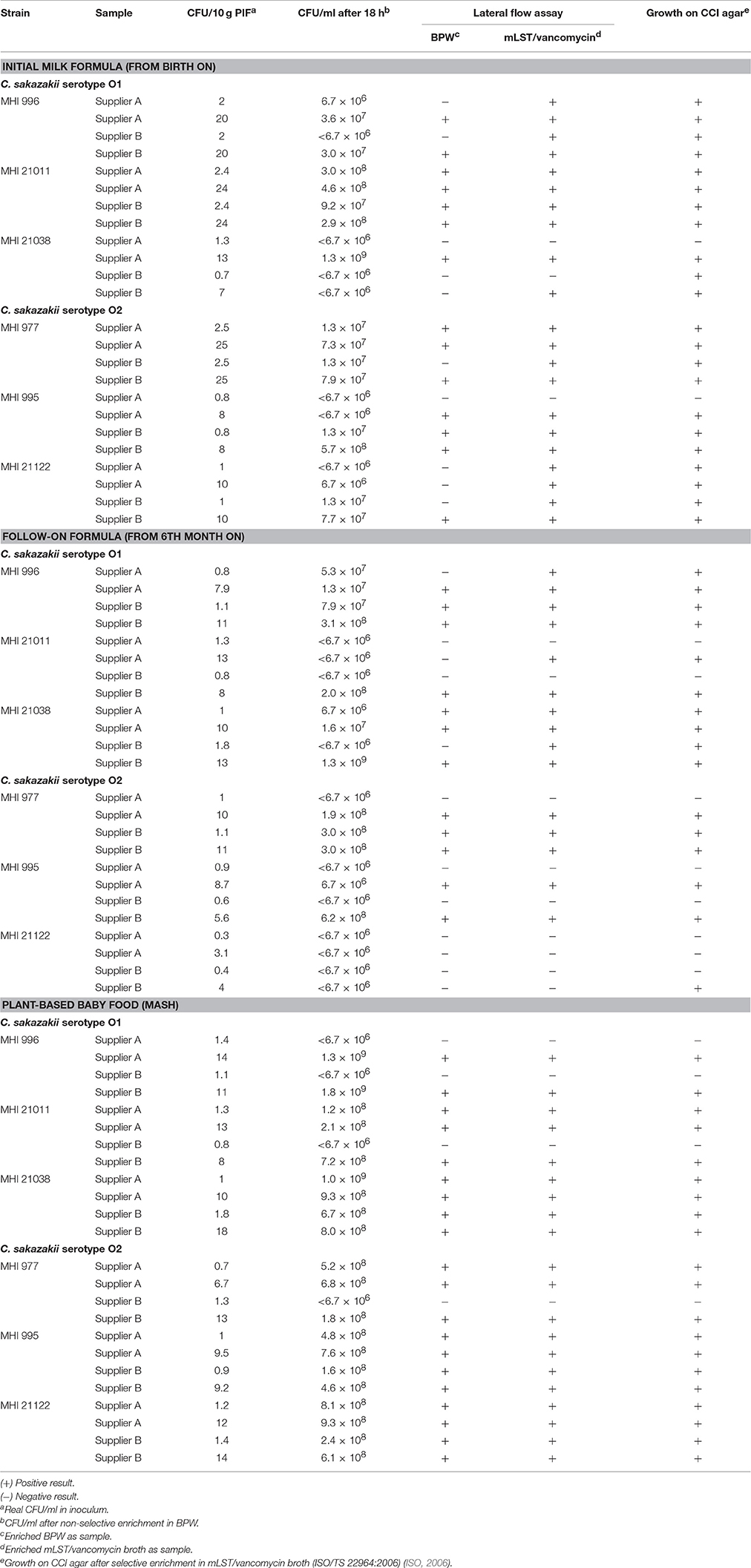
Table 3. Lateral flow test results of artificially contaminated dried baby food (n = 72) after non-selective and selective enrichment in comparison to the ISO detection method (ISO/TS 22964:2006).
Applying samples of follow-on formula spiked with different C. sakazakii serotype O1-strains to the test strip, the following results were obtained: 70% (n = 7) tested positive after non-selective enrichment and 100% (n = 10) after selective enrichment in mLST/vancomycin broth. In contrast, C. sakazakii serotype O2 was detected in 83.3% (n = 5) of artificially contaminated samples after non-selective enrichment and no improvement was observed after the additional selective enrichment. Bacterial cell counts as low as 6.7 × 10⁶ CFU/ml were detectable by the LFA in follow-on formula (Table 3). One sample, inoculated with low bacterial cell counts (Table 3, Figure 3) reacted positive using the ISO standard method but negative in the LFA. In this case, the CFU number of ≥108, characteristically obtained on average in mLST/vancomycin broth (Table 2), was not reached even after the selective enrichment.
For baby food samples such as plant-based mashes, that were spiked with the C. sakazakii serotypes O1 and O2, the LFA detected Cronobacter in 100% of the samples and in all cases already after non-selective enrichment (Figure 3). The lowest cell counts for Cronobacter in enriched baby food that could be detected by the developed LFA ranged from 1.2 to 1.6 × 10⁸ CFU/ml (Table 3). In contrast to the above described milk formula, a consistent growth of Cronobacter was observed in these plant-based baby foods (mash). In all spiked mash samples growth of Cronobacter could be confirmed, the CFU ranged from 1.2 × 10⁸ to 1.8 × 10⁹ CFU/ml after 18 h of non-selective enrichment (Table 3). Thus, identical results were obtained for the LFA and the ISO method (Figure 3).
Detection of C. sakazakii in Naturally Contaminated Factory Samples
Naturally contaminated environment and food samples (n = 5) were obtained from a PIF-producing factory and analyzed to test the applicability of the multiplexed LFA in routine testing. The commercial diagnostic system Assurance GDS™ E. sakazakii, which is routinely used in the PIF-producing factory, gave Cronobacter spp. positive results for two of three environmental swabs (floor samples before cleaning) and both PIF samples. The third environmental swab, taken from the floor after the cleaning, did not test positive. Identical results were obtained with the developed multiplexed LFA using the same samples enriched in Mossel-Bouillon. In all positive samples C. sakazakii serotype O1 could be identified by the developed LFA.
Discussion
With respect to the severe infections caused by Cronobacter spp. and manifested as necrotizing enterocolitis, sepsis or meningitis, a zero tolerance for the presence of Cronobacter spp. in PIF has been laid down within EU (FAO/WHO, 2007; VO(EU)365/2010, 2010). However, due to the ubiquitous character of this pathogen, PIF contamination during manufacture process, in households and hospitals cannot be fully avoided.
The distribution of the five C. sakazakii serotypes varies geographically and depends also in part on the sources of the isolation. In general, C. sakazakii serotypes O1 and O2 are the most frequently occurring C. sakazakii serotypes isolated from food and other different environments. For the identification of C. sakazakii conventional microbiological cultivation methods consisting of various enrichment procedures and biochemical confirmation are applied (ISO, 2006). These methods are laborious and time-consuming generally requiring 6 days for definitive identification of C. sakazakii. Although progress has been made during the last years in the development of highly sensitive and specific, but often complicated and expensive diagnostic methodologies, there is still a lack of a simple and rapid detection method for this pathogen in PIF. Thus, the aim of this study was to establish a multiplexed lateral flow assay, based on specific MAbs, to rapidly and reliably detect the most frequent serotypes of C. sakazakii.
In a previous study, we produced a set of specific and sensitive MAbs against C. sakazakii and developed a highly specific sandwich-EIA with excellent sensitivity ranging from 2 × 10³ to 3 × 10⁵ CFU/ml for the most commonly C. sakazakii serotypes O1, O2 as well as the serotype O3. Compared to the ISO standard method, this immunoassay represent a great improvement reducing the time to detect and simultaneous serotype C. sakazakii from 6 days to 18 h (Scharinger et al., 2016). Thus, this sandwich-EIA is well-suited for analytical or clinical laboratories but remains a multi-step and laboratory-based procedure. The development of a multiplexed lateral flow detection system to identify C. sakazakii contamination would bring additional key advantages, such as (i) lower sample volumes requirement, (ii) portability to enable point-of-care testing, and (iii) easier judgement of results by naked eye. Using the C. sakazakii specific MAbs 1C4 (O1) and 2F8 (O2) (Scharinger et al., 2016), we established a novel serotype specific LFA based on the sandwich EIA format. The direct reaction scheme is typically utilized when larger analytes with multiple antigenic sites are tested (Raeisossadati et al., 2016), as it is the case for the surface-located LPS recognized by the C. sakazakii serotype specific MAbs. In addition, the developed multiplexed LFA offers the great benefit of high diagnostic efficiency and generates conclusive information relating to C. sakazakii serotype. With the new test strip, C. sakazakii of the serotypes O1 and O2 can be detected and simultaneously serotyped within 15 min in pure bacterial culture or after an enrichment step of 18 h for environmental and clinical samples or food products.
The practical applicability of the established LFA was thoroughly examined by testing inclusivity and exclusivity, optimal incubation conditions and sensitivity in different baby food matrices. In pure bacterial culture, the lateral flow test strip showed 100% detection for C. sakazakii serotypes O1 and O2 and all tested bacteria listed in Table 1. None of the other Enterobacteriaceae that often simultaneously occur in the same PIF hampered the analyses and no cross-reactivity with other Cronobacter species and C. sakazakii serotypes tested was observed, demonstrating the specificity of the developed test system.
For the validation study inoculated PIF samples were prepared at a level which mimics naturally C. sakazakii contamination ranges found in infant food formulas. As the level of Cronobacter spp. in naturally contaminated samples is with <1 CFU/ml generally very low (Osaili and Forsythe, 2009), the sufficient enrichment of spiked samples up to at least 107 CFU/ml had to be ensured. Prolonged enrichment time increases the final bacterial cell count of the sample and, thus, the detection range of the LFA. According to the time lapses of the ISO standard method, incubation times of up to 20 h in buffered peptone water (BPW) were tested with respect to correlation between found bacterial cell counts and obtained intensity of the LFA test lines. The applicability of the LFA in routine testing was further investigated by the detection of C. sakazakii in commercially available PIF reference material and by a comprehensive validation study using three different kinds of infant food formulas for inoculation and three different C. sakazakii strains of each serotype at two bacterial cell count levels. In total, 72 different samples (3 baby food types × 2 producer brands × 2 serotypes × 3 strains × 2 CFU levels) were analyzed by LFA. The test strip allowed to detect up to 1–10 CFU in 10 g of infant formula, follow-on formula as well as in baby mash. After subtraction of the samples (n = 14), that were negative from the start (i.e., not contaminated), 79.3% (n = 46) tested positive for C. sakazakii serotypes O1 and O2 already after the initial non-selective enrichment. Negative samples were further enriched in mLST/vancomycin broth for 20 h according to the ISO standard method and were subsequently tested again using the LFA. After this additional enrichment, 96.6% (n = 56) of the artificially contaminated samples tested positive. False negative results were obtained only for two samples and in both samples a reduced Cronobacter growth was observed, i.e., CFU in non-selective enrichment broth were <6.7 × 10⁶ per ml.
This shows that in the vast majority of cases the LFA successfully detected C. sakazakii in PIF samples, inoculated with low (1 CFU/10 g PIF) or high (10 CFU/10 g PIF) pathogen counts, after non-selective or selective enrichment. Thus, the detection for C. sakazakii could be shortened from 6 days (ISO standard method) to 1 day (positive test result after non-selective enrichment in BPW) or to 2 days (positive test result after selective enrichment in mLST/vancomycin broth). The detection range of LFA for C. sakazakii in artificially contaminated PIF ranged from 106 CFU/ml (in some cases) to 107 CFU/ml for the detection of C. sakazakii serotypes O1 and O2, indicating that the LFA retains their sensitivity even in complex matrices such as PIF. Moreover, the detectability was comparable for both serotypes. In comparison to the LOD (≤103 CFU/ml) of the previously described highly specific and sensitive sandwich-EIA (Scharinger et al., 2016), the new multiplexed LFA shows a significantly lower sensitivity, however, the detection range of 107 CFU/ml is comparable to other lateral flow test systems developed for Cronobacter (Chen et al., 2014; Song et al., 2016) or other pathogenic bacteria such as Listeria, Salmonella, and Streptococcus (Raeisossadati et al., 2016; Eltzov and Marks, 2017). It is also noticeable, that after selective enrichment in mLST/vancomycin broth for 10 samples (17.2%, n = 10) a C. sakazakii contamination could be confirmed which reacted in the first test of the non-selective enrichment negative. This could be simply explained by the fact that, generally, the average of Cronobacter cell count in mLST/vancomycin broth was ≥108 CFU/ml, while non-selective enrichment in BPW often showed lower bacterial levels (<6.7 × 10⁶ CFU/ml), which is below the LOD of the developed LFA. Only in plant-based baby food (mash) samples, growth of both C. sakazakii serotypes to a higher cell count (108–109 CFU/ml) was consistently observed perhaps because Cronobacter is naturally associated with the plant environment as a possible reservoir of this pathogen (Grim et al., 2013; Singh et al., 2015; Vojkovska et al., 2016).
In the validation study, a relatively high percentage (19.4%, n = 14) of Cronobacter inoculated PIF samples gave negative results both in the LFA and the ISO standard method despite the fact that reconstituted infant formula is considered as a good medium for growth (Parra-Flores et al., 2016). This might be a consequence of spiking PIF samples with very low bacterial cell counts (1 CFU and 10 CFU per 10 g PIF). On the other hand, bacterial cultures are often heterogeneous and characterized by the presence of fast growing and different types of subpopulations–i.e., sensitive, less sensitive, and resistant to different environmental stresses–but not all cells within a population have the same probability to grow. Consequently, the ability of pathogenic microorganisms to grow to a high cell count from a low inoculum size could be affected by individual cell variability (Koutsoumanis, 2008). This variability was recently observed for C. sakazakii in single cell and micropopulations of ≤50 cells (Parra-Flores et al., 2016) and could explain false negatives as well as “not contaminated” results in PIF samples spiked with C. sakazakii while in pure bacterial cultures (~109 CFU/ml) a 100% detection for C. sakazakii serotypes O1 and O2 was achieved.
Advances in developing cost-effective and rapid bacterial test systems using a LFA format, have opened a new field of research in food safety and quality control. Many different LFAs have been developed recently to detect food contaminant compounds such as toxins, bacterial pathogens, or chemical contaminants such as additive, antibiotics (Law et al., 2014; Raeisossadati et al., 2016). During the last years, several immunochromatographic detection strips were developed for Cronobacter spp. as well: (i) the detection method of Blažková et al. (2011) combines nucleic acid amplification and the LFA principle but requires two additional steps, namely template DNA isolation and PCR amplification; (ii) Chen et al. (2014) used a silica-coated magnetic nanoparticles separation for developing an immunochromatographic strip; (iii) only Song et al. (2016) developed an immunochromatographic strip assay to specifically identify C. sakazakii. The assay was based on rabbit anti-C. sakazakii IgG-tagged liposome (immunoliposome) with a LOD of 107 CFU/ml, but the applicability of this test strip for food products was not described. In comparison to these previously described identification methods, the multiplexed LFA realized in this study is significantly easier to perform, less expensive and applicable in food products such as infant food formula. Additionally, the assay allows the simultaneous detection and serotyping of the most frequent C. sakazakii serotypes O1 and O2 with a detectability of less than 107 CFU/ml in pure bacterial culture or less than 10 Cronobacter cells in 10 g of PIF. In addition, the selected flexible LFA format offers the possibility to expand the number of detection lines so that the detection of other C. sakazakii serotypes could be included in future studies. For instance, a MAb enabling the specific detection of C. sakazakii serotype O3 has been described recently (Scharinger et al., 2016). Though the LFA was successfully validated in this study and enabled the sensitive detection of Cronobacter, particularly in baby food samples, a further increase of the assay sensitivity would be desirable to further improve the detectability of Cronobacter in PIF. As the LFA implemented MAbs 1C4 and 2F8 against C. sakazakii serotypes O1 and O2 are characterized by a very high affinity (Scharinger et al., 2016), test improvement is not a matter of antibody optimization but rather of signal amplifying strategies (Urusov et al., 2016). For instance, it is possible to considerable increase intensity of test lines and thus reduce the detection range by using gold/silver nanoparticles (Raeisossadati et al., 2016; Oliveira-Rodriguez et al., 2017). An increased sensitivity might improve the applicability of the LFA for on-site analyses.
Overall, the developed LFA can easily be used to routinely test dairy products for Cronobacter contamination, and thus ultimately minimize the risk of infant exposure to Cronobacter through contaminated PIF. Especially at 35–37°C, Cronobacter grows rapidly in PIF. For instance, the pathogen needs 5.2 h at room temperature, 1.8 h at 35°C and ca. 1.3 h at 37°C to grow from 100 to 1,000 CFU (Parra-Flores et al., 2015; K. Schauer, unpublished data). Despite detailed recommendations of the FAO/WHO for the preparation of milk based baby food, e.g., (1) using water at 70°C for PIF reconstitution, (2) the immediate use after being diluted, or (3) if not used, storage at <5°C (FAO/WHO, 2007; Silano et al., 2016), the reconstituted PIF is often stored in a bottle and baby food warmer or at room temperature, which is particularly problematic in geographic areas with high ambient temperature. Here, contaminated PIF with virulent Cronobacter could continue to grow and reach high cell densities, which are detectable with the developed LFA. This test strip may be also useful in epidemiological and clinical applications such as monitoring neonatal stations in hospitals for the presence of C. sakazakii and analyzing reconstituted PIF or Cronobacter isolates for C. sakazakii serotypes O1 and O2.
In conclusion, this study presents the first multiplexed LFA suitable for rapid and serotype specific detection of C. sakazakii serotypes O1 and O2. This method permits pathogen detection within 15 min in pure culture or after 18 h in PIF after a non-selective enrichment. Given a complete panel of mAbs and further modifications of the system to increase detection sensitivity, all relevant C. sakazakii serotypes could be detected with this approach in parallel. Low analysis cost, simplicity and quick performance are making this multiplexed lateral flow test strip a useful tool for point-of-care detection and attractive for fields such as food or health monitoring both to ensure the health of newborns and enhance microbiological safety of PIF.
Author Contributions
KS designed the experiments and wrote the manuscript. ES and KS performed the experiments. R-Biopharm AG and TW developed and prepared lateral flow test strip. KS and RD analyzed and interpreted the data. EM, ES, RD, and TW contributed to the drafting of the manuscript.
Conflict of Interest Statement
The authors declare that the research was conducted in the absence of any commercial or financial relationships that could be construed as a potential conflict of interest.
Acknowledgments
This work was supported in part by the Federal Ministry of Education and Research (BMBF) of Germany (Food supply and analysis (LEVERA), funding code 13N12611). We thank Gabi Acar for excellent technical assistance.
References
Almeida, C., Azevedo, N. F., Iversen, C., Fanning, S., Keevil, C. W., and Vieira, M. J. (2009). Development and application of a novel peptide nucleic acid probe for the specific detection of Cronobacter genomospecies (Enterobacter sakazakii) in powdered infant formula. Appl. Environ. Microbiol. 75, 2925–2930. doi: 10.1128/AEM.02470-08
Anomymous (2007). The Commission of the European Communities. Commission Regulation (EC) No. 1441/2007 of 5 December 2007 Amending Regulation (EC) No. 2073/2005 on Microbiological Criteria for Foodstuffs.
Barron, J. C., and Forsythe, S. J. (2007). Dry stress and survival time of Enterobacter sakazakii and other Enterobacteriaceae in dehydrated powdered infant formula. J. Food Prot. 70, 2111–2117. doi: 10.4315/0362-028X-70.9.2111
Blažková, M., Javurková, B., Fukal, L., and Rauch, P. (2011). Immunochromatographic strip test for detection of genus Cronobacter. Biosens. Bioelectron. 26, 2828–2834. doi: 10.1016/j.bios.2010.10.001
Blažková, M., Javurková, B., Vlach, J., Göselová, S., Karamonová, L., Ogrodzki, P., et al. (2015). Diversity of O antigens within the genus Cronobacter: from disorder to order. Appl. Environ. Microbiol. 81, 5574–5582. doi: 10.1128/AEM.00277-15
Bowen, A. B., and Braden, C. R. (2006). Invasive enterobacter sakazakii disease in infants. Emerg. Infect. Dis. 12, 1185–1189. doi: 10.3201/eid1208.051509
Carter, L., Lindsey, L. A., Grim, C. J., Sathyamoorthy, V., Jarvis, K. G., Gopinath, G., et al. (2013). Multiplex PCR assay targeting a diguanylate cyclase-encoding gene, cgcA, to differentiate species within the genus Cronobacter. Appl. Environ. Microbiol. 79, 734–737. doi: 10.1128/AEM.02898-12
Chen, F., Ming, X., Chen, X., Gan, M., Wang, B., Xu, F., et al. (2014). Immunochromatographic strip for rapid detection of Cronobacter in powdered infant formula in combination with silica-coated magnetic nanoparticles separation and 16S rRNA probe. Biosens. Bioelectron. 61, 306–313. doi: 10.1016/j.bios.2014.05.033
Cox, C. R., Jensen, K. R., Mondesire, R. R., and Voorhees, K. J. (2015). Rapid detection of Bacillus anthracis by gamma phage amplification and lateral flow immunochromatography. J. Microbiol. Methods 118, 51–56. doi: 10.1016/j.mimet.2015.08.011
Craven, H. M., Mcauley, C. M., Duffy, L. L., and Fegan, N. (2010). Distribution, prevalence and persistence of Cronobacter (Enterobacter sakazakii) in the nonprocessing and processing environments of five milk powder factories. J. Appl. Microbiol. 109, 1044–1052. doi: 10.1111/j.1365-2672.2010.04733.x
Drudy, D., Mullane, N. R., Quinn, T., Wall, P. G., and Fanning, S. (2006). Enterobacter sakazakii: an emerging pathogen in powdered infant formula. Clin. Infect. Dis. 42, 996–1002. doi: 10.1086/501019
Eltzov, E., and Marks, R. S. (2017). Colorimetric stack pad immunoassay for bacterial identification. Biosens. Bioelectron. 87, 572–578. doi: 10.1016/j.bios.2016.08.044
Fang, Z., Wu, W., Lu, X., and Zeng, L. (2014). Lateral flow biosensor for DNA extraction-free detection of Salmonella based on aptamer mediated strand displacement amplification. Biosens. Bioelectron. 56, 192–197. doi: 10.1016/j.bios.2014.01.015
FAO/WHO (2007). Safe Preparation, Storage and Handling of Powdered Infant Formula. Guidelines. Available online at http://www.who.int/foodsafety/publications/micro/pif2007/en
Grim, C. J., Kotewicz, M. L., Power, K. A., Gopinath, G., Franco, A. A., Jarvis, K. G., et al. (2013). Pan-genome analysis of the emerging foodborne pathogen Cronobacter spp. suggests a species-level bidirectional divergence driven by niche adaptation. BMC Genomics 14:366. doi: 10.1186/1471-2164-14-366
Hochel, I., and Škvor, J. (2009). Characterisation of antibodies for the immunochemical detection of Enterobacter sakazakii. Czech. J. Food Sci. 27, 66–74.
ISO (2006). ISO/TS 22964:2006 (IDF/RM 210:2006). Milk and Milk Products – Detection of Enterobacter Sakazakii. International Organization for Standardization, Geneva.
Iversen, C., and Forsythe, S. (2004). Isolation of Enterobacter sakazakii and other Enterobacteriaceae from powdered infant formula milk and related products. Food Microbiol. 21, 771–777. doi: 10.1016/j.fm.2004.01.009
Iversen, C., Lehner, A., Mullane, N., Bidlas, E., Cleenwerck, I., Marugg, J., et al. (2007a). The taxonomy of Enterobacter sakazakii: proposal of a new genus Cronobacter gen. nov. and descriptions of Cronobacter sakazakii comb. nov. Cronobacter sakazakii subsp. sakazakii, comb. nov., Cronobacter sakazakii subsp. malonaticus subsp. nov., Cronobacter turicensis sp. nov., Cronobacter muytjensii sp. nov., Cronobacter dublinensis sp. nov. and Cronobacter genomospecies 1. BMC Evol. Biol. 7:64. doi: 10.1186/1471-2148-7-64
Iversen, C., Lehner, A., Mullane, N., Marugg, J., Fanning, S., Stephan, R., et al. (2007b). Identification of “Cronobacter” spp. (Enterobacter sakazakii). J. Clin. Microbiol. 45, 3814–3816. doi: 10.1128/JCM.01026-07
Iversen, C., Mullane, N., Mccardell, B., Tall, B. D., Lehner, A., Fanning, S., et al. (2008). Cronobacter gen. nov., a new genus to accommodate the biogroups of Enterobacter sakazakii, and proposal of Cronobacter sakazakii gen. nov., comb. nov., Cronobacter malonaticus sp. nov., Cronobacter turicensis sp. nov., Cronobacter muytjensii sp. nov., Cronobacter dublinensis sp. nov., Cronobacter genomospecies 1, and of three subspecies, Cronobacter dublinensis subsp. dublinensis subsp. nov., Cronobacter dublinensis subsp. lausannensis subsp. nov. and Cronobacter dublinensis subsp. lactaridi subsp. nov. Int. J. Syst. Evol. Microbiol. 58, 1442–1447. doi: 10.1099/ijs.0.65577-0
Jarvis, K. G., Grim, C. J., Franco, A. A., Gopinath, G., Sathyamoorthy, V., Hu, L., et al. (2011). Molecular characterization of Cronobacter lipopolysaccharide O-antigen gene clusters and development of serotype-specific PCR assays. Appl. Environ. Microbiol. 77, 4017–4026. doi: 10.1128/AEM.00162-11
Jason, J. (2012). Prevention of invasive Cronobacter infections in young infants fed powdered infant formulas. Pediatrics 130, 1076–1084. doi: 10.1542/peds.2011-3855
Joseph, S., Cetinkaya, E., Drahovska, H., Levican, A., Figueras, M. J., and Forsythe, S. J. (2012). Cronobacter condimenti sp. nov., isolated from spiced meat, and Cronobacter universalis sp. nov., a species designation for Cronobacter sp. genomospecies 1, recovered from a leg infection, water and food ingredients. Int. J. Syst. Evol. Microbiol. 62, 1277–1283. doi: 10.1099/ijs.0.032292-0
Kawatsu, K., Kumeda, Y., Taguchi, M., Yamazaki-Matsune, W., Kanki, M., and Inoue, K. (2008). Development and evaluation of immunochromatographic assay for simple and rapid detection of Campylobacter jejuni and Campylobacter coli in human stool specimens. J. Clin. Microbiol. 46, 1226–1231. doi: 10.1128/JCM.02170-07
Koutsoumanis, K. (2008). A study on the variability in the growth limits of individual cells and its effect on the behavior of microbial populations. Int. J. Food Microbiol. 128, 116–121. doi: 10.1016/j.ijfoodmicro.2008.07.013
Lai, K. K. (2001). Enterobacter sakazakii infections among neonates, infants, children, and adults. case reports and a review of the literature. Medicine 80, 113–122. doi: 10.1097/00005792-200103000-00004
Law, J. W., Ab Mutalib, N. S., Chan, K. G., and Lee, L. H. (2014). Rapid methods for the detection of foodborne bacterial pathogens: principles, applications, advantages and limitations. Front. Microbiol. 5:770. doi: 10.3389/fmicb.2014.00770
Lehner, A., Nitzsche, S., Breeuwer, P., Diep, B., Thelen, K., and Stephan, R. (2006). Comparison of two chromogenic media and evaluation of two molecular based identification systems for Enterobacter sakazakii detection. BMC Microbiol. 6:15. doi: 10.1186/1471-2180-6-15
Mullane, N. R., Ryan, M., Iversen, C., Murphy, M., O'gaora, P., Quinn, T., et al. (2008). Development of multiple-locus variable-number tandem-repeat analysis for the molecular subtyping of Enterobacter sakazakii. Appl. Environ. Microbiol. 74, 1223–1231. doi: 10.1128/AEM.01726-07
Nazarowec-White, M., and Farber, J. M. (1997). Incidence, survival, and growth of Enterobacter sakazakii in infant formula. J. Food Prot. 60, 226–230. doi: 10.4315/0362-028X-60.3.226
Oliveira-Rodriguez, M., Serrano-Pertierra, E., Garcia, A. C., Lopez-Martin, S., Yanez-Mo, M., Cernuda-Morollon, E., et al. (2017). Point-of-care detection of extracellular vesicles: sensitivity optimization and multiple-target detection. Biosens. Bioelectron. 87, 38–45. doi: 10.1016/j.bios.2016.08.001
Osaili, T., and Forsythe, S. (2009). Desiccation resistance and persistence of Cronobacter species in infant formula. Int. J. Food Microbiol. 136, 214–220. doi: 10.1016/j.ijfoodmicro.2009.08.006
Park, S., Shukla, S., Kim, Y., Oh, S., Hun Kim, S., and Kim, M. (2012). Development of sandwich enzyme-linked immunosorbent assay for the detection of Cronobacter muytjensii (formerly called Enterobacter sakazakii). Microbiol. Immunol. 56, 472–479. doi: 10.1111/j.1348-0421.2012.00466.x
Parra-Flores, J., Juneja, V., Garcia De Fernando, G., and Aguirre, J. (2016). Variability in cell response of Cronobacter sakazakii after mild-heat treatments and its impact on food safety. Front. Microbiol. 7:535. doi: 10.3389/fmicb.2016.00535
Parra-Flores, J., Rodriguez, A., Riffo, F., Arvizu-Medrano, S. M., Arias-Rios, E. V., and Aguirre, J. (2015). Investigation on the factors affecting Cronobacter sakazakii contamination levels in reconstituted powdered infant formula. Front. Pediatr. 3:72. doi: 10.3389/fped.2015.00072
Raeisossadati, M. J., Danesh, N. M., Borna, F., Gholamzad, M., Ramezani, M., Abnous, K., et al. (2016). Lateral flow based immunobiosensors for detection of food contaminants. Biosens. Bioelectron. 86, 235–246. doi: 10.1016/j.bios.2016.06.061
Scharinger, E. J., Dietrich, R., Kleinsteuber, I., Märtlbauer, E., and Schauer, K. (2016). Simultaneous rapid detection and serotyping of Cronobacter sakazakii serotypes O1, O2 and O3 using specific monoclonal antibodies. Appl. Environ. Microbiol. 82, 2300–2311. doi: 10.1128/AEM.04016-15
Silano, M., Paganin, P., and Davanzo, R. (2016). Time for the 70°C water precautionary option in the home dilution of powdered infant formula. Ital. J. Pediatr. 42:17. doi: 10.1186/s13052-016-0228-9
Singh, N., Goel, G., and Raghav, M. (2015). Prevalence and characterization of Cronobacter spp. from various foods, medicinal plants, and environmental samples. Curr. Microbiol. 71, 31–38. doi: 10.1007/s00284-015-0816-8
Song, X., Shukla, S., Lee, G., and Kim, M. (2016). Immunochromatographic strip assay for detection of Cronobacter sakazakii in pure culture. J. Microbiol. Biotechnol. 26, 1855–1862. doi: 10.4014/jmb.1606.06004
Stephan, R., Grim, C. J., Gopinath, G. R., Mammel, M. K., Sathyamoorthy, V., Trach, L. H., et al. (2014). Re-examination of the taxonomic status of Enterobacter helveticus, Enterobacter pulveris and Enterobacter turicensis as members of the genus Cronobacter and their reclassification in the genera Franconibacter gen. nov. and Siccibacter gen. nov. as Franconibacter helveticus comb. nov., Franconibacter pulveris comb. nov. and Siccibacter turicensis comb. nov., respectively. Int. J. Syst. Evol. Microbiol. 64, 3402–3410. doi: 10.1099/ijs.0.059832-0
Stephan, R., Lehner, A., Tischler, P., and Rattei, T. (2011). Complete genome sequence of Cronobacter turicensis LMG 23827, a food-borne pathogen causing deaths in neonates. J. Bacteriol. 193, 309–310. doi: 10.1128/JB.01162-10
Stoop, B., Lehner, A., Iversen, C., Fanning, S., and Stephan, R. (2009). Development and evaluation of rpoB based PCR systems to differentiate the six proposed species within the genus Cronobacter. Int. J. Food Microbiol. 136, 165–168. doi: 10.1016/j.ijfoodmicro.2009.04.023
Sun, Y., Wang, M., Wang, Q., Cao, B., He, X., Li, K., et al. (2012). Genetic analysis of the Cronobacter sakazakii O4 to O7 O-antigen gene clusters and development of a PCR assay for identification of all C. sakazakii O serotypes. Appl. Environ. Microbiol. 78, 3966–3974. doi: 10.1128/AEM.07825-11
Tothova, L., Celec, P., Babickova, J., Gajdosova, J., Al-Alami, H., Kamodyova, N., et al. (2011). Phage therapy of Cronobacter-induced urinary tract infection in mice. Med. Sci. Monit. 17, BR173–BR178. doi: 10.12659/MSM.881844
Urusov, A. E., Petrakova, A. V., Zherdev, A. V., and Dzantiev, B. B. (2016). “Multistage in one touch” design with a universal labelling conjugate for high-sensitive lateral flow immunoassays. Biosens. Bioelectron. 86, 575–579. doi: 10.1016/j.bios.2016.07.027
VO(EU)365/2010 (2010). Commission Regulation (EU) No 365/2010 of 28 April 2010 Amending Regulation (EC) NO 2073/2005 on Microbiological Criteria for Foodstuffs as Regards Enterobacteriaceae in Pasteurised Milk and Other Pasteurised Liquid Dairy Products and Listeria monocytogenes in Food Grade Salt. OJ L 107 (29.4.2010), 9–11.
Vojkovska, H., Karpiskova, R., Orieskova, M., and Drahovska, H. (2016). Characterization of Cronobacter spp. isolated from food of plant origin and environmental samples collected from farms and from supermarkets in the Czech Republic. Int. J. Food Microbiol. 217, 130–136. doi: 10.1016/j.ijfoodmicro.2015.10.017
Wang, M., Cao, B., Gao, Q., Sun, Y., Liu, P., Feng, L., et al. (2009). Detection of Enterobacter sakazakii and other pathogens associated with infant formula powder by use of a DNA microarray. J. Clin. Microbiol. 47, 3178–3184. doi: 10.1128/JCM.00366-09
Wu, W., Zhao, S., Mao, Y., Fang, Z., Lu, X., and Zeng, L. (2015). A sensitive lateral flow biosensor for Escherichia coli O157:H7 detection based on aptamer mediated strand displacement amplification. Anal. Chim. Acta 861, 62–68. doi: 10.1016/j.aca.2014.12.041
Xu, X., Zhang, Y., Shi, M., Sheng, W., Du, X. J., Yuan, M., et al. (2014). Two novel analytical methods based on polyclonal and monoclonal antibodies for the rapid detection of Cronobacter spp.: development and application in powdered infant formula. LWT Food Sci. Technol. 56, 335–340. doi: 10.1016/j.lwt.2013.11.028
Yan, Q., and Fanning, S. (2015). Strategies for the identification and tracking of Cronobacter species: an opportunistic pathogen of concern to neonatal health. Front. Pediatr. 3:38. doi: 10.3389/fped.2015.00038
Yan, Q., Jarvis, K. G., Chase, H. R., Hebert, K., Trach, L. H., Lee, C., et al. (2015a). A proposed harmonized LPS molecular-subtyping scheme for Cronobacter species. Food Microbiol. 50, 38–43. doi: 10.1016/j.fm.2015.03.003
Yan, Q., Wang, J., Gangiredla, J., Cao, Y., Martins, M., Gopinath, G. R., et al. (2015b). Comparative genotypic and phenotypic analysis of Cronobacter species cultured from four powdered infant formula production facilities: Indication of pathoadaptation along the food chain. Appl. Environ. Microbiol. 81, 4388–4402. doi: 10.1128/AEM.00359-15
Zhou, B., Chen, B., Wu, X., Li, F., Yu, P., Aguilar, Z. P., et al. (2016). A new application of a sodium deoxycholate-propidium monoazide-quantitative PCR assay for rapid and sensitive detection of viable Cronobacter sakazakii in powdered infant formula. J. Dairy Sci. 99, 9550–9559. doi: 10.3168/jds.2016-11538
Keywords: Cronobacter sakazakii, multiplexed lateral flow immunoassay, PIF, food analysis and safety, serotyping
Citation: Scharinger EJ, Dietrich R, Wittwer T, Märtlbauer E and Schauer K (2017) Multiplexed Lateral Flow Test for Detection and Differentiation of Cronobacter sakazakii Serotypes O1 and O2. Front. Microbiol. 8:1826. doi: 10.3389/fmicb.2017.01826
Received: 20 June 2017; Accepted: 06 September 2017;
Published: 20 September 2017.
Edited by:
Julio Parra-Flores, University of the Bío Bío, ChileReviewed by:
Ariadnna Cruz-Córdova, Hospital Infantil de México Federico Gómez, MexicoJuan Aguirre, Universidad de Chile, Chile
Copyright © 2017 Scharinger, Dietrich, Wittwer, Märtlbauer and Schauer. This is an open-access article distributed under the terms of the Creative Commons Attribution License (CC BY). The use, distribution or reproduction in other forums is permitted, provided the original author(s) or licensor are credited and that the original publication in this journal is cited, in accordance with accepted academic practice. No use, distribution or reproduction is permitted which does not comply with these terms.
*Correspondence: Kristina Schauer, kristina.schauer@mh.vetmed.uni-muenchen.de
†Present Address: Eva J. Scharinger, Milchprüfring Bayern e.V., Wolnzach, Germany