- 1Centre for Tropical Diseases, Sacro Cuore Don Calabria Hospital, Verona, Italy
- 2Medical Oncology, Sacro Cuore Don Calabria Hospital, Verona, Italy
- 3Department of Computer Science, University of Verona, Verona, Italy
For many years microscopy has been considered the mainstay of the diagnosis of parasitic infections. In our laboratory, before the advent of molecular biology, the approach for the identification of parasitic infections in stools was the microscopic exam of three samples. Once we adopted molecular biology, a real-time PCR on one single sample was added to the classical coproparasitological exam of three samples. Given the high sensitivity of real-time PCR (Rt-PCR), we then decided to evaluate if a change of our routine was justified. In detail, we intended to assess if a much more practical routine, based on the analysis of a single fecal sample, was sufficiently sensitive to replace the routine described above. The new approach to be evaluated included, on the same and unique fecal sample, a classical coproparasitological exam plus Rt-PCR. The data obtained showed that the sensitivity of the new proposed approach remains very high, despite the reduction of coproparasitological exams from three to one, with the advantage of reducing costs and saving time, both for patients and for the laboratory.
Introduction
Microscopy is the classical procedure for diagnosing parasitic infections, including the identification of protozoan trophozoites and cysts in feces, and is still the primary, often only, test offered by most routine diagnostic services.
Because several intestinal parasites are shed intermittently, patients are usually asked to deliver multiple stool samples for examination (Danciger and Lopez, 1975; Cartwright, 1999). Although this procedure is relatively simple, and allows for the identification of both helminth eggs and larvae, as well as protozoan cysts and vegetative forms, microscopy clearly has its limitations and is a time consuming procedure (Utzinger et al., 2010). Some species are difficult or even impossible to differentiate (e.g., the complex Entamoeba histolytica-E. dispar-E. moshkovskii), moreover the identification of any parasite highly depends on the skills and accuracy of the microscopist (Visser et al., 2006). Many laboratories perform fecal concentration techniques and/or staining to improve the diagnostic yield, thus further increasing the workload.
Alternative approaches have been developed to improve the diagnosis of enteric parasitic infections, and recently molecular approaches have been described, in particular gene amplification methods (Bretagne and Costa, 2006; Murray and Cappello, 2008). In the last decades, real-time PCR (Rt-PCR) has become widely used in many laboratories. This technique allows highly sensitive and specific identification of parasite DNA. With the use of different fluorescent labels, multiple targets can be identified simultaneously within a single reaction tube (Multiplex Rt-PCR) (ten Hove et al., 2007). Many studies have shown good accuracy of Rt- PCR on parasites such as Giardia duodenalis (Verweij et al., 2003b), E. histolytica, E. dispar (Verweij et al., 2000), Blastocystis sp. (Stensvold et al., 2012), Dientamoeba fragilis (Verweij et al., 2007), Strongyloides stercoralis (Rocca et al., 2016), Schistosoma sp. (ten Hove et al., 2008), and many other. A number of parasitology reference laboratories have introduced, in addition (or as an alternative) to stool microscopy (multiplex) Rt-PCR as their frontline test for the diagnosis of enteric protozoa, following the trend previously observed in clinical virology and bacteriology (Amar et al., 2007; Gunson et al., 2008; Liu, 2008; Bruijnesteijn van Coppenraet et al., 2009; Muldrew, 2009; Weile and Knabbe, 2009).
In our laboratory, before the advent of molecular biology, the approach for the identification of a parasitic infections in stools was the microscopic exam of three samples, collected on alternate days and submitted to formol-ether concentration (Buonomini et al., 1956). The collection of three samples on alternate days was laborious for the patients and time consuming for the laboratory as well. Once we adopted molecular biology, a real time PCR on one single sample was added to the classical coproparasitological exam of three samples. This new routine was adopted in January, 2014 and basically comprised two multiplex Rt-PCR for protozoa (E. histolytica/E. dispar/Cryptosporidium sp., and D. fragilis/Giardia duodenalis/Blastocystis sp.), developed according to the literature (Verweij et al., 2000, 2003b, 2007; Jothikumar et al., 2008; Stensvold et al., 2012) and substantially increasing the diagnostic yield for the targeted protozoa. Based on epidemiological considerations, a few multiplex Rt-PCR for helminths were also used in selected cases. Given the high sensitivity of Rt-PCR, we then decided to evaluate if a change of our routine was justified. In detail, we intended to assess if a much more practical routine, based on the analysis of a single fecal sample, was sufficiently sensitive to replace the routine described above. The new approach to be evaluated included, on the same and unique fecal sample, a classical coproparasitological exam plus Rt-PCR. In the present study, we compare (for intestinal protozoan infections) this new diagnostic approach with the previous routine based on the coproparasitological exam of three samples plus Rt-PCR on one sample.
Materials and Methods
Study Design
This observational retrospective study was conducted on the clinical records of all the patients of the Centre for Tropical Diseases (CTD), Negrar, Verona, Italy, who were requested stool microcopy and Rt-PCR for intestinal protozoa between 01.01.2014 and 31.12.2015.
Ethics Statement
The study protocol received ethical clearance by the local competent Ethics Committee (Comitato Etico per la Sperimentazione Clinica delle Province di Verona e Rovigo, protocol number 18526).
Participants
Eligibility Criteria
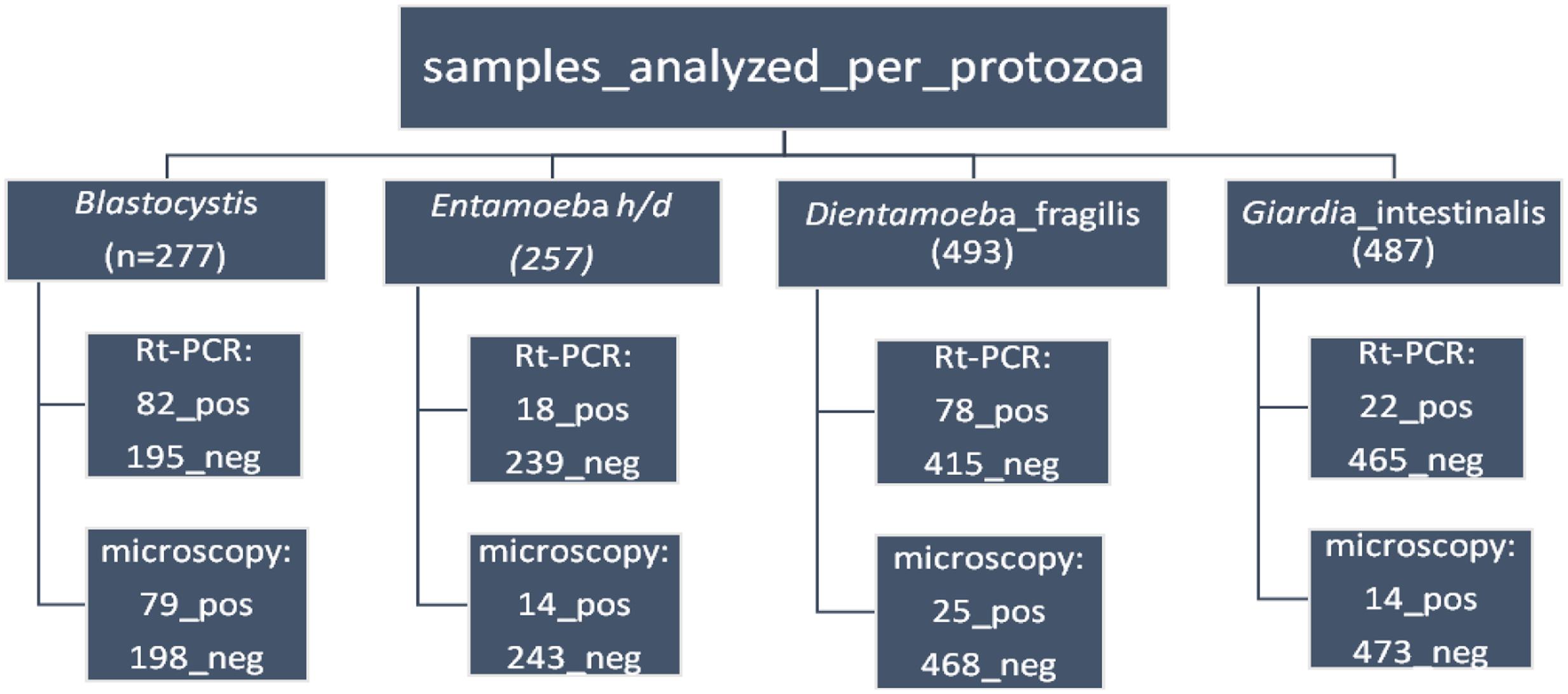
All consecutive patients having provided written informed consent for the use of their biological samples by the CTD, Negrar, Verona, Italy, who were requested a stool microcopy exam and a Rt-PCR for intestinal protozoa between 01.01.2014 and 31.12.2015 (Figure 1).

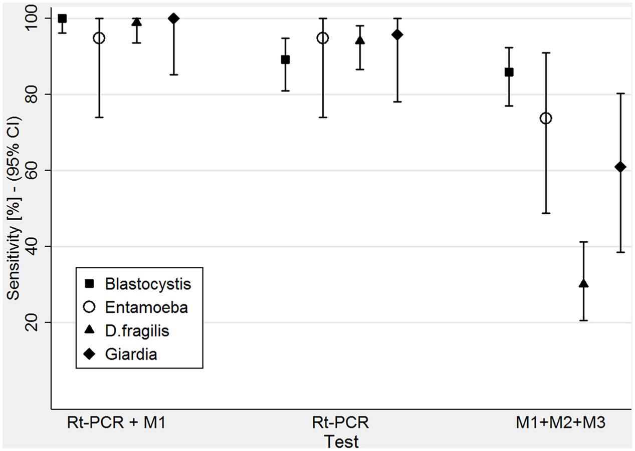
FIGURE 1. Comparison of the sensitivity of the three tests used: Rt-PCR + M1 (microscopy on one sample), Rt-PCR alone and M1 + M2 + M3 (microscopy on all three samples).
Test Methods
Stool microscopy
Following the protocol of our laboratory, stool microscopy was performed on three stool samples collected in 10% formalin on consecutive or alternate days. The samples were concentrated according to a modified Ritchie’s method (Buonomini et al., 1956) before the microscopic analysis.
DNA extraction and Rt-PCR
Stool specimens were collected as described previously (Formenti et al., 2015) according to the protocol procedure of our laboratory. In detail, 200 mg of stool were stored at -20°C overnight in a solution of 1X PBS with 2% polyvinylpolypyrrolidone (PvPP) (Sigma–Aldrich, Milan, Italy). In each sample, Phocine Herpes Virus type-1 (PhHV-1, kindly provided by Dr. Pas S., Erasmus MC, Department of Virology, Rotterdam) was added to the S.T.A.R. buffer (Roche), serving as an internal control for the isolation and amplification steps. Prior to DNA extraction, all the samples were frozen and then boiled for 10 min at 100°C. The DNA was extracted using the MagnaPure LC.2 instrument (Roche Diagnostic, Monza, Italy), following the protocol “DNA I Blood_Cells High performance II,” using the kit “DNA isolation kit I” (Roche). The DNA was eluted in a final volume of 100 ul.
The Rt-PCR targets are shown in Table 1. In brief, amplification reactions for all the Rt-PCR were performed in 25 ul volumes containing PCR buffer (SsoFast master mix, Bio-Rad Laboratories, Milan, Italy), 2.5 ug of BSA (Sigma–Aldrich), 80 nM of each of the PhHV-1 specific primers, and 200 nM of PhHV-1 CY5-BHQ2 labeled probe. Depending on the multiplex performed, we had the following protocol of primers/probes concentration:
1. 300 nM of each Giardia intestinalis specific primers, 200 nM of G. intestinalis CY5.5-BHQ3 labeled probe. 100 nM of each D. fragilis specific primers and 100 nM of D. fragilis VIC-MGB labeled probe. 300 nM of each Blastocystis sp. specific primers and 100 nM of Blastocystis sp. FAM-MGB labeled probe.
2. 60 nM of each E. histolytica/E. dispar specific primers and 200 nM of E. histolytica FAM-MGB labeled probe and E. dispar VIC-MGB labeled probe. 200 nM of each Cryptosporidium sp. specific primers and 100 nM of Cryptosporidium sp. CY5.5-BHQ3 labeled probe.
The Rt-PCR cycle protocol consists of 3 min at 95°C followed by 40 cycles of 15 s at 95°C and 30 s at 60°C, and 30 s at 72°C. The reactions, detection and data analyses were performed with the CFX 96 detection system (Biorad Laboratories) using white plates. Positive and negative controls were included in all the experiments; in detail as positive control we used two pool of positive DNA for the targets included in the multiplex. One had a low Ct (30 < Ct < 36) and the other a high Ct (37 < Ct < 39.9). For all the Rt-PCR analysis, the threshold was set at 200. As a control for Rt-PCR inhibitors and amplification, the exogenous PhHV-1 DNA was amplified with the appropriate primers/probe mix.
Statistical Analysis
In studies of diagnostic accuracy, the results of one or more tests under evaluation (index tests) are compared with those obtained with the reference standard, both measured in subjects who are suspected of having the condition of interest. In this framework, we considered as the reference standard the extended routine (coproparasitological exam of three samples plus Rt-PCR) while the index test was represented by the new proposed, restricted routine (coproparasitological exam of one sample plus Rt-PCR). For the assessment of microscopic accuracy of one single sample, we conventionally considered the first of each series of three samples (Supplementary Information File). We assessed the sensitivity of the index test and its 95% confidence interval [as the specificity of the restricted routine could not logically be lower than that of the extended routine, moreover Rt-PCR specificity was proven to be 100% (Stark et al., 2006; Stensvold et al., 2011) and so is conventionally the specificity of microscopy].
The analysis was performed with STATA vers. 14 (StataCorp, 4905 Lakeway Dr., College Station, TX 77845, United States) and a p-value of 5% was considered statistically significant.
Results
Blastocystis sp.
A total of 277 samples were analyzed by microscopy and Rt-PCR for Blastocystis sp.
The new diagnostic approach resulted in 100.0% sensitivity (CI 96.1–100.0%) (Table 2). The microscopy-only performed on three samples resulted in 85.9% sensitivity (CI 77.0–92.3%) (Table 3). Rt-PCR (Table 4) without microscopy resulted in 89.1% sensitivity (CI 80.9–94.7%).
Entamoeba histolytica/dispar
A total of 257 samples were analyzed by microscopy and Rt-PCR for E. histolytica/dispar.
The sensitivity of the new diagnostic approach was 94.7% (CI 74.0–99.9%) (Table 2). That of microscopy-only performed on three samples was 73.7% (CI 48.8–90.9%) (Table 3). Rt-PCR (Table 4) without microscopy had a sensitivity of 94.7% (CI 74.0–99.9%).
Dientamoeba fragilis
A total of 493 samples were analyzed by microscopy and Rt-PCR for D. fragilis.
The new diagnostic approach resulted 98.8% sensitive (CI 93.5–100.0%) (Table 2). The sensitivity of microscopy-only (three samples) was 30.1% (CI 20.5–41.2%) (Table 3), that of Rt-PCR (Table 4) without microscopy was 94.0% (CI 86.5–98.0%).
Giardia intestinalis
A total of 487 samples were analyzed by microscopy and Rt-PCR for G. intestinalis.
The sensitivity of the new diagnostic approach was 100.0% (CI 85.2–100.0%) (Table 2), that of microscopy-only was 60.9% (CI 38.5–80.3%) (Table 3), that of Rt-PCR alone (Table 4) resulted 95.7% (CI 78.1–99.9%).
Discussion
The comparison of microscopy with Rt-PCR for protozoan infection has already been considered by other studies (Stark et al., 2006; ten Hove et al., 2007; Bruijnesteijn van Coppenraet et al., 2009; Verweij, 2014). These studies reported the higher sensitivity of Rt-PCR compared to the microscopy technique. The originality of our work is the assessment of a possible new approach to the routine laboratory approach to parasite diagnosis. Infections by protozoa are still common in Italy, and are very frequent in immigrants and travelers. Helminth ova or larvae are less frequently found in stools, and we did not have a sufficient number of positive samples to do an appropriate analysis. On the other hand, alternative diagnostic tools are available for the most important helminth infections: serology in particular has been shown to be more sensitive than fecal techniques for helminth infections of primary medical impostance such as strongyloidiasis and schistosomiasis, for example (Buonfrate et al., 2015; Beltrame et al., 2017).
In this work we retrospectively analyzed and focused on the data of the patients who had been requested a coproparasitological exam on three samples and Rt-PCR for protozoa. The reference standard considered in this study was the extended routine adopted by our laboratory once molecular biology was available, that added a Rt-PCR on one sample to the previous routine (coproparasitological exam on three samples). The “index test” was the proposed, restricted routine (both exams on one single sample). As illustrated in Table 2, the new protocol showed an excellent sensitivity for the protozoa analyzed (Figure 2), reaching 100% for Blastocystis sp. and for G. intestinalis and approaching this target for the other two protozoa analyzed, missing just one positive result, respectively, for E. histolytica/dispar and for Dientamoeba.
The sensitivity of the “old” routine coproparasitological exam on three samples without Rt-PCR (Table 3) gave different results depending on the target analyzed, but was invariably lower (exceedingly so for D. fragilis and G. intestinalis) than that of the new, restricted routine. Conversely, Rt-PCR without microscopy showed an excellent sensitivity for all the targets analyzed, although slightly lower (with the notable exception of the target E. histolytica/dispar) than that of the new proposed, restricted routine.
Conclusion
The present study shows that the sensitivity of the new proposed approach for the diagnosis of intestinal protozoa infection remains very high despite the reduction of fecal samples from three to one. Furthermore, it has the advantage of reducing costs and saving time of laboratory personnel. It is also, obviously, a much more practical approach for patients, allowing the sample collection at the moment of the outpatient contact with no need of further visits. It could be argued that Rt-PCR, given the higher sensitivity confirmed by our study, could completely replace microscopy. We disagree for four good reasons: (a) our data show that adding microscopy has the potential of enhancing the sensitivity of the diagnostic approach (although only for Blastocystis the difference was statistically significant); (b) microscopy can detect other parasitic infections that may not be included in the Rt-PCR targets requested; (c) residual DNA may persist and be detected by Rt-PCR after parasite clearance, potentially causing false-negative results (Frickmann et al., 2015; Meurs et al., 2017) [others authors obtained conflicting results, showing a fast DNA clearance after treatment (Mejia et al., 2013; van den Bijllaardt et al., 2014)]; (d) last but perhaps most important, good microscopists are becoming a species in danger of extinction and it would be a pity to disperse their precious skills and know-how, not only for protozoa of course, but also for helminths that were not targeted by this study for the reasons explained above.
Author Contributions
FF conducted the work and wrote the paper; MV performed the statistical analysis; MG contributed to perform the statistical analysis; FP and ZB revised the article; BP performed the molecular analysis; MM, ST, and MD performed the microscopy analysis.
Conflict of Interest Statement
The authors declare that the research was conducted in the absence of any commercial or financial relationships that could be construed as a potential conflict of interest.
Acknowledgment
The authors thank Dr. Jaco Verweij for advice in setting-up the molecular diagnostic assays and Dr. Jessica Malzahn and Dr. Federico Formenti for revising the manuscript.
Supplementary Material
The Supplementary Material for this article can be found online at: https://www.frontiersin.org/articles/10.3389/fmicb.2017.02191/full#supplementary-material
References
Amar, C. F., East, C. L., Gray, J., Iturriza-Gomara, M., Maclure, E. A., and McLauchlin, J. (2007). Detection by PCR of eight groups of enteric pathogens in 4,627 faecal samples: re-examination of the English case-control Infectious Intestinal Disease Study (1993-1996). Eur. J. Clin. Microbiol. Infect. Dis. 26, 311–323. doi: 10.1007/s10096-007-0290-8
Beltrame, A., Guerriero, M., Angheben, A., Gobbi, F., Requena-Mendez, A., Zammarchi, L., et al. (2017). Accuracy of parasitological and immunological tests for the screening of human schistosomiasis in immigrants and refugees from African countries: an approach with latent class analysis. PLOS Negl. Trop. Dis. 11:e0005593. doi: 10.1371/journal.pntd.0005593
Bretagne, S., and Costa, J. M. (2006). Towards a nucleic acid-based diagnosis in clinical parasitology and mycology. Clin. Chim. Acta 363, 221–228. doi: 10.1016/j.cccn.2005.05.051
Bruijnesteijn van Coppenraet, L. E., Wallinga, J. A., Ruijs, G. J., Bruins, M. J., and Verweij, J. J. (2009). Parasitological diagnosis combining an internally controlled real-time PCR assay for the detection of four protozoa in stool samples with a testing algorithm for microscopy. Clin. Microbiol. Infect. 15, 869–874. doi: 10.1111/j.1469-0691.2009.02894.x
Buonfrate, D., Formenti, F., Perandin, F., and Bisoffi, Z. (2015). Novel approaches to the diagnosis of Strongyloides stercoralis infection. Clin. Microbiol. Infect. 21, 543–552. doi: 10.1016/j.cmi.2015.04.001
Buonomini, G., Ricciardi, M. L., and Carli, G. (1956). English title not available. Ig. Mod. 49, 971–982.
Cartwright, C. P. (1999). Utility of multiple-stool-specimen ova and parasite examinations in a high-prevalence setting. J. Clin. Microbiol. 37, 2408–2411.
Danciger, M., and Lopez, M. (1975). Numbers of Giardia in the feces of infected children. Am. J. Trop. Med. Hyg. 24, 237–242. doi: 10.4269/ajtmh.1975.24.237
Formenti, F., Perandin, F., Bonafini, S., Degani, M., and Bisoffi, Z. (2015). Evaluation of the new ImmunoCard STAT!(R) CGE test for the diagnosis of Amebiasis. Bull. Soc. Pathol. Exot. 108, 171–174. doi: 10.1007/s13149-015-0434-5
Frickmann, H., Schwarz, N. G., Rakotozandrindrainy, R., May, J., and Hagen, R. M. (2015). PCR for enteric pathogens in high-prevalence settings. What does a positive signal tell us? Infect. Dis. 47, 491–498. doi: 10.3109/23744235.2015.1022212
Gunson, R. N., Bennett, S., Maclean, A., and Carman, W. F. (2008). Using multiplex real time PCR in order to streamline a routine diagnostic service. J. Clin. Virol. 43, 372–375. doi: 10.1016/j.jcv.2008.08.020
Jothikumar, N., da Silva, A. J., Moura, I., Qvarnstrom, Y., and Hill, V. R. (2008). Detection and differentiation of Cryptosporidium hominis and Cryptosporidium parvum by dual TaqMan assays. J. Med. Microbiol. 57(Pt 9), 1099–1105. doi: 10.1099/jmm.0.2008/001461-0
Liu, Y. T. (2008). A technological update of molecular diagnostics for infectious diseases. Infect. Disord. Drug Targets 8, 183–188. doi: 10.2174/1871526510808030183
Mejia, R., Vicuna, Y., Broncano, N., Sandoval, C., Vaca, M., Chico, M., et al. (2013). A novel, multi-parallel, real-time polymerase chain reaction approach for eight gastrointestinal parasites provides improved diagnostic capabilities to resource-limited at-risk populations. Am. J. Trop. Med. Hyg. 88, 1041–1047. doi: 10.4269/ajtmh.12-0726
Meurs, L., Polderman, A. M., Vinkeles Melchers, N. V., Brienen, E. A., Verweij, J. J., Groosjohan, B., et al. (2017). Diagnosing polyparasitism in a high-prevalence setting in Beira, Mozambique: detection of intestinal parasites in fecal samples by microscopy and real-time PCR. PLOS Negl. Trop. Dis. 11:e0005310. doi: 10.1371/journal.pntd.0005310
Muldrew, K. L. (2009). Molecular diagnostics of infectious diseases. Curr. Opin. Pediatr. 21, 102–111. doi: 10.1097/MOP.0b013e328320d87e
Murray, T. S., and Cappello, M. (2008). The molecular diagnosis of parasitic diseases. Pediatr. Infect. Dis. J. 27, 163–164. doi: 10.1097/INF.0b013e3181658af0
Rocca, P., Galderisi, S., Rossi, A., Bertolino, A., Rucci, P., Gibertoni, D., et al. (2016). Social cognition in people with schizophrenia: a cluster-analytic approach. Psychol. Med. 46, 2717–2729. doi: 10.1017/S0033291716001100
Stark, D., Beebe, N., Marriott, D., Ellis, J., and Harkness, J. (2006). Evaluation of three diagnostic methods, including real-time PCR, for detection of Dientamoeba fragilis in stool specimens. J. Clin. Microbiol. 44, 232–235. doi: 10.1128/JCM.44.1.232-235.2006
Stensvold, C. R., Ahmed, U. N., Andersen, L. O., and Nielsen, H. V. (2012). Development and evaluation of a genus-specific, probe-based, internal-process-controlled real-time PCR assay for sensitive and specific detection of Blastocystis spp. J. Clin. Microbiol. 50, 1847–1851. doi: 10.1128/JCM.00007-12
Stensvold, C. R., Lebbad, M., and Verweij, J. J. (2011). The impact of genetic diversity in protozoa on molecular diagnostics. Trends Parasitol. 27, 53–58. doi: 10.1016/j.pt.2010.11.005
ten Hove, R., Schuurman, T., Kooistra, M., Moller, L., van Lieshout, L., and Verweij, J. J. (2007). Detection of diarrhoea-causing protozoa in general practice patients in The Netherlands by multiplex real-time PCR. Clin. Microbiol. Infect. 13, 1001–1007. doi: 10.1111/j.1469-0691.2007.01788.x
ten Hove, R. J., Verweij, J. J., Vereecken, K., Polman, K., Dieye, L., and van Lieshout, L. (2008). Multiplex real-time PCR for the detection and quantification of Schistosoma mansoni and S. haematobium infection in stool samples collected in northern Senegal. Trans. R. Soc. Trop. Med. Hyg. 102, 179–185. doi: 10.1016/j.trstmh.2007.10.011
Utzinger, J., Botero-Kleiven, S., Castelli, F., Chiodini, P. L., Edwards, H., Kohler, N., et al. (2010). Microscopic diagnosis of sodium acetate-acetic acid-formalin-fixed stool samples for helminths and intestinal protozoa: a comparison among European reference laboratories. Clin. Microbiol. Infect. 16, 267–273. doi: 10.1111/j.1469-0691.2009.02782.x
van den Bijllaardt, W., Overdevest, I. T., Buiting, A. G., and Verweij, J. J. (2014). Rapid clearance of Giardia lamblia DNA from the gut after successful treatment. Clin. Microbiol. Infect. 20, O972–O974. doi: 10.1111/1469-0691.12626
Verweij, J. J. (2014). Application of PCR-based methods for diagnosis of intestinal parasitic infections in the clinical laboratory. Parasitology 141, 1863–1872. doi: 10.1017/S0031182014000419
Verweij, J. J., Mulder, B., Poell, B., van Middelkoop, D., Brienen, E. A., and van Lieshout, L. (2007). Real-time PCR for the detection of Dientamoeba fragilis in fecal samples. Mol. Cell. Probes 21, 400–404. doi: 10.1016/j.mcp.2007.05.006
Verweij, J. J., Oostvogel, F., Brienen, E. A., Nang-Beifubah, A., Ziem, J., and Polderman, A. M. (2003a). Short communication: prevalence of Entamoeba histolytica and Entamoeba dispar in northern Ghana. Trop. Med. Int. Health 8, 1153–1156. doi: 10.1046/j.1360-2276.2003.01145.x
Verweij, J. J., Schinkel, J., Laeijendecker, D., van Rooyen, M. A., van Lieshout, L., and Polderman, A. M. (2003b). Real-time PCR for the detection of Giardia lamblia. Mol. Cell. Probes 17, 223–225. doi: 10.1016/s0890-8508(03)00057-4
Verweij, J. J., van Lieshout, L., Blotkamp, C., Brienen, E. A., van Duivenvoorden, S., van Esbroeck, M., et al. (2000). Differentiation of Entamoeba histolytica and Entamoeba dispar using PCR-SHELA and comparison of antibody response. Arch. Med. Res. 31(Suppl. 4), S44–S46. doi: 10.1016/S0188-4409(00)00221-6
Visser, L. G., Verweij, J. J., Van Esbroeck, M., Edeling, W. M., Clerinx, J., and Polderman, A. M. (2006). Diagnostic methods for differentiation of Entamoeba histolytica and Entamoeba dispar in carriers: performance and clinical implications in a non-endemic setting. Int. J. Med. Microbiol. 296, 397–403. doi: 10.1016/j.ijmm.2006.03.001
Keywords: real-time PCR, protozoa, diagnosis, molecular biology, laboratory
Citation: Formenti F, Valerio M, Guerriero M, Perandin F, Pajola B, Mistretta M, Tais S, Degani M and Bisoffi Z (2017) Molecular Biology Can Change the Classic Laboratory Approach for Intestinal Protozoan Infections. Front. Microbiol. 8:2191. doi: 10.3389/fmicb.2017.02191
Received: 07 August 2017; Accepted: 25 October 2017;
Published: 07 November 2017.
Edited by:
José Roberto Mineo, Federal University of Uberlandia, BrazilReviewed by:
Ricardo Silvestre, Instituto de Pesquisa em Ciências da Vida e da Saúde (ICVS), PortugalOscar Campetella, National University of General San Martín, Argentina
Copyright © 2017 Formenti, Valerio, Guerriero, Perandin, Pajola, Mistretta, Tais, Degani and Bisoffi. This is an open-access article distributed under the terms of the Creative Commons Attribution License (CC BY). The use, distribution or reproduction in other forums is permitted, provided the original author(s) or licensor are credited and that the original publication in this journal is cited, in accordance with accepted academic practice. No use, distribution or reproduction is permitted which does not comply with these terms.
*Correspondence: Fabio Formenti, fabio.formenti@sacrocuore.it
 Fabio Formenti
 Matteo Valerio2


