 Jayasekara M. K. J. K. Premarathne1,2*
 Aimi S. Anuar1
 Tze Young Thung1
 Dilan A. Satharasinghe3,4Nuzul Noorahya Jambari5Noor-Azira Abdul-Mutalib5
 John Tang Yew Huat6Dayang F. Basri7
 Yaya Rukayadi1
 Yoshitsugu Nakaguchi8
 Mitsuaki Nishibuchi8Son Radu1,9*
- 1Faculty of Food Science and Technology, Food Safety Research Center, Universiti Putra Malaysia, Seri Kembangan, Malaysia
- 2Department of Livestock and Avian Science, Faculty of Livestock, Fisheries and Nutrition, Wayamba University of Sri Lanka, Kuliyapitiya, Sri Lanka
- 3Institute of Bio Science, Universiti Putra Malaysia, Seri Kembangan, Malaysia
- 4Department of Basic Veterinary Science, Faculty of Veterinary Medicine and Animal Science, University of Peradeniya, Peradeniya, Sri Lanka
- 5Food Safety Research Center (FOSREC), Faculty of Food Science and Technology, Universiti Putra Malaysia, Seri Kembangan, Malaysia
- 6Faculty of Food Technology, Universiti Sultan Zainal Abidin, Kuala Terengganu, Malaysia
- 7Faculty of Health Sciences, School of Diagnostic and Applied Health Sciences, Universiti Kebangsaan Malaysia, Kuala Lumpur, Malaysia
- 8Center for Southeast Asian Studies, Kyoto University, Kyoto, Japan
- 9Laboratory of Food Safety and Food Integrity, Institute of Tropical Agriculture and Food Security, Universiti Putra Malaysia, Seri Kembangan, Malaysia
Campylobacter is a major foodborne pathogen frequently associated with human bacterial gastroenteritis in the world. This study was conducted to determine the prevalence and antibiotic resistance of Campylobacter spp. in the beef food system in Malaysia. A total of 340 samples consisting of cattle feces (n = 100), beef (n = 120) from wet markets and beef (n = 120) from hypermarkets were analyzed for Campylobacter spp. The overall prevalence of Campylobacter was 17.4%, consisting of 33% in cattle fecal samples, 14.2% in raw beef from wet market and 7.5% in raw beef from the hypermarket. The multiplex-polymerase chain reaction (PCR) identified 55% of the strains as C. jejuni, 26% as C. coli, and 19% as other Campylobacter spp. A high percentage of Campylobacter spp. were resistant to tetracycline (76.9%) and ampicillin (69.2%), whilst low resistance was exhibited to chloramphenicol (7.6%). The MAR Index of Campylobacter isolates from this study ranged from 0.09 to 0.73. The present study indicates the potential public health risk associated with the beef food system, hence stringent surveillance, regulatory measures, and appropriate interventions are required to minimize Campylobacter contamination and prudent antibiotic usage that can ensure consumer safety.
Introduction
Thermophilic Campylobacter is a major bacterial pathogen that causes foodborne infections around the world (EFSA, 2011; WHO, 2012). C. jejuni and C. coli have been identified as the most common species that lead to campylobacteriosis (Silva et al., 2011). The high incidence rate of campylobacteriosis can be associated with the low minimal infective dose of thermophilic Campylobacter which is around 500–800 cells (Nachamkin et al., 2008). Campylobacter spp. colonize the enteric tract of birds, sheep, cattle and pigs (Stanley and Jones, 2003; Humphrey et al., 2007). Food originated from animals act as the primary source of Campylobacter infection (Doorduyn et al., 2010).
Reduce susceptibility of foodborne pathogens to antimicrobials significantly affect global public health (Chatre et al., 2010; Aarestrup, 2015). Increasing resistance in Campylobacter to antimicrobials particularly to tetracycline, erythromycin, and (fluoro)quinolones (Gaudreau and Gilbert, 2003; Zhu et al., 2006) was associated with reduce response to therapy leading to higher morbidity and mortality rates in humans (Zhu et al., 2006).
Campylobacter a major public health concern around the world, especially in developed countries and these countries have Campylobacter surveillance systems (Scallan et al., 2011; EFSA and ECDC, 2014). Even in the South-East Asia, Campylobacter is becoming a key foodborne pathogen (Premarathne et al., 2017). According to a hospital-based study conducted by Lee and Puthucheary (2002), Campylobacter spp. was reported at 5% prevalence level and placed among the first five causative agents associated with child's diarrhea. Furthermore, Campylobacter were reported in humans from Indonesia (Tjaniadi et al., 2003), Lao People's Democratic Republic (Yamashiro et al., 1998), Singapore (Chau et al., 2016), and Thailand (Bodhidatta et al., 2013). Singapore is the only South-East Asian country that performs national surveillance of Campylobacter infection in humans. Estimation and control of campylobacteriosis in South-East Asian countries, including Malaysia, is hindered due to insufficient regulations for food safety and inadequate data on epidemiology of Campylobacter cases (Premarathne et al., 2017). Therefore, prevalence and antimicrobial resistance of Campylobacter are important to be assessed in these countries.
In Malaysia, beef is sold in traditional wet markets and modern hypermarkets (Chamhuri and Batt, 2013). Beef available in wet markets were supplied by locally slaughtered and dressed cattle at the municipal abattoir or none-abattoir premises (Marimuthu et al., 2015). The wet markets were operated for a short time around 6 h per day and were able to provide fresh meat every day (Tang et al., 2009). The hypermarkets offered chilled or frozen meat that locally produced or from imported beef (Ariff et al., 2015). According to a consumer survey, Malaysians considered that fresh meat is more succulent, healthier and assure the halalness. Therefore, they preferred purchasing meat from wet markets over hypermarkets (Chamhuri and Batt, 2013).
Naturally Campylobacter are present in cattle; therefore, feces can contaminate the beef carcasses during slaughtering (Hakkinen et al., 2007). Further, a study conducted in Malaysisa found that C. jejuni can survive in food processing environments through formation of biofilms (Teh et al., 2014). The prevalence of Campylobacter species in poultry (Tang et al., 2009; Mansouri-Najand et al., 2012; Rejab et al., 2012), duck (Nor Faiza et al., 2013) and salad vegetables (Chai et al., 2007) have been reported in Malaysia. However, limited data exist on the prevalence of Campylobacter in other food commodities, including beef food system. Impelled by scarcity of data, this study was conducted to determine the prevalence of Campylobacter in the beef food system together with assessing the antibiogram profiles of Campylobacter spp.
Materials and Methods
Bacterial Strains
In this study C. jejuni ATCC 33221 and C. coli ATCC 33559 were used as the positive controls.
Sample Collection
A total of 100 fecal samples were collected from apparently healthy beef cattle in three different farms, while purchased 120 chilled beef samples from hypermarkets (n = 6) and 120 fresh beef samples from wet markets (n = 6) in Selangor, Malaysia. All collected samples were immediately transported to the laboratory in ice and processed within the same day of sample collection.
Sample Processing
The standardized and published protocol by Chai et al. (2007) and Tang et al. (2009) was used for sample processing. A 10 g portion of the sample was aseptically placed in a stomacher bag containing 90 mL Bolton broth base (Merck KGaA, Darmstadt, Germany) and homogenized using a stomacher lab-blender (Interscience, France) at normal speed for 60 s. The Bolton broth was supplemented with Bolton broth selective supplement contained Vancomycin, Cefoperazone, Trimethoprim Lactate and Amphotericin B (Merck KGaA) and 5% lysed horse blood.
Control samples were prepared only with supplemented Bolton broth and inoculated with C. jejuni ATCC 33221 and C. coli ATCC 33559. The controls and samples were incubated at 42°C for 48 h under microaerophilic conditions comprised of 8–10% (v/v) CO2 and 5–6% (v/v) O2 generated by the Anaerocult C system (Merck KGaA) in an anaerobic jar.
Microbiological Isolation
After incubation, a loop full of the enriched sample was streaked in duplicates onto modified charcoal-cefoperazone-deoxycholate agar (mCCDA; Merck KGaA) with CCDA selective supplement contained Cefoperazone and Amphotericin B (Merck KGaA) and incubated microaerobically (Anaerocult C system; Merck KGaA) for 48 h at 42°C. Presumptive identification of Campylobacter colonies was based on ISO method that includes (ISO, 2006a,b); oxidase test, catalase test, gram-staining and typical microscopic Campylobacter morphology. The presumptive colonies were confirmed by using PCR (Polymerase Chain Reaction) assay.
MPN-Enrichment
Campylobacter cells were counted using the MPN method with triplicate of three 10-fold dilution series prepared by transferring 100 μL of homogenized sample into 900 μL of Bolton broth base (Merck KGaA) supplemented with Bolton broth selective supplements. A control tube was prepared with supplemented Bolton broth and inoculated with C. jejuni ATCC 33221 and C. coli ATCC 33559. The MPN tubes were incubated at 42°C for 48 h under microaerophilic conditions generated by the Anaerocult C system (Merck KGaA) in an anaerobic jar.
DNA Extraction
Bacterial DNA was extracted from the enriched sample tubes and presumptive bacterial colonies using boiled-cell method (Tang et al., 2009). Briefly, enriched samples in the tubes were pelleted by centrifuging at 10,000 × g for 10 min. The supernatant was carefully removed, next the pellet was washed once with 500 μL sterile distilled water and harvested bacterial cells were suspended in 500 μL of sterile TE buffer (pH 8.0). Bacterial cells were lysed by subjecting to boiling for 10 min, followed by rapid cooling at −20°C for 10 min. Next the sample was centrifuged at 10,000 × g for 10 min. Finally, the supernatant containing bacterial DNA was stored at −20°C until used in PCR.
PCR Identification
All MPN tubes were subjected to identification of Campylobacter spp. by multiplex PCR reaction using primers specific for Campylobacter genes, C. jejuni and C. coli (Table 1).
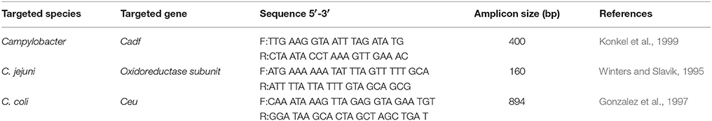
Table 1. PCR primers set used for detection of Campylobacter spp., C. jejuni and C. coli.
The PCR was carried out in 25 μL reactions, and each reaction contained 5 μL of 5 × PCR buffer (Promega, USA); 3 μL of 25 mM MgCl2 (Promega, USA); 1 U Taq Polymerase (Promega, USA); 1 μL of 10 mM deoxynucleoside triphosphate (dNTP) mix (Promega, USA); 1 μM of forward and reverse primer (Sigma, UK) and 5 μL of template DNA.
The cycling conditions were set as follows on the VeritiTM 96-Well Thermal Cycler (Applied Biosystems, USA), initial denaturing at 94°C for 4 min followed by 33 cycles of denaturing at 94°C for 1 min, annealing at 50°C for 1 min and extension at 72°C for 1 min followed by final extension at 72°C for 5 min.
A 5 μL of all PCR amplicons were horizontally electrophoresed through a 1.25% agarose gel stained with ethidium bromide in 1 × Tris-acetate-EDTA (TAE) (1 mM EDTA, 40 mM Tris-acetate) buffer at 90 V for 40 min. The agarose gel was visualized under ultraviolet (UV) light transilluminator (SynGene, Frederick, USA) and photographed. In each gel a positive control, non-template control and a DNA-molecular marker (100-bp ladder) (Vivantis Technologies, Malaysia) were included. The DNA extracted from C. coli (ATCC 33559) and C. jejuni (ATCC 33560) were used as the positive controls, while PCR mixture without DNA template was used as the non-template control.
Antibiotic Susceptibility Testing
Antimicrobial susceptibility tests were conducted using following antimicrobial impregnated disks (Oxoid, England, UK); ampicillin (AMP; 10 μg), cephalothin (CEF; 30 μg), chloramphenicol (CHL; 30 μg), ciprofloxacin (CIP; 5 μg), enrofloxacin (ENR; 5 μg), erythromycin (ERY; 15 μg), gentamicin (GEN; 10 μg), norfloxacin (NORr; 10 μg), nalidixic acid (NAL; 30 μg), sterptomycin (STR; 25 μg), and tetracycline (TET; 30 μg) according to the standard Kirby- Bauer disk diffusion method (Bauer et al., 1966) and performed according to the recommendations of the Clinical Laboratory Standards Institute (Clinical and Laboratory Standards Institute, 2012).
All isolates were revived from glycerol stocks using Bolton broth supplemented with 5% lysed horse blood. The suspensions were incubated for 48 h at 42°C under microaerophilic conditions. Revived cultures were grown in Brain heart infusion (BHI: Merck KGaA) broth at 42°C under microaerophilic conditions for 24 h and the turbidity of the suspension was adjusted to 0.5 McFarland standard (Clinical and Laboratory Standards Institute, 2012). Then the culture was swabbed uniformly using sterile cotton swabs onto MH agar plates (Merck KGaA) supplemented with 5% horse blood. The plates were incubated under microaerophilic conditions (Anaerocult C system; Merck KGaA) at 37°C for 48 h. C. jejuni ATCC 33560 and C. coli ATCC 33559 were used as reference strains.
The diameters of the inhibition zones around the antibiotic disk were measured. The breakpoints used to categorize isolates as susceptible (s), intermediate (i) and resistant (r) were based on CLSI recommendations (Clinical and Laboratory Standards Institute, 2012). As there were no CLSI recommendations for Campylobacter strains, the standards assigned to the Enterobacteriaceae family were used as breakpoints to interpret Campylobacter resistance.
Multiple Antimicrobial Resistance (MAR) Indexing
Multi-resistance of Campylobacter isolates were quantified using the MAR indexing.
where “a” indicate the number of antimicrobials to which the particular isolate was resistant and “b” indicate the total number of antimicrobials to which the particular isolate was tested (Krumperman, 1983).
Statistical Analysis
The difference in prevalence level between the two detection methods (MPN_plating and MPN_PCR) and various sample types obtained from cattle, wet market and hypermarket were analyzed using Pearson chi-square test (X2 test). The results were considered statistically significant at P < 0.05 at 95% confidence level.
Results
Culture-Based Detection and Colony Confirmation Applying PCR Method
Presumptive Campylobacter spp. were detected in 8% (8 of 100 samples) of the cattle feces samples and 1.6% (4 of 240) of the beef meat samples and all presumptive isolates were confirmed as Campylobacter spp. by PCR.
MPN-Plating Media Based Enumeration
The prevalence of Campylobacter spp. in cattle and beef meat samples determined by using the MPN-plating is presented in Table 2. The MPN-plating detected Campylobacter spp. in 3.5% of the total samples (Table 2). Campylobacter was detected only in 8 out of 100 fecal samples, including C. jejuni in 5% and C. coli in 2% of the fecal samples. The prevalence of C. jejuni was significantly higher (P ≤ 0.05) than the C. coli in fecal samples.

Table 2. Culture-based detection of Campylobacter spp. in cattle and beef.
The majority (66%) of isolates were identified as C. jejuni and the remainder as C. coli (34%). Campylobacter spp. were detected only in 2% of the beef samples from wet and hypermarket by the MPN-plating method. The MPN-plating found C. jejuni and C. coli in 0.8% of beef samples from wet and hypermarket (Table 2). The prevalence of Campylobacter spp. was statistically not significant (P ≤ 0.05) between the wet and hypermarkets. However, the prevalence of Campylobacter spp. in fecal samples was significantly higher (P ≤ 0.05) than that in beef samples.
MPN-PCR-Based Enumeration
The prevalence of Campylobacter spp determined by using the MPN-PCR method is presented in Table 3. Based on the MPN-PCR results Campylobacter spp. was detected in 59 (17.4%) of the total 340 examined samples. Detection of Campylobacter spp. by MPN-PCR method was significantly higher (P ≤ 0.05) comparative to the MPN-plating (Table 3).
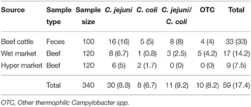
Table 3. MPN-PCR based detection of Campylobacter spp. in cattle and beef.
The MPN-PCR method detected Campylobacter in 33 out of 100 cattle fecal samples. According to the MPN-PCR, fecal samples contained 16, 8, and 5% of C. jejuni, both Campylobacter spp. and C. coli respectively. The prevalence of C. jejuni was significantly higher (P ≤ 0.05) than the C. coli in fecal samples.
Campylobacter was detected in a total of 17/120 (14.2%) beef from wet markets and in 9/120 (7.5%) beef from hypermarkets (Table 3). The predominant species of Campylobacter detected in beef samples from wet market (55%) was C. jejuni and remainder was C. coli (20%) and other Campylobacter spp. (25%). Similarly, at the hypermarket, C. jejuni was the most prevalent (67%) while C. coli and other Campylobacter spp. were detected in 22 and 11% of the beef samples respectively. The prevalence of Campylobacter spp. was statistically not significant (P ≤ 0.05) between the wet and hypermarkets.
Campylobacter concentration in cattle and beef was enumerated using the MPN method (Table 4). The highest number of Campylobacter found in fecal samples from cattle, including 3-460 MPN/g of C. jejuni and 3-43 MPN/g of C. coli and 3-75 MPN/g of other thermophilic Campylobacter spp. Beef from wet markets observed to carry Campylobacter spp. in the range of 3-75 MPN/g while the beef samples from hypermarkets harbored low Campylobacter spp. concentration ranged from 3 to 15 MPN/g.

Table 4. Concentration of Campylobacter spp. in cattle and beef.
Antimicrobial Resistance Testing
Eleven antimicrobials named under the different antimicrobial groups were employed in this study to determine the resistance of Campylobacter isolates. All the isolates were resistant at least one or more antimicrobials (Table 5). Antimicrobial resistance pattern among the tested Campylobacter spp. Indicated that majority of the isolates were resistant to tetracycline (76.9%) and ampicillin (69.2%). Least resistance was observed for chloramphenicol (7.6%), cephalothin (15.4%), ciprofloxacin (15.4%) and gentamicin (15.4%).
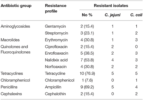
Table 5. The frequency of antibiotic drug resistance in Campylobacter spp.
The MAR index of Campylobacter species that were isolated from the current study indicate in Table 6. Campylobacter species exhibited 7 different antibiotic resistant patterns with MAR index ranging from 0.09 to 0.73. The highest MAR index was 0.73 and showed S, E, En, Na, Nor, Te, C, Amp resistant pattern. Meanwhile, lowest MAR index of 0.09 was demonstrated in isolates that indicate resistance to Te or Amp (Table 5). Moreover, 53.8% of the isolates were resistant to the three or more antimicrobials and demonstrated the Multi Drug Resistance (MDR).
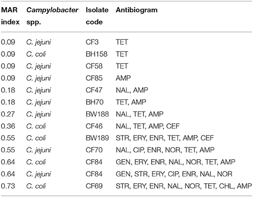
Table 6. Multiple antibiotic resistance (MAR) index of Campylobacter spp.
Discussion
Though the prevalence of Campylobacter in chicken and broilers was well documented in Malaysia; no or limited information was available for Campylobacter in other animals and food commodities, particularly for cattle and beef in the country. Therefore, the present study aimed to address the prevalence, concentration and antimicrobial resistant profiles of thermophilic Campylobacter species present in cattle fecal samples and beef.
The overall prevalence of thermophilic Campylobacter spp. in fecal samples of apparently healthy cattle was 33% (Table 2). Findings of this study was similar to the previous findings reported on prevalence level of Campylobacter spp. in cattle from countries including; Japan (39.6%) (Haruna et al., 2013) and Chile (35.9%) (Fernández and Hitschfeld, 2009). Sanad et al. (2011) found only 19.2% prevalence level in samples collected from feedlot, mature cows and bulls presented for slaughter across USA. Meanwhile, low mean Campylobacter prevalence level of 5.6% (Nonga et al., 2010) and 13.2% (Karikari et al., 2017) was reported from Tanzania and Ghana respectively. However, a study conducted in 5 different states in the U.S. reported a high prevalence (72.2%) of Campylobacter in feedlot cattle (Tang et al., 2017). While, 75–83.3% Campylobacter prevalence level was reported in dairy cattle from Lithuania (Ramonaite et al., 2013). Therefore, this study also contributes to prior discussions that cattle can act as a potential reservoir for transmitting Campylobacter spp. into the food system (Karenlampi et al., 2007; Cha et al., 2017). In the present study, C. jejuni (58.5%) was the predominantly isolated organism in cattle followed by C. coli (31.7%), and other thermophilic Campylobacter spp. The prevalence of C. jejuni and C. coli detected in this study was consistent with previous studies (Haruna et al., 2013). However, Karikari et al. (2017) reported higher prevalence level of C. coli (47.8%) comparatively C. jejuni was detected in 35.8% samples. Similarly, C. jejuni was detected in 35.2% cattle feacal samples with a high prevelance of C. coli (72.9%) (Sanad et al., 2011). However, Okunlade et al. (2015) reported low C. coli prevalence (20.4%) level in rectal swabs collected from cattle from Ibadan, Oyo State, Nigeria. A study conducted in Michigan, USA reported 69.2% prevalence level for C. jejuni (Cha et al., 2017). Other Campylobacter spp. (0.09%) detected in this study needs to be further characterized. Differences in the reported prevalence level can be resultant due to geographical location, sample size and method of analysis.
The low contamination frequency of Campylobacter species detected in beef in this study was in agreement with recent studies. The Campylobacter prevalence of fresh beef samples from Poland was 10.1% in Korsak et al. (2015), 16.2% in retail ground beef from Saskatchewan, Canada (Trokhymchuk et al., 2014) and 17.4% in whole beef cuts from retail shops in USA (Vipham et al., 2012). Meanwhile, some studies conducted previously could only detect very low Campylobacter contamination level in beef, including 1.9% in Tanzania (Nonga et al., 2010) and 3.3% in Belgium (Ghafir et al., 2007). Chilling reduce the surface humidity of red meat which can be related to low prevalence level Campylobacter in beef compared to chicken as Campylobacter is very sensitive to dehydration (Silva et al., 2011). The current study detected high Campylobacter prevalence in beef from wet markets (14.2%) comparative to beef from hypermarkets (7.5%) can be associated with the availability of fresh meat in the wet markets in Malaysia. Fresh meat is sold without chilling in the wet markets as it is considered fresh and succulent since it retains moisture. This may facilitate the survival of Campylobacter on raw red meat. Further, the low hygienic conditions in the wet markets (Chamhuri and Batt, 2013) may also contribute to the comparatively higher Campylobacter contamination rate and increase the potential for cross contamination. Moreover, no-abattoir slaughtering procedures (Marimuthu et al., 2015) can also result in high contamination of beef in wet markets with Campylobacter. We speculate that the main reason for the difference of Campylobacter prevalence in cattle and beef from various countries can be the result of differences in sampling methods, storage duration, microbiological and molecular methods employed. The results of this study indicate that not only cattle but also beef can be a potential reservoir for Campylobacter infection.
Findings of this study reported that Campylobacter spp. in cattle range from 460-3 MPN/g, while a lower concentration was detected in beef (29-3 MPN/g). Further this study was in line with Nielsen (2002) who reported that mean Campylobacter concentration in cattle feces was 126 MPN/g. A study conducted by Stanley et al. (1998) observed a maximum of Campylobacter concentration at the level of 6.3 × 10⁷ MPN/g in cattle. Campylobacter concentration in cattle intestine can be lower than that of broilers (Stanley and Jones, 2003). However, owed to a very low infective dose of Campylobacter the reported prevalence and concentration indicate the potential of contaminating beef during slaughter procedure. Therefore, proper sanitary and food safety measures should be implemented to ensure safety of beef. Further, an upward trend was observed among Malaysian consumers toward higher value meat such as beef and mutton (Yeong-Sheng et al., 2008). Therefore, beef can be a potential source of Campylobacter infection and need more attention focused on improving safety.
Isolation and identification of Campylobacter using conventional culture based methods can be challenging due to the fastidious growth requirements and discrepancies in biochemical tests (Nachamkin et al., 2008). Application of molecular methods for identification of foodborne pathogens has increasingly used for food samples to complement conventional microbiological methods owing to its rapid turnaround time and sensitivity (Inglis and Kalischuk, 2003). In this study, detection of Campylobacter spp. using PCR method was higher than plating on mCCDA agar. Previous studies on the prevalence of foodborne pathogens in food indicate a difference between the findings through the molecular biological methods comparative to findings of culturing methods (Inglis and Kalischuk, 2003; Chai et al., 2007; Tang et al., 2009). When growth conditions are not favorable Campylobacter cells can transition from vegetative stage into a viable but non-culturable (VBNC) state, in which the bacteria cannot be cultured using conventional culture methods (Oliver, 2010; Ramamurthy et al., 2014). The molecular amplification techniques can overcome the limitation of detecting VBNC cells with providing high specificity and sensitivity (Singh et al., 2011).
The high resistance detected in Campylobacter isolates against tetracycline in the current study was consistent with previously reported work. (Hong et al., 2007; Kashoma et al., 2015). Hong et al. (2007) reported that 93.4% Campylobacter strains from Korean beef samples were resistant to multiple antimicrobials and detected high resistance to tetracycline (94.6%). Campylobacter isolated from raw beef in Iran demonstrated the highest resistance to tetracycline (Rahimi et al., 2013). Campylobacter resistance to the tetracycline was mainly mediated through tet (O) plasmid (Pratt and Korolik, 2005) and worldwide 60–100% C. jejuni and C. coli were reported to carry tetracycline-resistant plasmids (Lee et al., 1994; Kim et al., 2010). High resistance to tetracycline in this study may be resultant due to tet (O) plasmid; however Campylobacter isolates from Malaysia need further investigation on prevalence and resistance profile of tet (O) plasmid. Similar to findings from the current study, the majority of the Campylobacter strains from food animals in Tanzania were resistant to ampicillin (70.3%) (Kashoma et al., 2015). Inherently resistance can be overserved in Campylobacter strains to β-lactams including ampicillin (Engberg, 2006; Li et al., 2007) which may be contributory to high ampicillin resistance observed in this study. The high resistance to tetracycline and ampicillin detected in this study could be associated with frequent usage of those antimicrobials in humans and animal husbandry (Chopra and Roberts, 2001; Hao et al., 2014).
Least resistance was observed for chloramphenicol, cephalothin, ciprofloxacin and gentamicin. Similarly low resistance was detected against gentamicin (1.8%) and chloramphenicol (4.5%) in the Campylobacter isolates from Tanzania (Kashoma et al., 2015). Further, Campylobacter spp. isolated from raw meat in Iran were resistant to gentamicin and chloramphenicol 3.2 and 6.5%, respectively (Rahimi et al., 2013). Contrary to the current study, Campylobacter isolates in beef from Korea showed high resistant to ciprofloxacin (95.9%) and nalidixic acid (94.6%) in Hong et al. (2007). Discrepancies and similarities in antibiotic resistance patterns can be attributed to variation in sample type, sampling procedure, type and frequency of antibiotic usage in animal husbandry practices and human therapy.
In the current study multiple antibiotic resistances was detected only in 53.8% of the isolates. Slightly higher multiple antibiotic resistance was observed in Campylobacter isolates from broilers in Malaysia (Saleha, 2002) and beef liver from USA (Noormohamed and Fakhr, 2014). The present study, 46.2% Campylobacter isolates reported to have a MAR less than 0.2. A bacterium that has a MAR index less than 0.2 has been identified to be isolated from animals that antimicrobials were seldom used. While, if the strain has a MAR index greater than 0.2 considered to be originated from producing animals that have a high potential for contamination (Marian et al., 2012). According to the National Pharmaceutical Control Bureau (NPCB) of the Ministry of Health, Malaysia, more than 97 antimicrobials have been registered for use in producing animals in Malaysia. However, comparative to poultry, a lower number of products has been registered to be used in cattle. The MAR index detected in Campylobacter isolates from cattle and beef of this study may be associated with low frequency of antibiotic usage in cattle compared to poultry in Malaysia. However, multiple antibiotic resistances associated with isolates from cattle and beef exacerbate the public health concern associated with Campylobacter infections.
Conclusion
Based on the findings of this study indicate the importance of considering the potential public health risk associated with Campylobacter in the beef food system in Malaysia. Therefore, implementation of good hygienic practices at the farm, slaughterhouse, and retail level can minimize the Campylobacter contamination. Furthermore, the presence of multiple antibiotic resistant Campylobacter spp. urge the prudent use of antimicrobials in animal husbandry, farmer awareness and application of good veterinary practices to minimize the likelihood of emerging superbugs.
Author Contributions
JP and AA conducted the experiment. TT and DS did the data analysis. JP prepared the manuscript. NJ, N-AA-M, JH, DB, YR, YN, MN, and SR supervised and assisted in the preparation of the manuscript. NJ and N-AA-M provided consumables.
Funding
The authors wish to acknowledge the Malaysian Ministry of Higher Education for the financial support through Fundamental Research Grant Scheme (FRGS-02-01-14-1475FR) under the project FRGS/1/2014/SG05/UPM/01/2 and Fund for Research on international cooperation in medical science, Research on global health issues, Health and Labor Science Research Grants, the Ministry of Health, Labor, Kyoto University Research Coordination Alliance, Japan, and by Kakenhi Grant-in-Aid for Scientific Research and from the Japan Society for the Promotion of Sciences.
Conflict of Interest Statement
The authors declare that the research was conducted in the absence of any commercial or financial relationships that could be construed as a potential conflict of interest.
References
Aarestrup, F. M. (2015). The livestock reservoir for antimicrobial resistance: a personal view on changing patterns of risks, effects of interventions and the way forward. Philos. Trans. R. Soc. B 370:20140085. doi: 10.1098/rstb.2014.0085
Ariff, O. M., Sharifah, N. Y., and Hafidz, A. W. (2015). Status of beef industry of Malaysia. Mal. J. Anim. 18, 1–21. Available online at: http://www.msap.my/pdf/mjas_18_2/1.Status-Ariff_r4-2.pdf
Bauer, A. W., Kirby, W. M. M., Sherris, J. C., and Turck, M. (1966). Antibiotic susceptibility testing by a standardized single disk method. Am. J. Clin. Path. 45, 493.
Bodhidatta, L., Srijan, A., Serichantalergs, O., Bangtrakulnonth, A., Wongstitwilairung, B., McDaniel, P., et al. (2013). Bacterial pathogens isolated from raw meat and poultry compared with pathogens isolated from children in the same area of rural Thailand. Southeast Asian J. Trop. Med. Public Health 44, 259–272. Available online at: https://pdfs.semanticscholar.org/b58b/d0c295026cbd8fb674f1a00db4ca80e3b498.pdf
Cha, W., Mosci, R. E., Wengert, S. L., Vargas, C. V., Rust, S. R., Bartlett, P. C., et al. (2017). Comparing the genetic diversity and antimicrobial resistance profiles of Campylobacter jejuni recovered from cattle and humans. Front. Microbiol. 8:818. doi: 10.3389/fmicb.2017.00818
Chai, L. C., Robin, T., Ragavan, U. M., Gunsalam, J. W., Bakar, F. A., Ghazali, F. M., et al. (2007). Thermophilic Campylobacter spp. in salad vegetables in Malaysia. Int. J. Food Microbiol. 117, 106–111. doi: 10.1016/j.ijfoodmicro.2007.02.014
Chamhuri, N., and Batt, P. J. (2013). Exploring the factors influencing consumers' choice of retail store when purchasing fresh meat in Malaysia. Int. Food Agribus. Man. 16, 99–122. Available online at: https://www.ifama.org/resources/Documents/v16i3/Chamhuri-Batt.pdf
Chatre, P., Haenni, M., Meunier, D., Botrel, M. A., Calavas, D., and Madec, J. (2010). Prevalence and antimicrobial resistance of Campylobacter jejuni and Campylobacter coli isolated from cattle between 2002 and 2006 in France. J. Food Prot. 73, 825–831. doi: 10.4315/0362-028X-73.5.825
Chau, M. L., Hartantyo, S. H. P., Yap, M., Kang, J. S. L., Aung, K. T., Gutiérrez, R. A., et al. (2016). Diarrheagenic pathogens in adults attending a hospital in Singapore. BMC Infect. Dis. 16:1. doi: 10.1186/s12879-016-1354-0
Chopra, I., and Roberts, M. (2001). Tetracycline antibiotics: mode of action, applications, molecular biology, and epidemiology of bacterial resistance. Microbiol. Mol. Biol. Rev. 65, 232–260. doi: 10.1128/MMBR.65.2.232-260.2001
Clinical and Laboratory Standards Institute (2012). Performance Standards for Antimicrobial Susceptibility Testing; Twenty-Second Informational Supplement M11- S22. Wayne, PA: CLSI.
Doorduyn, Y., Van Den Brandhof, W. E., Van Duynhoven, Y. T. H. P., Wannet, W. J. B., and Van Pelt, W. (2010). Risk factors for indigenous Campylobacter jejuni and Campylobacter coli infections in The Netherlands: a case-control study. Epidemiol. Infect. 138, 1391–1404. doi: 10.1017/S095026881000052X
Engberg, J. (2006). Contributions to the epidemiology of Campylobacter infections. Dan. Med. Bull. 53, 361–389. Available online at: http://www.danmedj.dk/dmb_2006/0406/0406-disputatser/DMB3853.pdf
European Food Safety Authority (EFSA) (2011). Analysis of the baseline survey on the prevalence of Campylobacter in broiler batches and of Campylobacter and Salmonella on broiler carcasses in the EU. EFSA J. 8, 1503 doi: 10.2903/j.efsa.2011.2017
European Food Safety Authority (EFSA) and European Centre for Disease Prevention and Control (ECDC) (2014). The European Union summary report on trends and sources of zoonoses, zoonotic agents and food-borne outbreaks in 2012. EFSA J. 12:3547. doi: 10.2903/j.efsa.2014.3547
Fernández, H., and Hitschfeld, M. (2009). Occurrence of Campylobacter jejuni and Campylobacter coli and their biotypes in beef and dairy cattle from the south of Chile. Braz. J. Microbiol. 40, 450–454. doi: 10.1590/S1517-83822009000300005
Gaudreau, C., and Gilbert, H. (2003). Antimicrobial resistance of Campylobacter jejuni subsp. jejuni strains isolated from humans in 1998 to 2001 in Montreal, Canada. Antimicrob. Agents Chemother. 47, 2027–2029. doi: 10.1128/AAC.47.6.2027-2029.2003
Ghafir, Y., China, B., Dierick, K., De Zutter, L., and Daube, G. (2007). Seven-year survey of Campylobacter contamination in meat at different production stages in Belgium. Int. J. Food Microbiol. 116, 111–120. doi: 10.1016/j.ijfoodmicro.2006.12.012
Gonzalez, I., Grant, K. A., Richardson, P. T., Park, S. F., and Collins, M. D. (1997). Specific identification of the enteropathogens Campylobacter jejuni and Campylobacter coli by using a PCR test based on the ceuE gene encoding a putative virulence determinant. J. Clin. Microbiol. 35, 759–763.
Hakkinen, M., Heiska, H., and Hanninen, M. L. (2007). Prevalence of Campylobacter spp. in cattle in Finland and antimicrobial susceptibilities of bovine Campylobacter jejuni strains. Appl. Environ. Microbiol. 73, 3232–3238. doi: 10.1128/AEM.02579-06
Hao, H., Cheng, G., Iqbal, Z., Ai, X., Hussain, H. I., Huang, L., et al. (2014). Benefits and risks of antimicrobial use in food-producing animals. Front. Microbiol. 5:288. doi: 10.3389/fmicb.2014.00288
Haruna, M., Sasaki, Y., Murakami, M., Mori, T., Asai, T., Ito, K., et al. (2013). Prevalence and antimicrobial resistance of Campylobacter isolates from beef cattle and pigs in Japan. J. Vet. Med. Sci. 75, 625–628. doi: 10.1292/jvms.12-0432
Hong, J., Berrang, M. E., Liu, T., Hofacre, C. L., Sanchez, S., Wang, L., et al. (2007). Prevalence and antibiotic resistance of Campylobacter spp. isolated from chicken meat, pork, and beef in Korea, from 2001 to 2006. J. Food Prot. 70, 860–866. doi: 10.4315/0362-028X-70.4.860
Humphrey, T., O'Brien, S., and Madsen, M. (2007). Campylobacter as zoonotic pathogens: a food production perspective. Int. J. Food Microbiol. 117, 237–257. doi: 10.1016/j.ijfoodmicro.2007.01.006
Inglis, G. D., and Kalischuk, L. D. (2003). Use of PCR for direct detection of Campylobacter species in bovine feces. Appl. Environ. Microbiol. 69, 3435–3447. doi: 10.1128/AEM.69.6.3435-3447.2003
ISO (2006a). Microbiology of Food and Animal Feeding Stuffs – Horizontal Method for Detection and Enumeration of Campylobacter spp. – Part 1: Detection Method. Geneva: International organization for standardization.
ISO (2006b). Microbiology of Food And Animal Feeding Stuffs – Horizontal Method for Detection and Enumeration of Campylobacter spp. – part 2: Colony-Count Technique. Geneva: International organization for standardization.
Karenlampi, R., Rautelin, H., and Hänninen, M. L. (2007). Longitudinal study of Finnish Campylobacter jejuni and C. coli isolates from humans, using multilocus sequence typing, including comparison with epidemiological data and isolates from poultry and cattle. Appl. Environ. Microbiol. 73, 148–155. doi: 10.1128/AEM.01488-06
Karikari, A. B., Obiri-Danso, K., Frimpong, E. H., and Krogfelt, K. A. (2017). Antibiotic resistance of campylobacter recovered from faeces and carcasses of healthy livestock. BioMed. Res. Int. 2017:4091856. doi: 10.1155/2017/4091856
Kashoma, I. P., Kassem, I. I., Kumar, A., Kessy, B. M., Gebreyes, W., Kazwala, R. R., et al. (2015). Antimicrobial resistance and genotypic diversity of Campylobacter isolated from pigs, dairy, and beef cattle in Tanzania. Front. Microbiol. 6:1240. doi: 10.3389/fmicb.2015.01240
Kim, J. M., Hong, J., Bae, W., Koo, H. C., Kim, S. H., and Park, Y. H. (2010). Prevalence, antibiograms, and transferable tet (O) plasmid of Campylobacter jejuni and Campylobacter coli isolated from raw chicken, pork, and human clinical cases in Korea. J. Food Prot. 73, 1430–1437. doi: 10.4315/0362-028X-73.8.1430
Konkel, M. E., Gray, S. A., Kim, B. J., Garvis, S. G., and Yoon, J. (1999). Identification of the enteropathogens Campylobacter jejuni and Campylobacter coli based on the cadF virulence gene and its product. J. Clin. Microbiol. 37, 510–517.
Korsak, D., Maćkiw, E., Rozynek, E., and Zyłowska, M. (2015). Prevalence of Campylobacter spp. in retail chicken, turkey, pork, and beef meat in Poland between 2009 and 2013. J. Food Prot. 78, 1024–1028. doi: 10.4315/0362-028X.JFP-14-353
Krumperman, P. H. (1983). Multiple antibiotic resistance indexing of Escherischia coli to identify high-risk sources of fecal contamination of food. Appl. Environ. Microbiol. 46, 165–170.
Lee, C. Y., Tai, C. L., Lin, S. C., and Chen, Y. T. (1994). Occurrence of plasmids and tetracycline resistance among Campylobacter jejuni and Campylobacter coli isolated from whole market chickens and clinical samples. Int. J. Food Microbiol. 24, 161–170. doi: 10.1016/0168-1605(94)90115-5
Lee, W. S., and Puthucheary, S. D. (2002). Bacterial enteropathogens isolated in childhood diarrhoea in Kuala Lumpur–the changing trend. Med. J. Malaysia, 57, 24–30. Available online at: http://www.e-mjm.org/2002/v57n1/Childhood_Diarrhoea.pdf
Li, X. Z., Mehrotra, M., Ghimire, S., and Adewoye, L. (2007). β-Lactam resistance and β-lactamases in bacteria of animal origin. Vet. Microbiol. 121, 197–214. doi: 10.1016/j.vetmic.2007.01.015
Mansouri-Najand, L., Saleha, A. A., and Wai, S. S. (2012). Prevalence of multidrug resistance Campylobacter jejuni and Campylobacter coli in chickens slaughtered in selected markets, Malaysia. Trop. Biomed. 29, 231–238. Available online at: http://www.msptm.org/files/231_-_238_Saleha_A_A.pdf
Marian, M. N., Aminah, S. S., Zuraini, M. I., Son, R., Maimunah, M., Lee, H. Y., et al. (2012). MPN-PCR detection and antimicrobial resistance of Listeria monocytogenes isolated from raw and ready-to-eat foods in Malaysia. Food Control 28, 309–314. doi: 10.1016/j.foodcont.2012.05.030
Marimuthu, M., Adamu, L., Abdullah, F. F. J., Abubakar, M., and Mohammed, K. (2015). Antimicrobial residues in beef animals slaughtered in abattoir and non-abattoir small holders slaughter houses in negeri sembilan, Malaysia. Alexandria J. Vet. Sci. 44, 1–8. doi: 10.5455/ajvs.167605
Nachamkin, I., Szymanski, M. C., and Blaser, J. M. (2008). Campylobacter, 3rd Edn. Washington, DC: ASM Press.
Nielsen, E. M. (2002). Occurrence and strain diversity of thermophilic campylobacters in cattle of different age groups in dairy herds. Lett. Appl. Microbiol. 35, 85–89. doi: 10.1046/j.1472-765X.2002.01143.x
Nonga, H. E., Sells, P., and Karimuribo, E. D. (2010). Occurrences of thermophilic Campylobacter in cattle slaughtered at Morogoro municipal abattoir, Tanzania. Trop Anim. Health Prod. 42, 73. doi: 10.1007/s11250-009-9387-7
Noormohamed, A., and Fakhr, M. (2014). Molecular Typing of Campylobacter jejuni and Campylobacter coli isolated from various retail meats by MLST and PFGE. Foods 3, 82–93. doi: 10.3390/foods3010082
Nor Faiza, S., Saleha, A. A., Jalila, A., and Fauziah, N. (2013). Research note occurrence of campylobacter and salmonella in ducks and duck eggs in Selangor, Malaysia. Trop. Biomed. 30, 155–158. Available online at: http://msptm.org/files/155_-_158_Saleha_A_A.pdf
Okunlade, A. O., Ogunleye, A. O., Jeminlehin, F. O., and Ajuwape, A. T. P. (2015). Occurrence of Campylobacter species in beef cattle and local chickens and their antibiotic profiling in Ibadan, Oyo State, Nigeria. Afr. J Microbiol. Res. 9, 1473–1479. doi: 10.5897/AJMR2014.7105
Oliver, J. D. (2010). Recent findings on the viable but nonculturable state in pathogenic bacteria. FEMS Microbiol. Rev. 34, 415–425. doi: 10.1111/j.1574-6976.2009.00200.x
Pratt, A., and Korolik, V. (2005). Tetracycline resistance of Australian Campylobacter jejuni and Campylobacter coli isolates. J. Antimicrob. Chemother 55, 452–460. doi: 10.1093/jac/dki040
Premarathne, J. M. K. J. K., Satharasinghe, D. A., Huat, J. T. Y., Basri, D. F., Rukayadi, Y., Nakaguchi, Y., et al. (2017). Impact of human Campylobacter infections in South-East Asia: the contribution of the poultry sector. Crit. Rev. Food Sci. Nutr. 57, 3971–3986. doi: 10.1080/10408398.2016.1266297
Rahimi, E., Ameri, M., Alimoradi, M., Chakeri, A., and Bahrami, A. R. (2013). Prevalence and antimicrobial resistance of Campylobacter jejuni and Campylobacter coli isolated from raw camel, beef, and water buffalo meat in Iran. Comp. Clin. Path. 22, 467–473. doi: 10.1007/s00580-012-1434-5
Ramamurthy, T., Ghosh, A., Pazhani, G. P., and Shinoda, S. (2014). Current perspectives on viable but non-culturable (VBNC) pathogenic bacteria. Front. Public Health 2:103. doi: 10.3389/fpubh.2014.00103
Ramonaite, S., Rokaityte, A., Tamulevičiene, E., Malakauskas, A., Alter, T., and Malakauskas, M. (2013). Prevalence, quantitative load and genetic diversity of Campylobacter spp. in dairy cattle herds in Lithuania. Acta. Vet. Scand. 55:87. doi: 10.1186/1751-0147-55-87
Rejab, S. B. M. K. H., Zessin, R., Fries, P., and Patchanee (2012). Campylobacter in chicken carcasses and slaughterhouses in Malaysia. Southeast Asian J. Trop. Med. Public Health 43, 96–104. Available online at: http://www.tm.mahidol.ac.th/seameo/2012-43-1/13-5196.pdf
Saleha, A. A. (2002). Isolation and characterization of Campylobacter jejuni from broiler chickens in Malaysia. Int. J. Poult. Sci. 1, 94–97. doi: 10.3923/ijps.2002.94.97
Sanad, Y. M., Kassem, I. I., Abley, M., Gebreyes, W., LeJeune, J. T., and Rajashekara, G. (2011). Genotypic and phenotypic properties of cattle-associated Campylobacter and their implications to public health in the USA. PLoS ONE 6:e25778. doi: 10.1371/journal.pone.0025778
Scallan, E., Griffin, P. M., Angulo, F. J., Tauxe, R. V., and Hoekstra, R. M. (2011). Foodborne illness acquired in the United States–unspecified agents. Emerg. Infect. Infect. Dis. 17, 16–22. doi: 10.3201/eid1701.P21101
Silva, J., Leite, D., Fernandes, M., Mena, C., Gibbs, P. A., and Teixeira, P. (2011). Campylobacter spp. as a foodborne pathogen: a review. Front. Microbiol. 2:200. doi: 10.3389/fmicb.2011.00200
Singh, H., Rathore, R. S., Singh, S., and Cheema, P. S. (2011). Comparative analysis of cultural isolation and PCR based assay for detection of Campylobacter jejuni in food and faecal samples. Braz. J. Microbiol. 42, 181–186. doi: 10.1590/S1517-83822011000100022
Stanley, K. N., Wallace, J. S., Currie, J. E., Diggle, P. J., and Jones, K. (1998). Seasonal variation of thermophilic campylobacters in lambs at slaughter. J. Appl. Microbiol. 84, 1111–1116.
Stanley, K., and Jones, K. (2003). Cattle and sheep farms as reservoirs of Campylobacter. J. Appl. Microbiol. 94, 104S–113S. doi: 10.1046/j.1365-2672.94.s1.12.x
Tang, J. Y. H., Mohamad Ghazali, F., Abdul Aziz, S., Nishibuchi, M., and Radu, S. (2009). Comparison of thermophilic Campylobacter spp. occurrence in two types of retail chicken samples. Int. Food Res. J. 16, 277–288. Available online at: http://www.ifrj.upm.edu.my/16%20(3)%202009/2[1]%20John.pdf
Tang, Y., Meinersmann, R. J., Sahin, O., Wu, Z., Dai, L., Carlson, J., et al. (2017). Wide but variable distribution of a hypervirulent Campylobacter jejuni clone in beef and dairy cattle in the United States. Appl. Environ. Microbiol. 83:AEM.01425-17. doi: 10.1128/AEM.01425-17
Teh, A. H. T., Lee, S. M., and Dykes, G. A. (2014). Does Campylobacter jejuni form biofilms in food-related environments? Appl. Environ. Microbiol. 80, 5154–5160. doi: 10.1128/AEM.01493-14
Tjaniadi, P., Lesmana, M., Subekti, D., Machpud, N., Komalarini, S., Santoso, W., et al. (2003). Antimicrobial resistance of bacterial pathogens associated with diarrheal patients in Indonesia. Am. J. Trop. Med. Hyg. 68, 666–670. Available online at: http://citeseerx.ist.psu.edu/viewdoc/download?doi=10.1.1.558.4922&rep=rep1&type=pdf
Trokhymchuk, A., Waldner, C., Chaban, B., Gow, S., and Hill, J. E. (2014). Prevalence and diversity of Campylobacter species in Saskatchewan retail ground beef. J. Food Prot. 77, 2106–2110. doi: 10.4315/0362-028X.JFP-14-247
Vipham, J. L., Brashears, M. M., Loneragan, G. H., Echeverry, A., Brooks, J. C., Chaney, W. E., et al. (2012). Salmonella and Campylobacter baseline in retail ground beef and whole-muscle cuts purchased during 2010 in the United States. J. Food Prot. 75, 2110–2115. doi: 10.4315/0362-028X.JFP-12-077
Winters, D. K., and Slavik, M. F. (1995). Evaluation of a PCR based assay for specific detection of Campylobacter jejuni in chicken washes. Mol. Cell. Probes 9, 307–310. doi: 10.1016/S0890-8508(95)91556-7
World Health Organization (WHO) (2012). The Global View of Campylobacteriosis. Report of Expert Consultation, World Health Organization. Utrecht; Geneva.
Yamashiro, T., Nakasone, N., Higa, N., Iwanaga, M., Insisiengmay, S., Phounane, T., et al. (1998). Etiological study of diarrheal patients in Vientiane, Lao People's Democratic Republic. J. Clin. Microbiol. 36, 2195–2199.
Yeong-Sheng, T., Mohamed Arshad, F., Shamsudin, M. N., Mohamed, Z., and Radam, A. (2008). Demand for Meat Quantitu and Quality in Malaysia: Implications to Australia. Munich: University Library of Munich.
Keywords: Campylobacter, prevalence, beef, antibiotic suspetibility, MPN-PCR
Citation: Premarathne JMKJK, Anuar AS, Thung TY, Satharasinghe DA, Jambari NN, Abdul-Mutalib N-A, Huat JTY, Basri DF, Rukayadi Y, Nakaguchi Y, Nishibuchi M and Radu S (2017) Prevalence and Antibiotic Resistance against Tetracycline in Campylobacter jejuni and C. coli in Cattle and Beef Meat from Selangor, Malaysia. Front. Microbiol. 8:2254. doi: 10.3389/fmicb.2017.02254
Received: 04 July 2017; Accepted: 31 October 2017;
Published: 04 December 2017.
Edited by:
Giovanna Suzzi, Università di Teramo, ItalyReviewed by:
Beatrix Stessl, Veterinärmedizinische Universität Wien, AustriaHeriberto Fernandez, Universidad Austral de Chile, Chile
Copyright © 2017 Premarathne, Anuar, Thung, Satharasinghe, Jambari, Abdul-Mutalib, Huat, Basri, Rukayadi, Nakaguchi, Nishibuchi and Radu. This is an open-access article distributed under the terms of the Creative Commons Attribution License (CC BY). The use, distribution or reproduction in other forums is permitted, provided the original author(s) or licensor are credited and that the original publication in this journal is cited, in accordance with accepted academic practice. No use, distribution or reproduction is permitted which does not comply with these terms.
*Correspondence: Jayasekara M. K. J. K. Premarathne, krissjayaruk@yahoo.com; Son Radu, son@upm.edu.my