Resting-State Brain Anomalies in Type 2 Diabetes: A Meta-Analysis
 Wenqing Xia
 Yu-Chen Chen
 Jianhua Ma1*
- 1Department of Endocrinology, Nanjing First Hospital, Nanjing Medical University, Nanjing, China
- 2Department of Radiology, Nanjing First Hospital, Nanjing Medical University, Nanjing, China
Resting-state functional magnetic resonance imaging (fMRI) studies have revealed abnormal neural activity in patients with type 2 diabetes mellitus (T2DM). Nonetheless, these findings are heterogeneous and have not been quantitatively reviewed. Thus, we aimed to conduct a meta-analysis that identified consistent results of existing resting-state fMRI studies to determine concordant resting-state neural brain activity alterations in T2DM patients. A systematic search was conducted for resting-state fMRI studies comparing T2DM patients with healthy controls. Coordinates were extracted from clusters with significant differences. The meta-analysis was performed using the activation likelihood estimation method, and nine studies were included. This meta-analysis identified robustly reduced resting-state brain activity in the whole brain of T2DM patients, including the bilateral lingual gyrus, left postcentral gyrus, right inferior temporal gyrus, right cerebellar culmen, right insula and right posterior cingulate cortex (PCC). The present study demonstrates a characteristic pattern of resting-state brain anomalies that will contribute to the understanding of neuropathophysiological mechanisms underlying T2DM.
Introduction
Type 2 diabetes mellitus (T2DM) is a common metabolic disorder that currently affects approximately 415 million people worldwide, and the prevalence is increasing (International Diabetes Federation, 2015). Patients with T2DM have an increased risk of dementia (Koekkoek et al., 2015), but the neuropathophysiological mechanisms underlying T2DM-related cognitive impairment are not entirely clear. In addition to studies that only rely on neuropsychological performance and experiments based on molecular biology, neuroimaging techniques provide important clues about brain structure, function and metabolism for documenting neurological involvement in T2DM.
Because resting-state functional magnetic resonance imaging (fMRI) is noninvasive and task-free, it has been widely used to investigate the pathogenesis of various neuropsychiatric disorders. Previous resting-state fMRI studies have identified the subtle brain changes located in widespread cortical and subcortical regions that are observed in T2DM patients, but these studies have reported relatively inconsistent results. For instance, several researchers observed reduced neural activity in the cerebellum of T2DM patients (Wang et al., 2014; Peng et al., 2016). In contrast, another study found increased activity in this region (Xia et al., 2013). Furthermore, only one study showed diminished activity in the precentral gyrus (Zhou et al., 2014). These reported discrepancies can potentially be attributed to the limited sample size, variable clinical demographics and use of different methods across studies. Thus, an urgent need exists for a meta-analysis to provide concordant results concerning the role of resting-state anomalies as common markers in T2DM patients.
As a coordinate-based meta-analytic method for meta-analyses of neuroimaging literature, activation likelihood estimation (ALE) can identify brain locations showing a consistent response across multiple experiments. This approach is based on the collection of peak coordinates from each study included in the meta-analysis rather than the input of raw images (Turkeltaub et al., 2002; Laird et al., 2005; Eickhoff et al., 2009). To date, the ALE technique has been successfully applied to neuroimaging studies of various neurological and psychiatric disorders (Shao et al., 2015; Weng et al., 2015). Hence, our present study aimed to quantitatively conduct a meta-analysis using the ALE algorithm to determine the resting-state brain anomalies underlying T2DM.
Materials and Methods
Search Strategies and Study Selection
This meta-analysis was performed according to the Meta-analysis of Observational Studies in Epidemiology (MOOSE) criteria (Stroup et al., 2000). A comprehensive literature search up to May 2016 was conducted in PubMed, Web of Knowledge and Embase using the following search terms: (1) “neuroimaging” <OR > “fMRI,” (2) “resting state”; and (3) “type 2 diabetes mellitus” <OR > “type 2 diabetes” <OR > “T2DM”. Our search was restricted to humans. In addition, we reviewed the references cited in articles that were retrieved.
Studies were selected according to the following inclusion criteria: (1) published as an article (not a letter or an abstract); (2) comparisons of T2DM patients with healthy control groups; and (3) clearly reported Montreal Neurological Institute (MNI) or Talairach coordinates of the activation areas (x, y, z). Studies reporting only findings for specific ROIs were not included in the present meta-analysis. For studies that contained two independent patient samples or used two independent methodologies, the appropriate coordinates were extracted as two separate experiments (Cui et al., 2014; Peng et al., 2016). We did not intentionally exclude studies that used a modality other than fMRI or focused on a particular analytic approach. Thus, the methods used in our included studies covered the following approaches: amplitude of low-frequency fluctuations (ALFF), regional homogeneity (ReHo), independent component analysis (ICA), arterial spin labeling (ASL) and degree centrality (DC). Overall, nine fMRI studies (11 experiments) were included in the ALE meta-analysis (Table 1). One study used the ASL perfusion MRI technique to measure resting-state abnormalities in cerebral blood flow in T2DM patients. Two independent reviewers (XW and CYC) evaluated the methodology and the risk of bias of the eligible studies. Any disagreements were assessed by the third reviewer (MJ). The majority opinion was used for the final analysis. We extracted demographic data from each article, including the first author’s name, year of publication, total patient number, sex distribution, patient mean age and range and statistical thresholds.
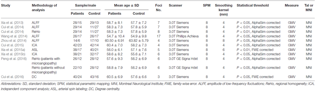
Table 1. List of all studies included in the meta-analysis: demographic and clinical characteristics of subjects.
Data Extraction and Coordinate-Based Meta-Analysis
The x, y and z peak activation coordinates of all eligible contrasts constituted the meta-analysis input. The MNI coordinates were recorded and implemented in GingerALE 2.3.31 (Research Imaging Institute of the University of Texas Health Science Center, San Antonio, TX, USA).
Ginger ALE software was used to compare the brain changes between T2DM patients and healthy controls. The reported loci of maximal anatomical differences were modeled as the peaks of three-dimensional Gaussian probability density functions defined by the full-width at half-maximum (FWHM), which was set according to a quantitative uncertainty model (Laird et al., 2005; Eickhoff et al., 2009). ALE values were calculated on a voxel-by-voxel basis by measuring the union model activation (MA) maps modeled above. This revised analysis tested for convergence by study (random effects) instead of foci (fixed effects). These maps were finally thresholded at P < 0.05 and corrected for multiple comparisons using the false-discovery rate (FDR; q; Genovese et al., 2002). The volume of the minimum cluster threshold was set at 200 mm3. The coordinates of the weighted center were generated for each cluster. The resulting significant anatomical areas were labeled based on probabilistic cytoarchitectonic maps of the human brain using the SPM Anatomy Toolbox v2.1 (Eickhoff et al., 2005). The results were visualized with Mango software2 using the Colin brain template in the MNI space3.
Results
We identified nine eligible studies (Xia et al., 2013, 2015a,b; Cui et al., 2014, 2015, 2016; Wang et al., 2014; Zhou et al., 2014; Peng et al., 2016) for the meta-analysis according to the search criteria mentioned above. In one study, the analysis was performed based on two different subgroups of T2DM patients who were then compared with the same healthy control group (Peng et al., 2016). In another study, the analysis was performed with two different methodologies (Wang et al., 2014). Therefore, we treated these studies as unique reports, with each patient subgroup included independently in the meta-analysis; therefore, a total of 11 datasets were ultimately included in the meta-analysis. Figure 1 shows the details of the literature search strategy and data extraction process. The clinical and demographic data of the participants from all recruited studies are presented in Table 1. The patients and controls from each study were generally matched by age, gender and education.
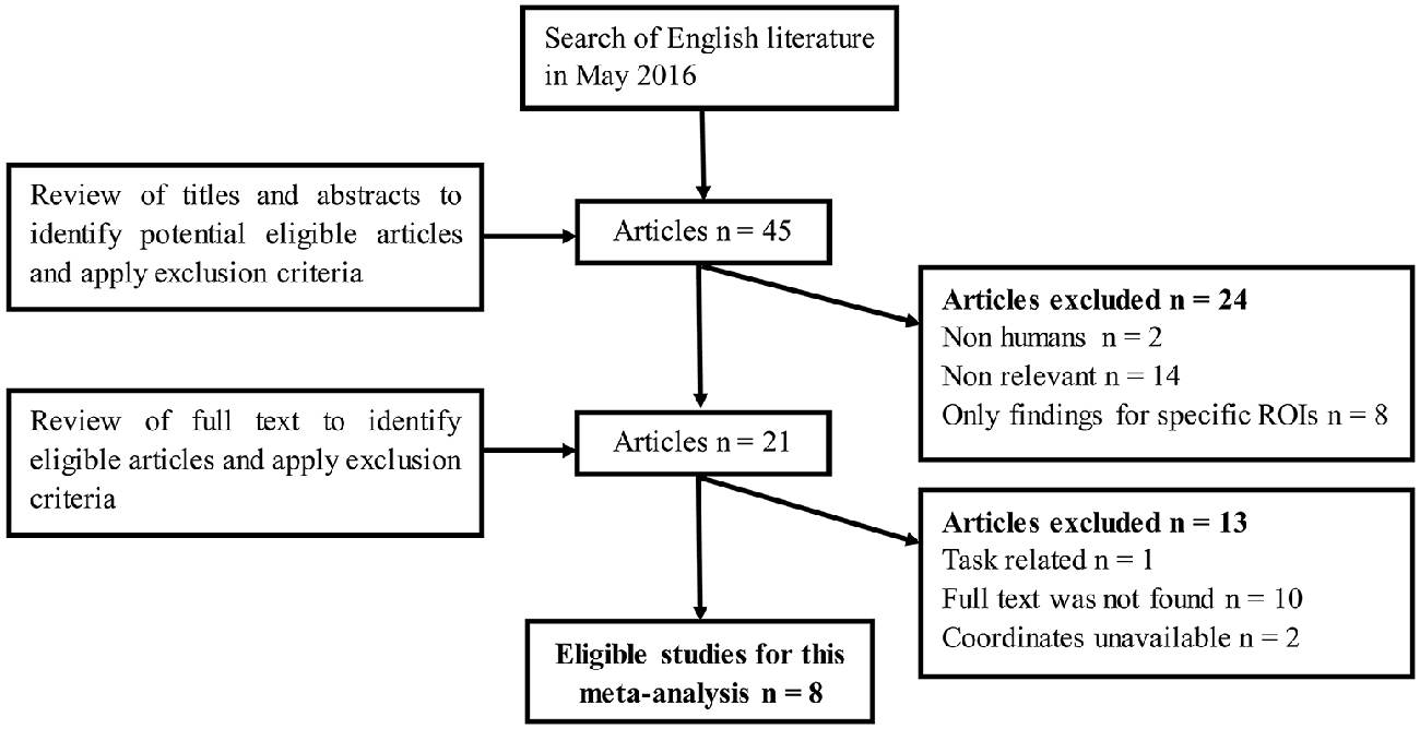
Figure 1. Flow diagram of the literature search. The flow diagram shows the results of the systematic search for the selected studies in this meta-analysis.
As illustrated in Figure 2, a total of 73 peak foci were reported in this meta-analysis. Compared to healthy controls, T2DM patients had widespread reduced resting-sate neural activity in the whole brain, including the bilateral lingual gyrus, left postcentral gyrus, right inferior temporal gyrus, right cerebellar culmen, right insula and right posterior cingulate cortex (PCC). However, enhancements were also observed in the right precuneus and left superior frontal gyrus. Table 2 displays the coordinates of the cluster maxima.
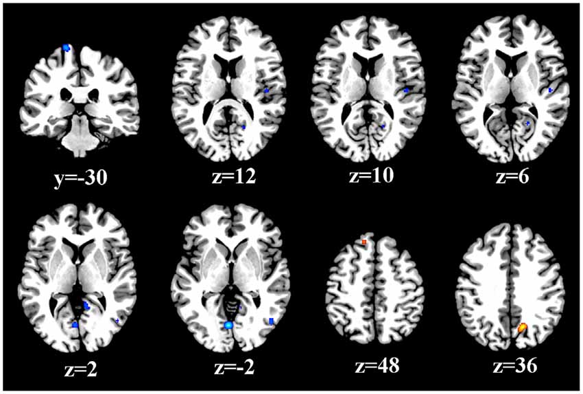
Figure 2. Resting-state brain activity alterations in T2DM patients compared to healthy controls. The results are from the ALE meta-analyses are shown. All activations are significant at P < 0.05 and corrected for multiple comparisons using FDR correction.
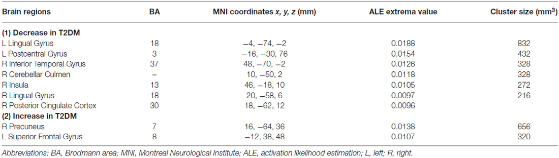
Table 2. Resting-state anomalies in T2DM patients compared with healthy controls.
Discussion
The current study is the first meta-analysis to explore resting-state brain abnormalities in T2DM patients. By analyzing nine eligible studies, this meta-analysis identified consistent regions of resting-state activity brain anomalies across almost the entire brain of T2DM patients, including the frontal, parietal, sensorimotor, temporal, occipital and insular cortices. Therefore, T2DM-related neuropsychiatric impairment is believed to be correlated with diffuse aberrant resting-state brain activity involving both cortical and subcortical structures, and these findings will allow for a more comprehensive understanding of neuropathological mechanisms of this disorder.
Consistent with previous brain perfusion studies without resultant coordinates (Novak et al., 2006), parietal and occipital regions were found to be significantly altered in this meta-analysis. As a key region of the visual cortex, the occipital gyrus is the dominant region that exhibited remarkable reduction of resting-state neural activity in T2DM patients, consistent with the idea that visuospatial dysfunction is a common manifestation in T2DM patients (Moran et al., 2013). Worse visuospatial performance has been observed in T2DM patients than in their matched controls (Bangen et al., 2015), which could be due to the deficits in this area. Furthermore, the postcentral gyrus is known to be the main sensory receptive region for the sense of touch, proprioception, pain and temperature, which could contribute to common diabetic limb pain and temperature sensation decline (Cauda et al., 2009). In fact, impaired sensorimotor function has been described in T2DM patients with peripheral neuropathy (Menz et al., 2004). Nevertheless, the cause-and-effect relationship between deactivation in the postcentral gyrus and diabetic neuropathy cannot be explained at this stage because all the included studies were cross-sectional.
The temporal lobe has been linked to memory, verbal fluency, language processing and speech production, which are impaired in T2DM patients (McCrimmon et al., 2012). T2DM patients showed an abnormal pattern in the inferior temporal gyrus in the current study, which is also consistent with a previous voxel-based morphometry (VBM) study that revealed that T2DM patients exhibited gray matter atrophy in the right temporal lobe (Chen et al., 2012). Although it has been assumed that functional activity changes could precede or lead to subtle brain structural abnormalities, the relationship between functional and structural alterations should be further clarified by longitudinal studies. Several hypothesis-driven MRI studies have also demonstrated the vulnerability of the temporal lobe in T2DM patients. By seeding the PCC during a resting-state fMRI scan, two different research groups reported decreased functional connectivity within the temporal lobe (Musen et al., 2012; Chen et al., 2014). Similarly, by seeding the bilateral hippocampus, abnormal connections within the temporal lobe have also been found. A subtle effect of T2DM on cortical atrophy has also been observed in this area (Brundel et al., 2010). These aforementioned findings provide evidence of disrupted temporal functioning in T2DM patients and suggest that temporal dysfunction should be evaluated when cognitive decline occurs in T2DM patients.
Reduced resting-state brain activity in the cerebellum and insula may also contribute to cognitive deficits in T2DM patients. The cerebellum controls not only motor coordination but also advanced cognitive function (Marklund et al., 2007). Given that the salience network has been found to be impaired in T2DM patients (Chen et al., 2015), and the insula acts as an integral area in the salience network (Menon and Uddin, 2010), decreased insula activity could lead to impaired generation of appropriate behavioral responses to salient stimuli in these patients. Interestingly, according to the results, the right hemisphere of T2DM patients is more seriously impaired than the left hemisphere. Likewise, the effects of T2DM on the cortical surface and volume were only significantly pronounced in the right hemisphere (Brundel et al., 2010). The right hemisphere has been determined to be dominant over the left in geometric analysis and visual-spatial perceptual functioning, including the perception of distance, direction, shape, position, orientation and the detection of complex and hidden figures (Joseph, 1988); therefore, visual-spatial perceptual disturbances, which have been described in T2DM-related cognitive decline, could arise due to right hemisphere impairment. Coincidentally, according to some results extracted from the studies included in this meta-analysis, performance on one neuropsychological test that reflects visual-spatial function, the Rey-Osterrieth Complex Figure Test, has been observed to be correlated with the neural activity in the right brain region (Xia et al., 2015a). Nonetheless, because most studies in this field observed brain alterations on both sides in T2DM patients, the lateralization of impairment of brain activity in T2DM patients requires further investigation.
An additional finding of the present meta-analysis was hyperactivity in the frontoparietal cortex. The precuneus is involved in visuospatial function and working memory, and the frontal lobe is considered as a critical region involved in executive function and attention that subserves cognitive and emotional processing (Radua et al., 2012). These enhanced activities could act in a compensatory or recruitment capacity in T2DM patients such that their cognitive performance could be retained to some extent.
Several limitations should be noted in this meta-analysis. First, the number of included studies was relatively small. Second, ALE can provide excellent control of false positive results, but false negatives are more difficult to avoid (Radua et al., 2012). Finally, heterogeneity of the included studies, such as the demographics of the patients and different imaging modalities that represented different aspects of resting-state abnormalities, could not be entirely excluded. For example, ALFF and ReHo are relevant to the intensity and temporal synchronization of regional spontaneous neuronal activity in the whole brain, respectively (Biswal et al., 1995; Zang et al., 2004). ICA decomposes the entire resting-state BOLD data set into spatially distributed components and automatically identifies meaningful brain networks (Mantini et al., 2007), and ASL perfusion MRI measures regional cerebral blood flow and reflects regional brain metabolism and neural activity (Detre and Alsop, 1999). Despite these differences, various analytic approaches might be complementary to each other and provide more comprehensive information. Similar patterns of resting-state anomalies could also be detected by different analytic modalities. For instance, reduced resting-state activity in the occipital lobe was found in most of our included studies, regardless of the imaging modality.
In summary, this meta-analysis demonstrated robust resting-state brain anomalies in T2DM patients compared to healthy controls that were widespread in the frontal, parietal, sensorimotor, temporal, occipital and insular cortex. Further large, multicenter, resting-state fMRI studies are required to comprehensively investigate whether this abnormal activity pattern is a valuable diagnostic and prognostic biomarker for T2DM-related neuropsychiatric impairment.
Author Contributions
WX and Y-CC performed the analysis and wrote the manuscript. JM designed the meta-analysis and revised the manuscript.
Conflict of Interest Statement
The authors declare that the research was conducted in the absence of any commercial or financial relationships that could be construed as a potential conflict of interest.
Acknowledgments
We would like to express our heartfelt gratitude to Prof. Ning Ma at School of Psychology, South China Normal University. This work was supported by the National Natural Science Foundation of China (No. 81600638, WX), the National Natural Science Foundation of China (No. 81601477, Y-CC).
Footnotes
References
Bangen, K. J., Gu, Y., Gross, A. L., Schneider, B. C., Skinner, J. C., Benitez, A., et al. (2015). Relationship between type 2 diabetes mellitus and cognitive change in a multiethnic elderly cohort. J. Am. Geriatr. Soc. 63, 1075–1083. doi: 10.1111/jgs.13441
Biswal, B., Yetkin, F. Z., Haughton, V. M., and Hyde, J. S. (1995). Functional connectivity in the motor cortex of resting human brain using echo-planar MRI. Magn. Reson. Med. 34, 537–541. doi: 10.1002/mrm.1910340409
Brundel, M., van den Heuvel, M., de Bresser, J., Kappelle, L. J., Biessels, G. J., and Utrecht Diabetic Encephalopathy Study Group (2010). Cerebral cortical thickness in patients with type 2 diabetes. J. Neurol. Sci. 299, 126–130. doi: 10.1016/j.jns.2010.08.048
Cauda, F., Sacco, K., Duca, S., Cocito, D., D’Agata, F., Geminiani, G. C., et al. (2009). Altered resting state in diabetic neuropathic pain. PLoS One 4:e4542. doi: 10.1371/journal.pone.0004542
Chen, Y. C., Jiao, Y., Cui, Y., Shang, S. A., Ding, J., Feng, Y., et al. (2014). Aberrant brain functional connectivity related to insulin resistance in type 2 diabetes: a resting-state fMRI study. Diabetes Care 37, 1689–1696. doi: 10.2337/dc13-2127
Chen, Z., Li, L., Sun, J., and Ma, L. (2012). Mapping the brain in type II diabetes: voxel-based morphometry using DARTEL. Eur. J. Radiol. 81, 1870–1876. doi: 10.1016/j.ejrad.2011.04.025
Chen, Y., Liu, Z., Zhang, J., Tian, G., Li, L., Zhang, S., et al. (2015). Selectively disrupted functional connectivity networks in type 2 diabetes Mellitus. Front. Aging Neurosci. 7:233. doi: 10.3389/fnagi.2015.00233
Cui, Y., Jiao, Y., Chen, H.-J., Ding, J., Luo, B., Peng, C.-Y., et al. (2015). Aberrant functional connectivity of default-mode network in type 2 diabetes patients. Eur. Radiol. 25, 3238–3246. doi: 10.1007/s00330-015-3746-8
Cui, Y., Jiao, Y., Chen, Y. C., Wang, K., Gao, B., Wen, S., et al. (2014). Altered spontaneous brain activity in type 2 diabetes: a resting-state functional MRI study. Diabetes 63, 749–760. doi: 10.2337/db13-0519
Cui, Y., Li, S.-F., Gu, H., Hu, Y. Z., Liang, X., Lu, C.-Q., et al. (2016). Disrupted brain connectivity patterns in patients with type 2 diabetes. AJNR Am. J. Neuroradiol. doi: 10.3174/ajnr.a4858 [Epub ahead of print].
Detre, J. A., and Alsop, D. C. (1999). Perfusion magnetic resonance imaging with continuous arterial spin labeling: methods and clinical applications in the central nervous system. Eur. J. Radiol. 30, 115–124. doi: 10.1016/s0720-048x(99)00050-9
Eickhoff, S. B., Laird, A. R., Grefkes, C., Wang, L. E., Zilles, K., and Fox, P. T. (2009). Coordinate-based activation likelihood estimation meta-analysis of neuroimaging data: a random-effects approach based on empirical estimates of spatial uncertainty. Hum. Brain Mapp. 30, 2907–2926. doi: 10.1002/hbm.20718
Eickhoff, S. B., Stephan, K. E., Mohlberg, H., Grefkes, C., Fink, G. R., Amunts, K., et al. (2005). A new SPM toolbox for combining probabilistic cytoarchitectonic maps and functional imaging data. Neuroimage 25, 1325–1335. doi: 10.1016/j.neuroimage.2004.12.034
Genovese, C. R., Lazar, N. A., and Nichols, T. (2002). Thresholding of statistical maps in functional neuroimaging using the false discovery rate. Neuroimage 15, 870–878. doi: 10.1006/nimg.2001.1037
International Diabetes Federation. (2015). IDF Diabetes Atlas. 7th Edn. Brussels: International Diabetes Federation.
Joseph, R. (1988). The right cerebral hemisphere: emotion, music, visual-spatial skills, body-image, dreams, and awareness. J. Clin. Psychol. 44, 630–673. doi: 10.1002/1097-4679(198809)44:5<630::AID-JCLP2270440502>3.0.CO;2-v
Koekkoek, P. S., Kappelle, L. J., van den Berg, E., Rutten, G. E., and Biessels, G. J. (2015). Cognitive function in patients with diabetes mellitus: guidance for daily care. Lancet Neurol. 14, 329–340. doi: 10.1016/s1474-4422(14)70249-2
Laird, A. R., Fox, P. M., Price, C. J., Glahn, D. C., Uecker, A. M., Lancaster, J. L., et al. (2005). ALE meta-analysis: controlling the false discovery rate and performing statistical contrasts. Hum. Brain Mapp. 25, 155–164. doi: 10.1002/hbm.20136
Mantini, D., Perrucci, M. G., Del Gratta, C., Romani, G. L., and Corbetta, M. (2007). Electrophysiological signatures of resting state networks in the human brain. Proc. Natl. Acad. Sci. U S A 104, 13170–13175. doi: 10.1073/pnas.0700668104
Marklund, P., Fransson, P., Cabeza, R., Larsson, A., Ingvar, M., and Nyberg, L. (2007). Unity and diversity of tonic and phasic executive control components in episodic and working memory. Neuroimage 36, 1361–1373. doi: 10.1016/j.neuroimage.2007.03.058
McCrimmon, R. J., Ryan, C. M., and Frier, B. M. (2012). Diabetes and cognitive dysfunction. Lancet 379, 2291–2299. doi: 10.1016/S0140-6736(12)60360-2
Menon, V., and Uddin, L. Q. (2010). Saliency, switching, attention and control: a network model of insula function. Brain Struct. Funct. 214, 655–667. doi: 10.1007/s00429-010-0262-0
Menz, H. B., Lord, S. R., St George, R., and Fitzpatrick, R. C. (2004). Walking stability and sensorimotor function in older people with diabetic peripheral neuropathy. Arch. Phys. Med. Rehabil. 85, 245–252. doi: 10.1016/j.apmr.2003.06.015
Moran, C., Phan, T. G., Chen, J., Blizzard, L., Beare, R., Venn, A., et al. (2013). Brain atrophy in type 2 diabetes: regional distribution and influence on cognition. Diabetes Care 36, 4036–4042. doi: 10.2337/dc13-0143
Musen, G., Jacobson, A. M., Bolo, N. R., Simonson, D. C., Shenton, M. E., McCartney, R. L., et al. (2012). Resting-state brain functional connectivity is altered in type 2 diabetes. Diabetes 61, 2375–2379. doi: 10.2337/db11-1669
Novak, V., Last, D., Alsop, D. C., Abduljalil, A. M., Hu, K., Lepicovsky, L., et al. (2006). Cerebral blood flow velocity and periventricular white matter hyperintensities in type 2 diabetes. Diabetes Care 29, 1529–1534. doi: 10.2337/dc06-0261
Peng, J., Qu, H., Luo, T. Y., Lv, F. J., Chen, L., Wang, Z. N., et al. (2016). Abnormal spontaneous brain activity in type 2 diabetes with and without microangiopathy revealed by regional homogeneity. Eur. J. Radiol. 85, 607–615. doi: 10.1016/j.ejrad.2015.12.024
Radua, J., Mataix-Cols, D., Phillips, M. L., El-Hage, W., Kronhaus, D. M., Cardoner, N., et al. (2012). A new meta-analytic method for neuroimaging studies that combines reported peak coordinates and statistical parametric maps. Eur. Psychiatry 27, 605–611. doi: 10.1016/j.eurpsy.2011.04.001
Shao, N., Yang, J., and Shang, H. (2015). Voxelwise meta-analysis of gray matter anomalies in Parkinson variant of multiple system atrophy and Parkinson’s disease using anatomic likelihood estimation. Neurosci. Lett. 587, 79–86. doi: 10.1016/j.neulet.2014.12.007
Stroup, D. F., Berlin, J. A., Morton, S. C., Olkin, I., Williamson, G. D., Rennie, D., et al. (2000). Meta-analysis of observational studies in epidemiology: a proposal for reporting. Meta-analysis of observational studies in epidemiology (MOOSE) group. JAMA 283, 2008–2012. doi: 10.1001/jama.283.15.2008
Turkeltaub, P. E., Eden, G. F., Jones, K. M., and Zeffiro, T. A. (2002). Meta-analysis of the functional neuroanatomy of single-word reading: method and validation. Neuroimage 16, 765–780. doi: 10.1006/nimg.2002.1131
Wang, C.-X., Fu, K.-L., Liu, H.-J., Xing, F., and Zhang, S.-Y. (2014). Spontaneous brain activity in type 2 diabetics revealed by amplitude of low-frequency fluctuations and its association with diabetic vascular disease: a resting-state fMRI study. PLoS One 9:e108883. doi: 10.1371/journal.pone.0108883
Weng, H. H., Chen, C. F., Tsai, Y. H., Wu, C. Y., Lee, M., Lin, Y. C., et al. (2015). Gray matter atrophy in narcolepsy: an activation likelihood estimation meta-analysis. Neurosci. Biobehav. Rev. 59, 53–63. doi: 10.1016/j.neubiorev.2015.03.009
Xia, W., Rao, H., Spaeth, A. M., Huang, R., Tian, S., Cai, R., et al. (2015a). Blood pressure is associated with cerebral blood flow alterations in patients with T2DM as revealed by perfusion functional MRI. Medicine (Baltimore) 94:e2231. doi: 10.1097/MD.0000000000002231
Xia, W., Wang, S., Rao, H., Spaeth, A. M., Wang, P., Yang, Y., et al. (2015b). Disrupted resting-state attentional networks in T2DM patients. Sci. Rep. 5:11148. doi: 10.1038/srep11148
Xia, W., Wang, S., Sun, Z., Bai, F., Zhou, Y., Yang, Y., et al. (2013). Altered baseline brain activity in type 2 diabetes: a resting-state fMRI study. Psychoneuroendocrinology 38, 2493–2501. doi: 10.1016/j.psyneuen.2013.05.012
Zang, Y., Jiang, T., Lu, Y., He, Y., and Tian, L. (2004). Regional homogeneity approach to fMRI data analysis. Neuroimage 22, 394–400. doi: 10.1016/j.neuroimage.2003.12.030
Keywords: type 2 diabetes, resting-state fMRI, meta-analysis, activation likelihood estimation
Citation: Xia W, Chen Y-C and Ma J (2017) Resting-State Brain Anomalies in Type 2 Diabetes: A Meta-Analysis. Front. Aging Neurosci. 9:14. doi: 10.3389/fnagi.2017.00014
Received: 29 August 2016; Accepted: 17 January 2017;
Published: 31 January 2017.
Edited by:
Aurel Popa-Wagner, University of Rostock, GermanyReviewed by:
Doris Doudet, University of British Columbia, CanadaAdonis Sfera, Loma Linda University, USA
Copyright © 2017 Xia, Chen and Ma. This is an open-access article distributed under the terms of the Creative Commons Attribution License (CC BY). The use, distribution and reproduction in other forums is permitted, provided the original author(s) or licensor are credited and that the original publication in this journal is cited, in accordance with accepted academic practice. No use, distribution or reproduction is permitted which does not comply with these terms.
*Correspondence: Jianhua Ma, majianhua@china.com
†These authors have contributed equally to this work.