 Katerina Akassoglou1,2*
 Dritan Agalliu3
 Christopher J. Chang4
 Dimitrios Davalos5
 Jaime Grutzendler6
 Elizabeth M. C. Hillman7
 Baljit S. Khakh8
 David Kleinfeld9
 Dorian B. McGavern10
 Sarah J. Nelson11
 Berislav V. Zlokovic12
- 1Gladstone Institute of Neurological Disease, University of California, San Francisco, San Francisco, CA, USA
- 2Department of Neurology, University of California, San Francisco, San Francisco, CA, USA
- 3Departments of Neurology, Pathology and Cell Biology and Pharmacology, Columbia University Medical Center, New York, NY, USA
- 4Departments of Chemistry and Molecular and Cell Biology, Howard Hughes Medical Institute, Helen Wills Neuroscience Institute, University of California, Berkeley, Berkeley, CA, USA
- 5Department of Neurosciences, Lerner Research Institute, Cleveland Clinic Foundation, Cleveland, OH, USA
- 6Department of Neurology, Yale University, New Haven, CT, USA
- 7Laboratory for Functional Optical Imaging, Departments of Biomedical Engineering and Radiology, Kavli Institute for Brain Science, Columbia University, New York, NY, USA
- 8Departments of Neurobiology and Physiology, David Geffen School of Medicine, University of California, Los Angeles, Los Angeles, CA, USA
- 9Department of Physics and Section of Neurobiology, University of California, San Diego, La Jolla, CA, USA
- 10Viral Immunology and Intravital Imaging Section, National Institute of Neurological Disorders and Stroke, National Institutes of Health, Bethesda, MD, USA
- 11Department of Radiology and Biomedical Imaging, University of California, San Francisco, San Francisco, CA, USA
- 12Department of Physiology and Biophysics, Keck School of Medicine, Zilkha Neurogenetic Institute, University of Southern California, Los Angeles, CA, USA
Breakthrough advances in intravital imaging have launched a new era for the study of dynamic interactions at the neurovascular interface in health and disease. The first Neurovascular and Immuno-Imaging Symposium was held at the Gladstone Institutes, University of California, San Francisco in March, 2015. This highly interactive symposium brought together a group of leading researchers who discussed how recent studies have unraveled fundamental biological mechanisms in diverse scientific fields such as neuroscience, immunology, and vascular biology, both under physiological and pathological conditions. These Proceedings highlight how advances in imaging technologies and their applications revolutionized our understanding of the communication between brain, immune, and vascular systems and identified novel targets for therapeutic intervention in neurological diseases.
Introduction
The presentations at the first Neurovascular and Immuno-Imaging Symposium covered a broad range of topics, from physiological neurovascular coupling in the brain to cellular and molecular responses at the blood brain barrier (BBB), to recent developments in molecular imaging, bioengineering, and the identification of novel therapeutic targets for human disease. A common thread among the presentations was the study of the nervous system in health and disease using imaging technologies and molecular tools for recording ongoing biological processes in living organisms, from mice to humans (Figure 1). Brief outlines of the presentations together with a discussion of the imaging approaches used and the implications of the findings for better understanding of normal brain function or pathological processes in disease, have been grouped in the following five thematic areas.
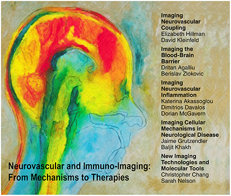
Figure 1. Five thematic areas discussed at the Neurovascular and Immuno-Imaging Symposium. Etching entitled “Mind on Fire” kindly provided by Elizabeth Jameson.
Imaging Neurovascular Coupling
Physiological brain function relies on a large number of interconnected cellular, metabolic, and vascular processes. The term neurovascular coupling describes the relationship between neural activity and metabolism with alterations in cerebral blood flow (CBF). Though the interaction among these processes is well established, the mechanisms linking them and their importance for brain function remain largely unknown. Functional neurovascular coupling refers to the local modulation of CBF that occurs at sites of neural activity in the brain (Kozberg et al., 2013; Hillman, 2014).
Many brain cell types link neural activity and vessel dilation, including astrocytes (Takano et al., 2005), interneurons (Cauli et al., 2004), and pericytes (Hall et al., 2014). However, many features of the cortical hemodynamic response to neural activity are unaccounted for by proposed models. Dr. Elizabeth Hillman presented work from her group, demonstrating that the vascular endothelium plays a role in this active coupling process by propagating vasodilatory signals upstream to pial arterioles (Chen et al., 2014). This pathway, which is known to provide local blood flow modulation elsewhere in the body, has many component mechanisms with different spatiotemporal properties and pharmacological sensitivities (e.g., COX and NO dependent and independent mechanisms; Wölfle et al., 2011) and can explain many of the previously anomalous results of earlier studies. Moreover, dependence of neurovascular coupling on healthy endothelial function implicates a far wider range of systemic cardiovascular disorders as having direct cerebrovascular impact. A state in which endothelial coupling is impaired could lead to neurodegeneration, or even acute cognitive deficiencies. Equally, drugs, such as those aimed at treating blood pressure, inflammation and pain, which act directly on pathways within the vascular endothelium, might affect neurovascular coupling (Bakalova et al., 2002).
Dr. David Kleinfeld presented optical imaging studies that could explain reported resting-state functional magnetic resonance imaging (fMRI) observations correlating neuronal activity in different parts of the brain and slow alterations in blood oxygen levels. fMRI is a powerful tool to probe the extent of activity in the human brain, and the blood-oxygenation-level-dependent (BOLD) fMRI signal (Ogawa et al., 1990) in particular, forms the central technology of modern cognitive neuroscience. An intriguing issue is that ultra-slow variations (~0.1 Hz) in brain tissue oxygenation appear mirrored across conjugate brain areas of the two hemispheres (Biswal et al., 1995). The discovery of this “resting-state” BOLD fMRI (Fox and Raichle, 2007) has been inverted in human cognition studies, so that ultra-slow co-fluctuations are interpreted as function connections (Sporns et al., 2005). Yet a mechanism explaining this observation has been lacking. The Kleinfeld group used ultra-large field two-photon laser scanning microscopy (Tsai et al., 2015) in mice with a thin-skull transcranial window (Drew et al., 2010), together with conventional techniques, to perform microscopic measurements of neuronal activity, vascular dynamics, and tissue oxygenation. They discovered evidence for a biophysical basis linking the co-activation of ongoing neuronal activity with ultra-slow oscillations in blood oxygenation that could justify inferring neuronal connections from synchronous ultra-slow vasodynamics across different brain areas.
Imaging the Blood-Brain Barrier
Neuronal computation and normal brain function requires tight control of the chemical composition of the neuronal “milieu” that is maintained by the BBB (Zlokovic, 1995; Zlokovic et al., 2010). Brain endothelial cells tightly interconnected through adherens and tight junctions (TJs) are the building blocks of the barrier, while astrocytes and pericytes are essential for BBB formation and maintenance. The BBB plays a pivotal role for the healthy central nervous system (CNS) as it regulates the access of blood-borne solutes to the brain and spinal cord, and limits the entry of neurotoxic plasma-derived proteins, circulating metals, red blood cells and leukocytes into the brain.
Dr. Berislav Zlokovic discussed the role of pericytes for BBB breakdown, particularly in the context of neurodegenerative disease. Studies from his group in transgenic murine models have shown that chronic BBB breakdown caused by pericyte degeneration or aberrant signaling to pericytes from endothelial cells or astrocytes leads to accumulation of blood-derived neurotoxic proteins in the CNS (e.g., fibrin, thrombin, hemoglobin, free iron, plasmin; Bell et al., 2010, 2012). This causes progressive neurodegeneration mediated by direct neuronal toxicity, oxidant stress and/or detachment of neurons from the extracellular matrix (Bell et al., 2010, 2012). Pericytes degenerate in Alzheimer's' disease (AD) that is associated with BBB breakdown in human post-mortem studies (Sengillo et al., 2013; Halliday et al., 2016). Accelerated pericyte degeneration in a murine model of AD leads to BBB breakdown and increased accumulation of Alzheimer's toxin amyloid-β (Sagare et al., 2013). Using a high resolution dynamic contrast-enhanced magnetic resonance (MR) imaging protocol to quantify regional BBB permeability in the human brain, they showed BBB breakdown during normal aging that begins in the hippocampus (a region critical for learning and memory), worsens with mild cognitive impairment, and correlates with pericyte injury (Montagne et al., 2015).
Although BBB breakdown is a hallmark of many neurological disorders like AD, ischemic stroke and multiple sclerosis (MS; Zlokovic, 2008), the cellular mechanism of BBB disruption in different diseases is not established. In the healthy CNS, endothelial cells regulate both paracellular and transcellular access through the BBB via the presence of TJs and scarce endocytotic vesicles, respectively. Dr. Dritan Agalliu presented his group's studies of structural and functional BBB changes using in vivo two-photon microscopy in animal models of ischemic stroke and neuroinflammatory disease. Using a novel transgenic mouse strain with eGFP-labeled TJs (Tg eGFP-Claudin5) they found that TJs break down between 24 and 48 h after occlusion in the transient Middle Cerebral Artery Occlusion animal model (t-MCAO). However, BBB function was impaired as early as 6 h after stroke, a time point at which they found an increased rate of transcytosis within endothelial cells (Knowland et al., 2014). These findings suggest a stepwise impairment in transcellular followed by paracellular pathways that contribute to BBB breakdown in ischemic stroke. Moreover, Caveolin-1 deficient mice, that have reduced transcellular permeability, display a normal increase in paracellular permeability after t-MCAO, suggesting that these two mechanisms are independent (Knowland et al., 2014). In similar studies in the MS animal model Experimental Autoimmune Encephalomyelitis (EAE), they found that recycling of TJ proteins was faster in the spinal cord than in the cortex, which may underlie the preferential vulnerability of the spinal cord to EAE. Moreover, dynamic changes in TJs occur prior to EAE onset and throughout the disease. In contrast, BBB transcytosis increases at peak EAE and Caveolin1−∕− mice exhibit decreased peak severity and demyelination. These findings suggest that TJ disruption is necessary for the onset of EAE, whereas transcellular permeability enhances disease severity. Moreover, the kinetics of paracellular vs. transcellular impairment in BBB function is quite different between ischemic stroke and EAE.
Maging Neurovascular Inflammation
BBB disruption, microglial activation and neurodegeneration are hallmarks of MS (Lassmann et al., 2001; Dutta and Trapp, 2014). However, whether plasma proteins contribute to neuroinflammation and neuronal damage remains poorly understood. Studies in human MS have identified abundant deposition of the blood coagulation factor fibrinogen, not only in active and chronic plaques, but also in early lesions prior to demyelination (Kwon and Prineas, 1994; Claudio et al., 1995; Gay et al., 1997; Vos et al., 2005; Marik et al., 2007; Han et al., 2008). Dr. Katerina Akassoglou discussed her group's studies that identified fibrinogen-induced microglial activation as a critical mediator of axonal damage in neuroinflammation (Adams et al., 2007; Davalos et al., 2012, 2014; Bardehle et al., 2015). Using in vivo two-photon microscopy, they showed that in EAE, microglia specifically cluster around blood vessels with BBB disruption at sites of perivascular fibrin deposition even prior to disease onset (Davalos et al., 2012). By developing a molecular probe to detect coagulation activity in the CNS, they demonstrated early activation of coagulation and fibrin deposition even before onset of EAE (Davalos et al., 2014). Intriguingly, fibrin is sufficient to induce reactive oxygen species release by microglia, and thus contribute to neurodegeneration (Davalos et al., 2012). Pharmacologic or genetic disruption of the interaction between fibrin and the CD11b/CD18 integrin receptor (other names Mac-1, complement receptor 3) on microglia suppresses microglial cluster formation, neurological symptoms, inflammation, demyelination, and axonal damage in EAE (Adams et al., 2007; Davalos et al., 2012). Overall, their studies identified fibrinogen as a novel activator of CNS innate immunity that promotes neurodegeneration. Fibrin has the potential for selective drug targeting to suppress its damaging functions in the nervous system without affecting its beneficial effects in hemostasis (Adams et al., 2007; Davalos and Akassoglou, 2012; Davalos et al., 2012; Bardehle et al., 2015; Ryu et al., 2015). Fibrin-selective inhibition of innate immunity may offer novel strategies to combat axonal damage in neuroinflammatory disease.
BBB disruption through the compromise of the TJs of the non-fenestrated endothelium comprising the cerebral and meningeal blood vessels can also be the result of CNS infections and injuries. The resulting unregulated passage of blood-derived materials into the cerebrospinal fluid and CNS parenchyma can in turn give rise to neurological dysfunction, seizures, edema, and ultimately, fatal herniation if unrelieved. Dr. Dorian McGavern discussed recent studies showing that both mechanical and immunological factors can contribute to alterations in the integrity of CNS vasculature (Kim et al., 2009; Roth et al., 2014). Following traumatic brain injury (TBI), mechanical forces stemming from the injury itself can open and / or occlude CNS vasculature, even when the injury is mild (Roth et al., 2014; Corps et al., 2015). This can trigger regional hypoxia, edema, and cell death, resulting in the mobilization of a sterile immune reaction driven by purinergic receptor signaling. Moreover, CNS infection by viruses, bacteria, parasites, and fungi can similarly open cerebral vasculature, yet through different mechanisms (Kang and McGavern, 2010). Meningitis and encephalitis are commonly observed following CNS infection and result from the recruitment of immune cells into the meninges or parenchyma, respectively (Swanson and McGavern, 2015). Innate and adaptive immune cells are recruited to the CNS to clear invading pathogens, but sometimes the manner by which these cells are recruited promotes vascular injury. Intravital imaging studies by his group revealed that pathogen-specific CD8+ T cells can promote synchronous extravasation of innate myelomonocytic cells through release of chemoattractants (Kim et al., 2009; McGavern and Kang, 2011). This type of extravasation is incredibly injurious to CNS vasculature and contributes to seizures and fatal edema. Thus, while the mechanisms of vascular breakdown may differ between infections and injuries, the resultant neurological complications stemming from a loss in CNS barrier integrity are often similar.
Imaging Cellular Mechanisms in Neurological Disease
Dr. Jaime Grutzendler discussed novel methods for longitudinal imaging of the brain microvasculature and myelinated axons in vivo and presented new biological discoveries made with such techniques. The fibrinolytic system has been recognized as the principal mechanism in charge of clearing the vasculature from occluding thromboemboli. Two photon microscopy of cortical capillaries in vivo revealed an additional innate mechanism of microvascular recanalization capable of clearing emboli composed of a variety of substances including those not susceptible to fibrinolysis (Lam et al., 2010). This mechanism which they termed angiophagy involves rapid remodeling of the endothelium, engulfment of the occluding emboli and their translocation through the arteriolar vascular wall (Lam et al., 2010; Grutzendler et al., 2014). This discovery has the potential to greatly impact our understanding and treatment of thromboembolic disorders and the ischemia no-reflow phenomenon. His group also developed a label-free technique (Spectral Confocal Reflection Microscopy-SCoRe) to image myelinated axons in vivo (Hill and Grutzendler, 2014; Schain et al., 2014). This technique allowed for the first time high-resolution in vivo imaging of brain myelin development and pathology in mice and is being explored for potential in vivo diagnostic applications in peripheral nerve human disorders. This method opens new capabilities to study myelin development and pathology and functional interactions with neurovascular cells in living mice and other animals.
Huntington's disease (HD) is caused by intracellular accumulation and aggregation of a mutant form of the huntingtin protein (mHTT). Dr. Baljit Khakh discussed his group's recent findings that suggest neural circuit–specific astrocyte roles for the onset and progression of disease in HD mouse models. They found that a significant increase in astrocyte mHTT is associated with a reduction of important functional proteins like Glt1 (EAAT2) and Kir4.1, which alter astrocyte function without noticeable astrogliosis at least at the onset of neurological symptoms (Tong et al., 2014). Kir4.1 potassium channels are expressed primarily by astrocytes and regulate extracellular K+ levels (Kofuji and Newman, 2004) in the CNS, and loss of the glutamate transporter Glt1 (EAAT2) has been extensively studied in the context of HD (Estrada-Sánchez and Rebec, 2012). Indeed, loss of Kir4.1 in striatal astrocytes led to higher ambient extracellular K+ concentrations, which contributed to increased medium spiny neuron (MSN) excitability in HD mouse models (Tong et al., 2014). Moreover, selective expression of GFP-labeled Kir4.1 channels in astrocytes rescued these deficits, demonstrating that astrocyte Kir4.1 loss can phenocopy hallmark MSN changes of HD mouse models (Cepeda et al., 2010). Together these results support that key aspects of altered MSN excitability in HD are caused at least in part by the failure of astrocytes to maintain homeostatic levels of extracellular K+. This would imply an important yet diverse role for astrocytes in HD pathology, which changes with the disease progression. At early stages of the disease deficits in Kir4.1 and Glt1 (EAAT2) could contribute to underlying HD pathological mechanisms, which could further facilitate the progressive increase in astrocyte reactivity observed with disease progression in humans. Thus, therapeutic strategies targeting astrocytes need to take into account a range of potential astrocytic contributions to HD progression ranging from early homeostatic dysfunction to advanced reactive responses.
New Imaging Technologies and Molecular Tools
MR is a powerful non-invasive imaging modality that makes it possible to visualize anatomic and vascular structures in the brain, as well as physiological and metabolic properties that reflect changes in biological function. Dr. Sarah Nelson discussed recent advances in technology that allow whole body 7T MR scanners and high performance gradients, providing major improvements in the signal to noise ratio and sensitivity achieved in the human brain. High resolution time of flight angiography and susceptibility weighted imaging have been used to take advantage of these capabilities and are critical for monitoring changes in arterial and venous structures associated with vascular disease, aging, trauma and radiation-induced damage (Lupo et al., 2012). The contrast obtained with blood oxygenation level dependent (BOLD) and phase imaging is significantly higher at 7T, facilitating improved fMRI and the detection of abnormalities in deep gray matter structures that are associated with neurodegenerative diseases such as AD, HD (Apple et al., 2014) and MS (Bian et al., 2013). Metabolic imaging that uses multi-voxel H-1 spectroscopy has been applied to evaluate abnormalities caused by neurological and psychiatric diseases and has been shown to benefit from both the higher signal to noise ratio and increased spectral resolution that is possible at 7T (Li et al., 2015) MR imaging.
Dr. Christopher Chang focused his presentation on the chemistry/neuroscience interface, and in particular on the development of molecular imaging probes to enable discovery and study of new types of chemical signals based on metals and redox molecules that govern brain activity. The traditional view of metals in biology is that sodium, potassium, and calcium are the major dynamic signals whereas transition metals are solely static metabolic cofactors. However, invention of several classes of fluorescent probes for metals (Domaille et al., 2008; Que et al., 2008; Aron et al., 2015; Cotruvo et al., 2015) led to the identification of copper as an endogenous regulator of spontaneous activity (Dodani et al., 2011, 2014), representing a new paradigm of transition metal signaling (Chang, 2015). Along the same lines, Dr. Chang presented work from his group illustrating an alternative approach to sensing where reactive small-molecule analytes are sorted by their chemical reactivity as opposed to shape-based recognition and binding (Chan et al., 2012). New probes for emerging signaling / stress molecules like hydrogen peroxide (Lippert et al., 2011) and hydrogen sulfide (Lin et al., 2015) have been devised to identify the essential role of redox molecules in neural stem cells (Dickinson et al., 2011) and H2O2/H2S crosstalk (Lin et al., 2013). These examples illustrate the utility of chemoselective probes for selective molecular imaging of biologically relevant molecules in their native environments.
Concluding Remarks
The first Neurovascular and Immuno-Imaging Symposium provided a forum for the discussion of current research on the cellular and molecular mechanisms that regulate the interface between brain and periphery, both in physiology and in neurological disease. Imaging technologies have proven invaluable in facilitating new discoveries that have reshaped our understanding of cellular behaviors in the CNS (Davalos and Akassoglou, 2008; Merlini et al., 2012). Such discoveries have began to explain not only fundamental physiological functions like the coupling of neuronal activity to blood supply in the brain, but also underlying mechanisms for disease development, like those when the BBB is compromised and peripheral immune cells infiltrate the CNS. The continuous development of new imaging technologies and novel molecular tools that can target ongoing cellular and molecular events at the neurovascular interface will further expand our capabilities to decipher physiological and pathological mechanisms, and design better strategies for therapies in neurological diseases.
Author Contributions
All authors listed, have made substantial, direct and intellectual contribution to the work, and approved it for publication. KA organized and chaired the Symposium. DD and KA wrote the manuscript with contribution from all authors.
Conflict of Interest Statement
The authors declare that the research was conducted in the absence of any commercial or financial relationships that could be construed as a potential conflict of interest.
Acknowledgments
We are grateful to the artist Elizabeth Jameson for “Mind on Fire”, Lennart Mucke and Mark Ellisman for discussions, Linda Turney for administrative support, Chris Goodfellow and Teresa Roberts for graphics. The Symposium was supported by funds from the Gladstone Institutes of Neurological Disease and Lundbeck A/S. Work in the authors' labs was funded by the National Institutes of Health (NIH) NINDS grants NS052189, NS51470, NS082976, the National Multiple Sclerosis Society (NMSS) RG4985A3, and Conrad N. Hilton Foundation Multiple Sclerosis Innovation Award to KA; NIH NHLBI HL116995-01 and NMSS RG4673A1/1 and FG2035-A1 to DA; NIH NIGMS GM79465 to CC; NMSS RG4595A1/T, American Heart Association 13SDG17210051, and a Young Investigator Award from the Race to Erase Multiple Sclerosis Foundation to DD; NIH NIA R21AG048181, NHLBI R01HL106815, NINDS R21NS087511 to JG; NIH R01NS063226, R01NS076628, NSF CAREER 0954796, UL1 TR000040 and the Human Frontier Science Program to EH; CHDI Foundation and NIH NS060677 and MH104069 to BK; NIH NIBIB EB003832, NINDS NS082097, and NIMH MH108503 to DK; DM is supported by the NIH intramural program.; NIH R01CA127612 to SN; NIH R01NS34467, R01AG23084, R01AG039452 and R01NS090904 to BZ. CC is an Investigator with the Howard Hughes Medical Institute.
References
Adams, R. A., Bauer, J., Flick, M. J., Sikorski, S. L., Nuriel, T., Lassmann, H., et al. (2007). The fibrin-derived γ377−395 peptide inhibits microglia activation and suppresses relapsing paralysis in central nervous system autoimmune disease. J. Exp. Med. 204, 571–582. doi: 10.1084/jem.20061931
Apple, A. C., Possin, K. L., Satris, G., Johnson, E., Lupo, J. M., Jakary, A., et al. (2014). Quantitative 7T phase imaging in premanifest Huntington disease. AJNR Am. J. Neuroradiol. 35, 1707–1713. doi: 10.3174/ajnr.A3932
Aron, A. T., Ramos-Torres, K. M., Cotruvo, J. A. Jr., and Chang, C. J. (2015). Recognition- and reactivity-based fluorescent probes for studying transition metal signaling in living systems. Acc. Chem. Res. 48, 2434–2442. doi: 10.1021/acs.accounts.5b00221
Bakalova, R., Matsuura, T., and Kanno, I. (2002). The cyclooxygenase inhibitors indomethacin and Rofecoxib reduce regional cerebral blood flow evoked by somatosensory stimulation in rats. Exp. Biol. Med. 227, 465–473.
Bardehle, S., Rafalski, V. A., and Akassoglou, K. (2015). Breaking boundaries-coagulation and fibrinolysis at the neurovascular interface. Front. Cell. Neurosci. 9:354. doi: 10.3389/fncel.2015.00354
Bell, R. D., Winkler, E. A., Sagare, A. P., Singh, I., LaRue, B., Deane, R., et al. (2010). Pericytes control key neurovascular functions and neuronal phenotype in the adult brain and during brain aging. Neuron 68, 409–427. doi: 10.1016/j.neuron.2010.09.043
Bell, R. D., Winkler, E. A., Singh, I., Sagare, A. P., Deane, R., Wu, Z., et al. (2012). Apolipoprotein E controls cerebrovascular integrity via cyclophilin A. Nature 485, 512–516. doi: 10.1038/nature11087
Bian, W., Harter, K., Hammond-Rosenbluth, K. E., Lupo, J. M., Xu, D., Kelley, D. A., et al. (2013). A serial in vivo 7T magnetic resonance phase imaging study of white matter lesions in multiple sclerosis. Mult. Scler. 19, 69–75. doi: 10.1177/1352458512447870
Biswal, B., Yetkin, F. Z., Haughton, V. M., and Hyde, J. S. (1995). Functional connectivity in the motor cortex of resting human brain using echo-planar MRI. Magn. Reson. Med. 34, 537–541. doi: 10.1002/mrm.1910340409
Cauli, B., Tong, X.-K., Rancillac, A., Serluca, N., Lambolez, B., Rossier, J., et al. (2004). Cortical GABA interneurons in neurovascular coupling: relays for subcortical vasoactive pathways. J. Neurosci. 24, 8940–8949. doi: 10.1523/JNEUROSCI.3065-04.2004
Cepeda, C., Cummings, D. M., André, V. M., Holley, S. M., and Levine, M. S. (2010). Genetic mouse models of Huntington's disease: focus on electrophysiological mechanisms. ASN Neuro 2:e00033. doi: 10.1042/AN20090058
Chan, J., Dodani, S. C., and Chang, C. J. (2012). Reaction-based small-molecule fluorescent probes for chemoselective bioimaging. Nat. Chem. 4, 973–984. doi: 10.1038/nchem.1500
Chang, C. J. (2015). Searching for harmony in transition-metal signaling. Nat. Chem. Biol. 11, 744–747. doi: 10.1038/nchembio.1913
Chen, B. R., Kozberg, M. G., Bouchard, M. B., Shaik, M. A., and Hillman, E. M. (2014). A critical role for the vascular endothelium in functional neurovascular coupling in the brain. J. Am. Heart Assoc. 3:e000787. doi: 10.1161/JAHA.114.000787
Claudio, L., Raine, C. S., and Brosnan, C. F. (1995). Evidence of persistent blood-brain barrier abnormalities in chronic-progressive multiple sclerosis. Acta Neuropathol. 90, 228–238. doi: 10.1007/BF00296505
Corps, K. N., Roth, T. L., and McGavern, D. B. (2015). Inflammation and neuroprotection in traumatic brain injury. JAMA Neurol. 72, 355–362. doi: 10.1001/jamaneurol.2014.3558
Cotruvo, J. A. Jr., Aron, A. T., Ramos-Torres, K. M., and Chang, C. J. (2015). Synthetic fluorescent probes for studying copper in biological systems. Chem. Soc. Rev. 44, 4400–4414. doi: 10.1039/C4CS00346B
Davalos, D., and Akassoglou, K. (2008). “Imaging microglia in the central nervous system: past, present and future,” in Central Nervous System Diseases and Inflammation, eds T. E. Lane, M. Carson, C. Bergmann, and T. Wyss-Coray (Springer), 45–57.
Davalos, D., and Akassoglou, K. (2012). Fibrinogen as a key regulator of inflammation in disease. Semin. Immunopathol. 34, 43–62. doi: 10.1007/s00281-011-0290-8
Davalos, D., Baeten, K. M., Whitney, M. A., Mullins, E. S., Friedman, B., Olson, E. S., et al. (2014). Early detection of thrombin activity in neuroinflammatory disease. Ann. Neurol. 75, 303–308. doi: 10.1002/ana.24078
Davalos, D., Ryu, J. K., Merlini, M., Baeten, K. M., Le Moan, N., Petersen, M. A., et al. (2012). Fibrinogen-induced perivascular microglial clustering is required for the development of axonal damage in neuroinflammation. Nat. Commun. 3, 1227. doi: 10.1038/ncomms2230
Dickinson, B. C., Peltier, J., Stone, D., Schaffer, D. V., and Chang, C. J. (2011). Nox2 redox signaling maintains essential cell populations in the brain. Nat. Chem. Biol. 7, 106–112. doi: 10.1038/nchembio.497
Dodani, S. C., Domaille, D. W., Nam, C. I., Miller, E. W., Finney, L. A., Vogt, S., et al. (2011). Calcium-dependent copper redistributions in neuronal cells revealed by a fluorescent copper sensor and X-ray fluorescence microscopy. Proc. Natl. Acad. Sci. U.S.A. 108, 5980–5985. doi: 10.1073/pnas.1009932108
Dodani, S. C., Firl, A., Chan, J., Nam, C. I., Aron, A. T., Onak, C. S., et al. (2014). Copper is an endogenous modulator of neural circuit spontaneous activity. Proc. Natl. Acad. Sci. U.S.A. 111, 16280–16285. doi: 10.1073/pnas.1409796111
Domaille, D. W., Que, E. L., and Chang, C. J. (2008). Synthetic fluorescent sensors for studying the cell biology of metals. Nat. Chem. Biol. 4, 168–175. doi: 10.1038/nchembio.69
Drew, P. J., Shih, A. Y., Driscoll, J. D., Knutsen, P. M., Blinder, P., Davalos, D., et al. (2010). Chronic optical access through a polished and reinforced thinned skull. Nat. Methods 7, 981–984. doi: 10.1038/nmeth.1530
Dutta, R., and Trapp, B. D. (2014). Relapsing and progressive forms of multiple sclerosis: insights from pathology. Curr. Opin. Neurol. 27, 271–278. doi: 10.1097/WCO.0000000000000094
Estrada-Sánchez, A. M., and Rebec, G. V. (2012). Corticostriatal dysfunction and glutamate transporter 1 (GLT1) in Huntington's disease: interactions between neurons and astrocytes. Basal Ganglia 2, 57–66. doi: 10.1016/j.baga.2012.04.029
Fox, M. D., and Raichle, M. E. (2007). Spontaneous fluctuations in brain activity observed with functional magnetic resonance imaging. Nat. Rev. Neurosci. 8, 700–711. doi: 10.1038/nrn2201
Gay, F. W., Drye, T. J., Dick, G. W., and Esiri, M. M. (1997). The application of multifactorial cluster analysis in the staging of plaques in early multiple sclerosis. Identification and characterization of the primary demyelinating lesion. Brain 120(Pt 8), 1461–1483. doi: 10.1093/brain/120.8.1461
Grutzendler, J., Murikinati, S., Hiner, B., Ji, L., Lam, C. K., Yoo, T., et al. (2014). Angiophagy prevents early embolus washout but recanalizes microvessels through embolus extravasation. Sci. Transl. Med. 6, 226ra231. doi: 10.1126/scitranslmed.3006585
Hall, C. N., Reynell, C., Gesslein, B., Hamilton, N. B., Mishra, A., Sutherland, B. A., et al. (2014). Capillary pericytes regulate cerebral blood flow in health and disease. Nature 508, 55–60. doi: 10.1038/nature13165
Halliday, M. R., Rege, S. V., Ma, Q., Zhao, Z., Miller, C. A., Winkler, E. A., et al. (2016). Accelerated pericyte degeneration and blood-brain barrier breakdown in apolipoprotein E4 carriers with Alzheimer's disease. J. Cereb. Blood Flow Metab. 36, 216–227. doi: 10.1038/jcbfm.2015.44
Han, M. H., Hwang, S. I., Roy, D. B., Lundgren, D. H., Price, J. V., Ousman, S. S., et al. (2008). Proteomic analysis of active multiple sclerosis lesions reveals therapeutic targets. Nature 451, 1076–1081. doi: 10.1038/nature06559
Hill, R. A., and Grutzendler, J. (2014). In vivo imaging of oligodendrocytes with sulforhodamine 101. Nat. Methods 11, 1081–1082. doi: 10.1038/nmeth.3140
Hillman, E. M. (2014). Coupling mechanism and significance of the BOLD signal: a status report. Annu. Rev. Neurosci. 37, 161–181. doi: 10.1146/annurev-neuro-071013-014111
Kang, S. S., and McGavern, D. B. (2010). Microbial induction of vascular pathology in the CNS. J. Neuroimmune Pharmacol. 5, 370–386. doi: 10.1007/s11481-010-9208-9
Kim, J. V., Kang, S. S., Dustin, M. L., and McGavern, D. B. (2009). Myelomonocytic cell recruitment causes fatal CNS vascular injury during acute viral meningitis. Nature 457, 191–195. doi: 10.1038/nature07591
Knowland, D., Arac, A., Sekiguchi, K. J., Hsu, M., Lutz, S. E., Perrino, J., et al. (2014). Stepwise recruitment of transcellular and paracellular pathways underlies blood-brain barrier breakdown in stroke. Neuron 82, 603–617. doi: 10.1016/j.neuron.2014.03.003
Kofuji, P., and Newman, E. A. (2004). Potassium buffering in the central nervous system. Neuroscience 129, 1045–1056. doi: 10.1016/j.neuroscience.2004.06.008
Kozberg, M. G., Chen, B. R., DeLeo, S. E., Bouchard, M. B., and Hillman, E. M. C. (2013). Resolving the transition from negative to positive blood oxygen level-dependent responses in the developing brain. Proc. Natl. Acad. Sci. U.S.A. 110, 4380–4385. doi: 10.1073/pnas.1212785110
Kwon, E. E., and Prineas, J. W. (1994). Blood-brain barrier abnormalities in longstanding multiple sclerosis lesions. An immunohistochemical study. J. Neuropathol. Exp. Neurol. 53, 625–636. doi: 10.1097/00005072-199411000-00010
Lam, C. K., Yoo, T., Hiner, B., Liu, Z., and Grutzendler, J. (2010). Embolus extravasation is an alternative mechanism for cerebral microvascular recanalization. Nature 465, 478–482. doi: 10.1038/nature09001
Lassmann, H., Brück, W., and Lucchinetti, C. (2001). Heterogeneity of multiple sclerosis pathogenesis: implications for diagnosis and therapy. Trends Mol. Med. 7, 115–121. doi: 10.1016/S1471-4914(00)01909-2
Li, Y., Larson, P., Chen, A. P., Lupo, J. M., Ozhinsky, E., Kelley, D., et al. (2015). Short-echo three-dimensional H-1 MR spectroscopic imaging of patients with glioma at 7 Tesla for characterization of differences in metabolite levels. J. Magn. Reson. Imaging 41, 1332–1341. doi: 10.1002/jmri.24672
Lin, V. S., Chen, W., Xian, M., and Chang, C. J. (2015). Chemical probes for molecular imaging and detection of hydrogen sulfide and reactive sulfur species in biological systems. Chem. Soc. Rev. 44, 4596–4618. doi: 10.1039/C4CS00298A
Lin, V. S., Lippert, A. R., and Chang, C. J. (2013). Cell-trappable fluorescent probes for endogenous hydrogen sulfide signaling and imaging H2O2-dependent H2S production. Proc. Natl. Acad. Sci. U.S.A. 110, 7131–7135. doi: 10.1073/pnas.1302193110
Lippert, A. R., Van de Bittner, G. C., and Chang, C. J. (2011). Boronate oxidation as a bioorthogonal reaction approach for studying the chemistry of hydrogen peroxide in living systems. Acc. Chem. Res. 44, 793–804. doi: 10.1021/ar200126t
Lupo, J. M., Chuang, C. F., Chang, S. M., Barani, I. J., Jimenez, B., Hess, C. P., et al. (2012). 7-Tesla susceptibility-weighted imaging to assess the effects of radiotherapy on normal-appearing brain in patients with glioma. Int. J. Radiat. Oncol. Biol. Phys. 82, e493–e500. doi: 10.1016/j.ijrobp.2011.05.046
Marik, C., Felts, P. A., Bauer, J., Lassmann, H., and Smith, K. J. (2007). Lesion genesis in a subset of patients with multiple sclerosis: a role for innate immunity? Brain 130, 2800–2815. doi: 10.1093/brain/awm236
McGavern, D. B., and Kang, S. S. (2011). Illuminating viral infections in the nervous system. Nat. Rev. Immunol. 11, 318–329. doi: 10.1038/nri2971
Merlini, M., Davalos, D., and Akassoglou, K. (2012). In vivo imaging of the neurovascular unit in CNS disease. Intravital 1, 87–94. doi: 10.4161/intv.22214
Montagne, A., Barnes, S. R., Sweeney, M. D., Halliday, M. R., Sagare, A. P., Zhao, Z., et al. (2015). Blood-brain barrier breakdown in the aging human hippocampus. Neuron 85, 296–302. doi: 10.1016/j.neuron.2014.12.032
Ogawa, S., Lee, T.-M., Nayak, A. S., and Glynn, P. (1990). Oxygenation-sensitive contrast in magnetic resonance image of rodent brain at high fields. Magn. Reson. Med. 14, 68–78. doi: 10.1002/mrm.1910140108
Que, E. L., Domaille, D. W., and Chang, C. J. (2008). Metals in neurobiology: probing their chemistry and biology with molecular imaging. Chem. Rev. 108, 1517–1549. doi: 10.1021/cr078203u
Roth, T. L., Nayak, D., Atanasijevic, T., Koretsky, A. P., Latour, L. L., and McGavern, D. B. (2014). Transcranial amelioration of inflammation and cell death after brain injury. Nature 505, 223–228. doi: 10.1038/nature12808
Ryu, J. K., Petersen, M. A., Murray, S. G., Baeten, K. M., Meyer-Franke, A., Chan, J. P., et al. (2015). Blood coagulation protein fibrinogen promotes autoimmunity and demyelination via chemokine release and antigen presentation. Nat. Commun. 6, 8164. doi: 10.1038/ncomms9164
Sagare, A. P., Bell, R. D., Zhao, Z., Ma, Q., Winkler, E. A., Ramanathan, A., et al. (2013). Pericyte loss influences Alzheimer-like neurodegeneration in mice. Nat. Commun. 4, 2932. doi: 10.1038/ncomms3932
Schain, A. J., Hill, R. A., and Grutzendler, J. (2014). Label-free in vivo imaging of myelinated axons in health and disease with spectral confocal reflectance microscopy. Nat. Med. 20, 443–449. doi: 10.1038/nm.3495
Sengillo, J. D., Winkler, E. A., Walker, C. T., Sullivan, J. S., Johnson, M., and Zlokovic, B. V. (2013). Deficiency in mural vascular cells coincides with blood-brain barrier disruption in Alzheimer's disease. Brain Pathol. 23, 303–310. doi: 10.1111/bpa.12004
Sporns, O., Tononi, G., and Kötter, R. (2005). The human connectome: a structural description of the human brain. Public Library Sci. Comput. Biol. 1:e42. doi: 10.1371/journal.pcbi.0010042
Swanson, P. A. 2nd., and McGavern, D. B. (2015). Viral diseases of the central nervous system. Curr. Opin. Virol. 11, 44–54. doi: 10.1016/j.coviro.2014.12.009
Takano, T., Tian, G., Peng, W., Lou, N., Libionka, W., Han, X., et al. (2005). Astrocyte-mediated control of cerebral blood flow. Nat. Neurosci. 9, 260–267. doi: 10.1038/nn1623
Tong, X., Ao, Y., Faas, G. C., Nwaobi, S. E., Xu, J., Haustein, M. D., et al. (2014). Astrocyte Kir4.1 ion channel deficits contribute to neuronal dysfunction in Huntington's disease model mice. Nat. Neurosci. 17, 694–703. doi: 10.1038/nn.3691
Tsai, P. S., Mateo, C., Field, J. J., Schaffer, C. B., Anderson, M. E., and Kleinfeld, D. (2015). Ultra–large field-of-view two-photon laser scanning microscopy. Opt. Express 23, 13833–13847. doi: 10.1364/OE.23.013833
Vos, C. M., Geurts, J. J., Montagne, L., van Haastert, E. S., Bö, L., van der Valk, P., et al. (2005). Blood-brain barrier alterations in both focal and diffuse abnormalities on postmortem MRI in multiple sclerosis. Neurobiol. Dis. 20, 953–960. doi: 10.1016/j.nbd.2005.06.012
Wölfle, S. E., Chaston, D. J., Goto, K., Sandow, S. L., Edwards, F. R., and Hill, C. E. (2011). Non-linear relationship between hyperpolarisation and relaxation enables long distance propagation of vasodilatation. J. Physiol. 589, 2607–2623. doi: 10.1113/jphysiol.2010.202580
Zlokovic, B. V. (1995). Cerebrovascular permeability to peptides: manipulations of transport systems at the blood-brain barrier. Pharm. Res. 12, 1395–1406. doi: 10.1023/A:1016254514167
Zlokovic, B. V. (2008). The blood-brain barrier in health and chronic neurodegenerative disorders. Neuron 57, 178–201. doi: 10.1016/j.neuron.2008.01.003
Zlokovic, B. V., Deane, R., Sagare, A. P., Bell, R. D., and Winkler, E. A. (2010). Low-density lipoprotein receptor-related protein-1: a serial clearance homeostatic mechanism controlling Alzheimer's amyloid beta-peptide elimination from the brain. J. Neurochem. 115, 1077–1089. doi: 10.1111/j.1471-4159.2010.07002.x
Keywords: neuroinflammation, multiple sclerosis, traumatic brain injury, Alzheimer's disease, blood-brain barrier, microglia, myelin, two-photon microscopy
Citation: Akassoglou K, Agalliu D, Chang CJ, Davalos D, Grutzendler J, Hillman EMC, Khakh BS, Kleinfeld D, McGavern DB, Nelson SJ and Zlokovic BV (2016) Neurovascular and Immuno-Imaging: From Mechanisms to Therapies. Proceedings of the Inaugural Symposium. Front. Neurosci. 10:46. doi: 10.3389/fnins.2016.00046
Received: 28 December 2015; Accepted: 01 February 2016;
Published: 22 February 2016.
Edited by:
Astrid E. Cardona, The University of Texas at San Antonio, USAReviewed by:
Daniel A. Lawrence, University of Michigan Medical School, USADavid Pleasure, University of California, Davis, USA
Copyright © 2016 Akassoglou, Agalliu, Chang, Davalos, Grutzendler, Hillman, Khakh, Kleinfeld, McGavern, Nelson and Zlokovic. This is an open-access article distributed under the terms of the Creative Commons Attribution License (CC BY). The use, distribution or reproduction in other forums is permitted, provided the original author(s) or licensor are credited and that the original publication in this journal is cited, in accordance with accepted academic practice. No use, distribution or reproduction is permitted which does not comply with these terms.
*Correspondence: Katerina Akassoglou, kakassoglou@gladstone.ucsf.edu