 Yi Gou1
 Hongyu Zhou
 Gaoxing Su
- 1Jiangsu Province Key Laboratory of Inflammation and Molecular Drug Targets, School of Pharmacy, Nantong University, Nantong, China
- 2Guangzhou Key Laboratory of Environmental Exposure and Health and Guangdong Key Laboratory of Environmental Pollution and Health, School of Environment, Jinan University, Guangzhou, China
Over the past decade, more interests have been aroused in engineering protein-based nanoformulations for cancer treatment. This excitement originates from the success of FDA approved Abraxane (Albumin-based paclitaxel nanoparticles) in 2005. The new generation of biocompatible endogenous protein-based nanoformulations is currently constructed through delivering cancer therapeutic and diagnostic agents simultaneously, as named potential theranostics. Protein nanoformulations are commonly incorporated with dyes, contrast agents, drug payloads or inorganic nanoclusters, serving as imaging-guided combinatorial cancer therapeutics. Employing the nature identity of proteins, the theranostics, escape the clearance by reticuloendothelial cells and have a long blood circulation time. The nanoscale sizet allows them to be penetrated deeply into tumor tissues. In addition, stimuli release and targeted molecules are incorporated to improve the delivery efficiency. The ongoing advancement of protein-based nanoformulations for cancer theranostics in recent 5 years is reviewed in this paper. Fine-designed nanoformulations based on albumin, ferritin, gelatin, and transferrin are highlighted from the literature. Finally, the current challenges are identified in translating protein-based nanoformulations from laboratory to clinical trials.
Introduction
Cancer, a leading cause of death globally, reaching approximately 8.2 million mortalities yearly, poses an enormous burden on society (Torre et al., 2015; Mehra et al., 2017). Cancer counts as a multifactorial and refractory disease caused by the local tissue micro-environmental and genetic factors intertwined (Hanahan and Weinberg, 2011). Common treatment strategies, including radiotherapy and/or chemotherapy with surgery, result in high treatment failure rate (Aoun et al., 2015; Kouchakzadeh and Abbas Shojaosadati, 2016). The reasons of treatment failure are generally diverse: (1) cancer is commonly detected at a later stage, thus accuracy and susceptibility of diagnosis and monitoring methods for early-stage cancer require further improvement (Ge and Liu, 2013; Torre et al., 2015); (2) in most cases, the conventional chemotherapy have been disappointing in efficacy due to multidrug resistance (MDR) and severe side effects (Gelperina et al., 2005; Pérez-Tomás, 2006; Szakács et al., 2006; Ge and Liu, 2013); (3) the cancer therapeutic biological agents (inclusive of antibodies, proteins and nucleic acids), the new class of anticancer drugs, are commonly unstable in in vivo circulation, with rapid degradation and inactivation before reaching the target site (Panyam and Labhasetwar, 2003; Sinha and Trehan, 2003). Accordingly, early detection, effective diagnosis and effective treatment of cancer are needed to be optimized to increase the survival rate and decrease the cancer associated deaths.
“Theranostics,” the portmanteau of therapeutics and diagnostics, has incorporated diagnostic and therapeutic functions into a single nanoplatform (Pene et al., 2009; Chen and Liu, 2016). It is noteworthy that the theranostics have been proposed as a new and revolutionary therapeutic concept in cancer therapy, enabling simultaneous diagnosis and treatment response monitoring using personalized medicine with high accuracy and specificity (Janib et al., 2010; Bardhan et al., 2011). Additionally, it is likely to incorporate numerous different therapeutic drugs into a single theranostics nanoplatform through judicious design to reach synergistic treatment of cancer (Moon et al., 2015; Chen and Liu, 2016). In this regard, theranostics have become a research orientation arousing great concern and been promising in the field of cancer treatment.
Considerable efforts have been recently made to develop new systems for cancer theranostics (Opoku-Damoah et al., 2016; Chen et al., 2017b; Guo et al., 2017; Mohammadi et al., 2017; Tekade and Sun, 2017; Yue et al., 2017). Thus, far, numerous types of theranostic platforms have been reported, consisting of protein nanocarriers (Ng et al., 2011; Chen and Liu, 2016; Truffi et al., 2016), liposome nanocarriers (Wang et al., 2014b; Dai and Yue, 2017), inorganic nanocarriers (Huang et al., 2011; Sharma et al., 2015), polymer nanocarriers (Kamaly et al., 2012; Charron et al., 2015; Sk and Kojima, 2015) and inorganic/organic hybrid nanocarriers (Barreto et al., 2011; Zhu et al., 2015; Li et al., 2017). Among these platforms, protein-based nanoplatforms have aroused the greatest concern by virtue of their biodegradability, biocompatibility, no or low toxicity and ease of modification (Maham et al., 2009; Elzoghby, 2013; Yewale et al., 2013; Chen and Liu, 2016; Kouchakzadeh and Abbas Shojaosadati, 2016). Some proteins, e.g., transferrin and lactoferrin, can be specifically bound to receptor(s) highly expressed in considerable cancer cells via receptor-ligand interaction, enabling the construction of active targeted theranostic nanoplatforms (Kanwar et al., 2016; Wang et al., 2016a, 2017a). Furthermore, in the presence of reactive groups such as –COOH, –NH2, and –OH, protein-based theranostic platforms can be further decorated with functional molecules for different purposes. Numerous proteins as versatile platforms for delivering therapeutic agents have been elucidated in previous review articles (Yewale et al., 2013; Paliwal and Palakurthi, 2014; Kouchakzadeh and Abbas Shojaosadati, 2016).
This review article stresses the protein-based multifunctional theranostics progress in the past 5 years (Table 1) and summaries critical foundation for further studying theranostics. The therapeutic methods include photothermal therapy (PTT), photodynamic therapy (PDT) and chemotherapy. The diagnostic or imaging methods included magnetic resonance (MR) imaging, fluorescence imaging, computed tomography (CT) imaging, photoacoustic (PA) imaging, positron emission tomography (PET) imaging and so on. The combinatorial strategies between diagnostic and therapeutic methods are summarized in Figure 1. Properties of commonly used proteins, i.e., albumin, ferritin, gelatin, and transferrin, were introduced. The preparation methods and key outcomes of in vitro or in vivo studies of these protein-based nanoformulations for cancer theranostics were elaborated.
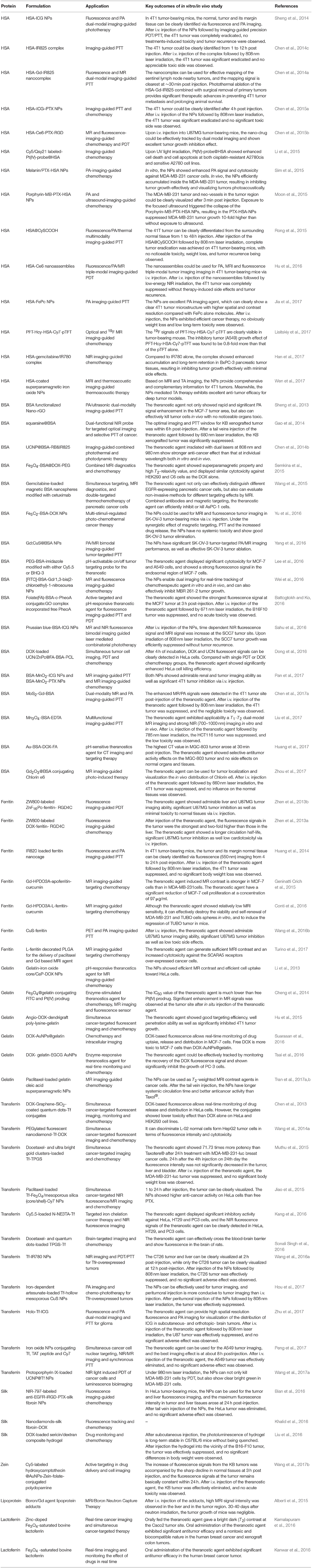
Table 1. Overview of the protein-based nanoformulations for cancer theranostics.
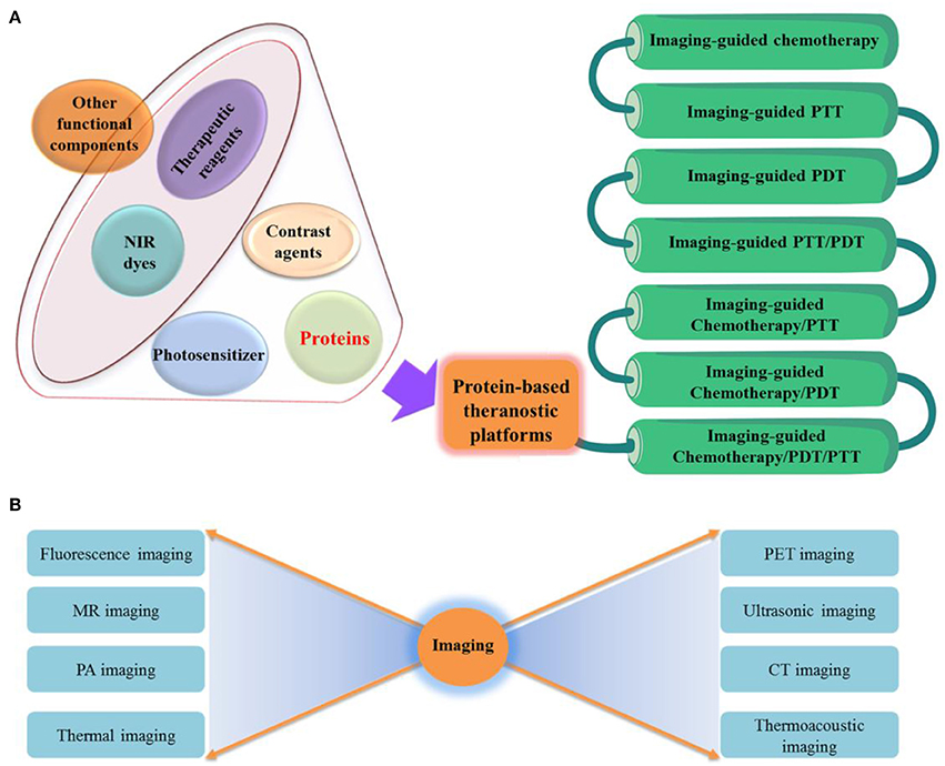
Figure 1. (A) Engineering of protein-based nanoformulations as cancer theranostic platforms. (B) A series of imaging technologies are incorporated in the theranostic platforms.
Albumin-Based Nanoformulations
Albumin and Its Properties
Albumins are obtained commercially in considerable amount from human serum (human serum albumin, HSA), bovine serum (bovine serum albumin, BSA), rat serum (RSA) and egg white (ovalbumin) (Karimi et al., 2016). Based on practicality, this review focus on HSA and BSA.
BSA shares ca. 76% sequence homology with HSA (Anand and Mukherjee, 2013), and is very soluble with a 69,323 Da molecular weight, consisting of 583 amino acid residues. BSA has an isoelectric point (pI) of 4.7 with a net charge of −18 mV (Anand and Mukherjee, 2013). BSA molecule is heart-shaped, consisting of three repeating domains (labeled I–III), with each of which falling into two sub-domains A and B (Majorek et al., 2012). BSA is extensively adopted for cancer theranostics by virtue of low cost, abundance, ease of purification and proper delivering properties.
HSA, the most abundant plasma protein (35–50 mg/mL human serum), is synthesized in the liver. HSA is a single-chain and non-glycosylated polypeptide with 66,500 Da in molecular weight, consisting of 585 amino acid residues, and heart-shaped with 80 × 80 × 30 Å in ca. dimensions (Sugio et al., 1999). From X-ray crystallographic analyses, the vital difference between BSA and HSA is that the former contains two tryptophan amino acid residues (Trp-135 and Trp-212), whereas the latter has merely one, Trp-214. HSA is very soluble, being extremely robust toward temperature (available at 60°C for 10 h), pH (stable in pH 4–9) and organic solvents. Besides, HSA protein has preferentially been uptaken to tumor interstitium via the pathway of SPARC glycoprotein and gp60 glycoprotein transcytosis. These properties as well as its deficient toxicity, immunogenicity, and biodegradability make it an ideal candidate for cancer theranostics.
HSA for Cancer Theranostics
HSA has broadly served as a natural carrier to isolate organic molecules or inorganic oxide, inclusive of IR780, superparamagnetic iron oxide, IR825, and chlorin e6 (Ce6), for synthesizing effective theranostic agents.
Individual HSA-Dye Complexes
HSA is well-known with multiple hydrophobic binding pockets, and able to be bound non-covalently with many organic dyes, forming HSA-dye complexes with a high fluorescence quantum yield. In recent years, near-infrared (NIR) dyes, e.g., IR780, indocyanine green (ICG) and IR825, are broadly employed for cancer theranostics because of their relatively deep penetration and low interference. IR825 can be bound to the hydrophobic domain of HSA (the molar ratio of 1:1) via hydrophobic interactions, which fabricates a HSA-IR825 complex (Figure 2A) (Chen et al., 2014c). The HSA-IR825 complex have a high fluorescence quantum yield at 600 nm excitation and an even high absorbance whereas low fluorescence quantum yield under 808 nm excitation, showing a great performance in NIR imaging and PTT at separated wavelengths. A gadolinium was further fabricated on HSA-IR825 for dual-modal imaging-guided PTT of tumor in a follow-up study (Figure 2B) (Chen et al., 2014a). In this work, HSA was conjugated with the Gd(III) compound of diethylenetriamine pentaacetic acid, and further complexed with IR825, to form HAS-Gd-IR825 complex. The HAS-Gd-IR825 complex has great fluorescence and NIR absorbance, impressive T1 relaxivity of 4.82 mM−1 s−1. More recently, a gemcitabine functionalized HSA-IR780 agent was reported for chemotherapy and imaging-guided PTT of tumor (Han et al., 2017). HSA was first conjugated with gemcitabine via cathepsin B cleavable peptide GFLG, and then mixed with IR780 dye at the molar ratio of 1:1. IR780 could bound to HSA via hydrophobic interactions.
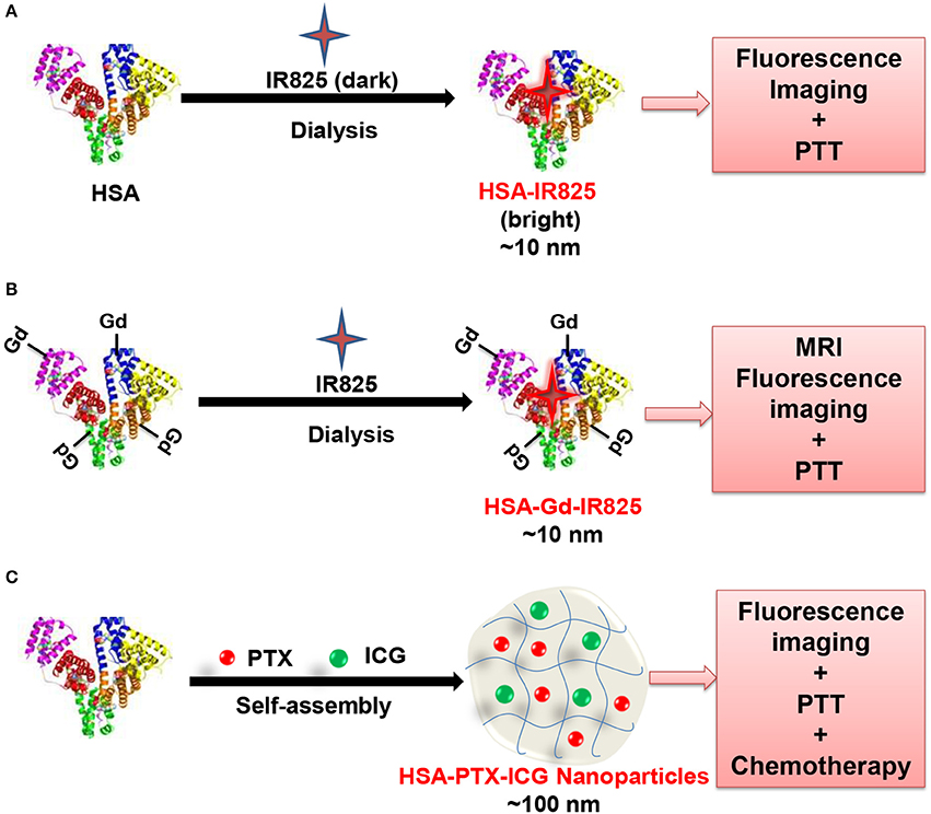
Figure 2. Schematic illustration of the fabrication of HAS-IR825 complex (A) (Chen et al., 2014c), HAS-Gd-IR825 complex (B) (Chen et al., 2014a), and HSA-ICG-PTX nanoparticles (C) (Chen et al., 2015a).
For above systems, the NIR dyes were bound to the HSA via non-covalent interactions, possibly causing dye leakage during in vivo circulation (Rong et al., 2015). To address this problem, heptamethine CySCOOH dye (a NIR cyanine dye) was covalently conjugated to the lysine residues of HSA (Rong et al., 2015) via a modified EDC/NHS reaction for effective photoacoustic (PA), NIR fluorescence, thermal multimodality imaging and PTT. Such conjugation, compared with free CySCOOH dye under the identical conditions, resulted in higher PTT efficacy, tumor accumulation and longer circulation. Moreover, the maleimide group can be rapidly and selectively bound to the Cys34 residue of HSA via a Michael addition reaction. Recently, the Michael addition reaction was employed to couple fluorescent dye Cy7 to Cys34 position of HAS (Lisitskiy et al., 2017). And meantime, the chemotherapeutic agent pTFT (5-trifluoromethyl-2′-deoxyuridine 5′-monophosphate) was coupled to lysine residues of HSA via a redox and pH dual-sensitive linker. The conjugates could not only serve as an optical and 19F MR imaging, but also be applied for delivery of chemotherapeutics.
HSA-Based Complexes
HSA has an effective diameter of 7.2 nm. Yet nanocarriers with sizes of 100-200 nm are well known to tend to accumulate in tumor tissues more efficiently via the enhanced permeability and retention (EPR) effect (Peer et al., 2007). Besides, theranostic agents usually require more functional ingredients to expand the application. Accordingly, great efforts have been devoted to design HSA-based complexes with appropriate sizes and more characteristics, to establish multifunctional HSA-based theranostics. For example, melanin and paclitaxel (PTX)-loaded HSA nanoparticles (HMP-NPs) with size of ca. 192 nm were fabricated with a desolvation-crosslinking method (Mo et al., 2007; Sim et al., 2015). The HMP-NPs showed effective PA signal intensity in the tumor site and the capability to tumor chemotherapy with long circulation time, as confirmed by in vivo experiments. In addition, the desolvation-crosslinking method was adopted to form HSA-based NPs (Pt(IV)-probe@HSA) for theranostic application (Li et al., 2015). In this work, the HSA NPs surface were conjugated with Pt(IV) antitumor prodrug, NIR fluorophore Cy5, and quencher Qsy21. The Pt(IV)-probe@HSA can not only selectively trigger the localized activation of Pt(IV) prodrug, but also enable real-time tumor cell imaging with high resolution.
For the above systems, glutaraldehyde is employed to stabilize the HSA NPs, whereas possible aldehyde residue may cause some side effects for in vivo applications (Fürst and Banerjee, 2005). To address this problem, some facile methods involving no toxic chemicals or exogenous cross-linkers have been broadly adopted in the preparation (Sheng et al., 2014; Hu et al., 2016). As an example, HSA-ICG NPs was developed with an average hydrodynamic diameter of nearly 75 nm based on the intermolecular disulfide bond cross-linking within HSA, for imaging-guided PDT and PTT treatments (Sheng et al., 2014). In this work, they first broke up the intramolecular disulfide bonds of HSA with the endogenous reducing agent glutathione, and then fabricated the HSA NPs with a desolvation method. A similar strategy was also adopted in their recent work to fabricate HSA nanoassemblies (NAs) with photosensitizer chlorin e6 (HSA-Ce6 NAs) for multi-modal imaging-guided PDT (Hu et al., 2016). The proposed HSA-Ce6 NAs had a diameter of ca. 100 nm, excellent tumor selectivity, promising triple-modal (fluorescence, PA and MR) imaging, and effective PDT properties.
Another method proposed by Liu's group not introducing exogenous cross-linkers is to fabricate HSA-based multifunctional theranostic NPs with drug-induced protein assembly strategy (Chen et al., 2015a). In this work, a multifunctional “Abraxane-like” theranostics agent was formulated through simply incorporating three clinical approved agents, ICG, PTX and HSA together (Figure 2C). In this formulation, the NIR fluorescence imaging, thermal imaging, PTT and chemotherapy were efficiently combined. Accordingly, a synergistic therapeutic effect is demonstrated in treating metastatic and subcutaneous breast tumors. As a result, the strategy is enriched to design tumor-targeted theranostics agent for multimodal imaging-guided therapy of tumors (Chen et al., 2015b). HSA is pre-modified in their design, with either a tumor-targeting acyclic Arg-Gly-Asp (RGD) peptide (HSA-RGD) or a photosensitizing agent Ce6 (HSA-Ce6). The anticancer drug PTX is then employed to induce the self-assembly of HSA-RGD and HSA-Ce6 to fall into two different NPs. After incorporation of manganese(II), both of NPs could be tracked by MR imaging and fluorescence imaging, which can be adopted for combinatorial cancer PDT and chemotherapy. Similarly, photosensitizer agent iron (II) phthalocyanine (FePc)-induced HSA self-assembly, is employed in a recent study to fabricate multifunctional HSA-FePc NPs for PA imaging-guided PTT (Jia et al., 2017). The as-prepared HSA-FePc NPs exhibited high stability, high PA imaging quality, efficient PTT treatment, and low long-term toxicity in vivo.
HSA-Coated Complexes
PTX-loaded HSA NPs were conjugated to the surface of porphyrin microbubbles for cancer theranostics (Moon et al., 2015). In this system, porphyrin microbubbles were fabricated using porphyrin-phospholipid conjugates to simultaneously intensify ultrasound and PA signal. The developed multifunctional theranostics agent is high sensitive in PA and ultrasound imaging, and effective in delivery of anticancer drug PTX to a tumor site. HSA-coated superparamagnetic iron oxide NPs were presented more recently, which can absorb pulsed microwave energy and transform efficiency into shockwave with the thermoelastic effect besides using as MR contrast agents (Wen et al., 2017).
BSA for Cancer Theranostics
Individual BSA–Dye Complexes
BSA consists of multiple hydrophobic binding sites, and can naturally serve as a carrier of numerous small NIR dyes agents. Squaraine (SQ) was selectively bound to hydrophobic domain of BSA via hydrogen bonding and hydrophobic interactions with 80-fold enhanced fluorescence intensity (Gao et al., 2014). Based on this, a supramolecular adducts of SQ and BSA (SQ@BSA) was constructed and served as PTT agent and effective bioimaging probe simultaneously. In addition, folic acid (FA) functionalized SQ@BSA (SQ@BSA-FA) has additional functions, e.g., monitoring the time-dependent bio-distribution of adducts and targeting tumor sites. ICG, an FDA-approved NIR dye, can be adsorbed on BSA for NIR fluorescence imaging, PTT and PDT as excited by single-wavelength (Chen and Liu, 2016). Besides, BSA-based theranostics system covalently modified with NIR dyes has been reported (Lee et al., 2016). For example, a zinc-coordinated pH-sensitive theranostics agent is reported, consisting of two types of polyethylene glycol-BSA-imidazole covalently modified with either BHQ-3 quencher (NIR dark quencher) or Cy5.5 dye (donor NIR dye). At pH 5.0 (e.g., endo/lysosomes in cancer cells), the theranostics agent was disassembled rapidly, and emitted strong NIR fluorescence (Lee et al., 2016).
BSA-Based Complexes
Besides bound to NIR dyes, BSA can be further engineered with other functional agents via complexation, such as gadolinium (Wei et al., 2016; Yang et al., 2016; Chen et al., 2017a; Zhou et al., 2017), manganese (Liu et al., 2017; Pan et al., 2017) and graphene's derivatives (Sheng et al., 2013). For example, hollow BSA was employed to fabricate a size-tunable Gd2O3@BSA conjugating Ce6 theranostics agent for MR imaging-guided PDT and PTT (Figure 3) (Zhou et al., 2017). Notably, the BSA nanoreactor can not only effectively regulate the longitudinal relaxivity of Gd2O3, but conjugate readily with photosensitizers. A facile strategy is presented, adopting BSA as a biotemplate at physiological temperature, to construct a biocompatible Gd-integrated CuS multifunctional theranostics agent (Gd:CuS@BSA) (Yang et al., 2016). The fabricated Gd:CuS@BSA theranostics agent possessed ultrasmall sizes (about 9 nm), acceptable longitudinal r1 relaxivity of 16.032 mM−1·s−1, impressive temperature rise and intense PA signals under NIR irradiation. Recently, a multifunctional MoS2-Gd-BSA theranostics agent was fabricated through incorporating the good photothermal effect of MoS2 nanoflakes with the high longitudinal proton relaxivity of BSA-Gd complex via the amine reaction between carboxyl groups of MoS2 nanoflakes and amino groups of BSA-Gd (Chen et al., 2017a). The MoS2-Gd-BSA theranostics agent exhibits a strong NIR absorbance and high r1 relaxivity, which are helpful for PA and T1-weighted MR dual-modal imaging-guided PTT of cancer. Besides, a chemotherapeutic drug carmustine-encapsulated FITC-BSA NPs were prepared by desolvation/denaturation method and conjugated with a MR gadolinium(III) salt to form nanomedicine with dual imaging modalities (Wei et al., 2016).
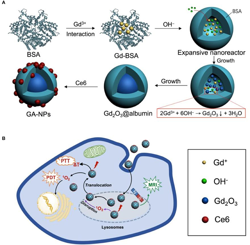
Figure 3. Schematic illustration the synthesis of core-shell Gd2O3@BSA conjugating Ce6 (A) for MRI-guided cancer photothermal therapy (PTT) and photodynamic therapy (PDT) (B). Reproduced with permission (Zhou et al., 2017).
At the identical stage, contrast agent Mn-based BSA complexes have been also reported for cancer theranostics (Liu et al., 2017; Pan et al., 2017). For example, a multifunctional BSA-MnO2 theranostics agent was fabricated via a mimicking drug-substrate interaction strategy, adopting BSA as both reductant agent and template (Pan et al., 2017). It is noteworthy that ICG and PTX can be loaded on BSA-MnO2 with a facile and energy-saving mixing. The ICG/PTX-loaded BSA-MnO2 theranostics agent could be adopted for MR imaging guided PTT and chemotherapy in vitro and in vivo.
Furthermore, a BSA-assisted synthesis route was developed to produce reduced graphene oxide (nano-rGO) theranostic agent (Sheng et al., 2013). The BSA functionalized nano-rGO displayed a high stability and low cytotoxicity, enabling PA imaging and PTT treatment without further surface modification. On that basis, a folate receptor active-targeted, pH-responsive BSA-pheophorbide-a (PheoA) photosensitizer incorporated GO nanocarrier (PheoA + GO:FA-BSA-c-PheoA NC) is developed as an theranostic agent (Battogtokh and Ko, 2016). The theranostic agent carries a good pH-responsive photosensitizer and generates a synergistic PTT and PDT effect against tumor by NIR irradiation.
BSA-Coated Complexes
BSA serves not only as a delivery platform to load various functional molecules, e.g., dyes and chemotherapy drugs, but also as a coating agent of other nanocarriers to increase the physiological stability, water-solubility and blood circulation time of theranostics systems. For example, BSA is employed to coat NaGdY4-based upconversion NPs (UCNPs), resulting in UCNP@BSA NPs possessing well stability and water-solubility in physiological environments (Chen et al., 2014b). And meantime, two different dyes, consisting of an IR825 dye and a Rose Bengal (RB) photosensitizer, can be effectively loaded onto the BSA shell layer of the UCNP@BSA system. It is noteworthy that the characteristic absorbance peak of RB complies well with the green emission peak of UCNPs (980 nm excitation), which effectively kills cancer cells by PDT. Therefore, the dual-dye loaded theranostics agent can be adopted for MR diagnostic, upconversion optical imaging, PDT as well as PTT both in vitro and in vivo. BSA-coated magnetite Fe3O4 core-shell structures with anticancer drug (gemcitabine) were developed, where BSA serving as the outer shell was further functionalized by the active targeted agent Anti-EGFR mab C225 (Wang et al., 2015). The theranostics agent can efficiently regulate double-targeted thermochemotherapy against pancreatic tumor, monitor different cellular targeting by MR imaging, and distinguish various EGFR-expressing pancreatic tumor cells. On that basis, a BSA-coated magnetite Fe5C2 theranostics agent was developed with a high loading of antitumor drug doxorubicin (DOX) (Yu et al., 2016). Notably, the DOX can be released in acidic condition and irradiated by NIR. In this regard, the theranostics agent serves as a smart nanoplatform for MR imaging, effective chemotherapy and PTT. A pH-responsive protein–polymer bioconjugate-coated theranostics agent consisting of a superparamagnetic magnetite Fe3O4 core, BSA–poly(ethylene glycol) (PEG) shell and anticancer drug DOX were presented for combined MR imaging diagnostics and chemotherapy (Semkina et al., 2015). A BSA–poly(ε-caprolactone) bioconjugate-coated upconversion theranostics agent is constructed with the similar preparation method for simultaneous cancer cell imaging, PDT, and chemotherapy (Dong et al., 2016). BSA stabilized Prussian blue (PB) NPs were fabricated, and ICG molecules were further attached non-covalently by a biocompatible and simple method (Sahu et al., 2016). Here, PB serves as a MR contrast enhancer. Accordingly, the multifunctional theranostics system could provide dual mode MR signal and NIR fluorescence imaging as well as combined therapy with PDT and PTT. A pH-responsive Au–BSA core/shell theranostics agent consisting of Au core, BSA shell that conjugated with DOX and FA was developed recently, which manifested tumor computed tomography (CT) imaging application and targeted cancer therapy (Huang et al., 2017).
Albumin-based NPs could be synthesized by using albumin as a scaffold, template, or stabilizer, and conjugating to polymers, drugs, and contrast agents. Covalent and non-covalent conjugation or assembly were employed. Due to the success of Abraxane, researchers preferred the non-convalent self-assembly. However, NPs prepared using the non-convalent strategy will vary batch to batch, which need to be overcome in the future studies (An and Zhang, 2017).
Ferritin-Based Nanoformulations
Ferritin and Its Properties
Ferritin counts as an abundant protein in circulation, existing in intracellular and extracellular compartments. Ferritin is a 450 kDa hollow nanocage with internal and external dimensions of 8 and 12 nm, respectively (Banyard et al., 1978). It accumulates and stores approximately 4,500 iron atoms in a non-toxic whereas bioavailable form (Alkhateeb and Connor, 2013). In mammals, each ferritin protein consists of 24 subunits self-assembled into a spherical symmetrical protein shell (Alkhateeb and Connor, 2013). The ferritins from eukaryotes are produced by self-assembly of two subunit types, i.e., L-ferritin chain (19 kDa) and H-ferritin chain (21 kDa). The H-chain is centered by an iron oxidase center, required to oxidize Fe(II) to Fe(III), whereas the L-chain without ferroxidase activity nucleates iron (Bellini et al., 2014). Ferritin counts as a multifunctional protein with iron storage and metabolism. Ferritin is critical for angiogenesis, proliferation and immunosuppression, as demonstrated by growing number of evidence (Alkhateeb and Connor, 2013).
Ferritin is a ubiquitous protein robust extremely: it can be reversibly disassembled in the extremely acidic pH (pH 2–3) or basic pH (pH 10–12) and can be heated at 80°C exerting no deleterious effects (Truffi et al., 2016). These properties as well as its biocompatibility and biodegradability make it a suitable candidate for cancer theranostics (Bhushan et al., 2014).
Ferritin for Cancer Theranostics
The ferritin not only provides a reaction vessel to fabricate numerous non-native metallic NPs inside its core, but also serves as a nanocarrier for various applications (Bhushan et al., 2014). For example, Zn hexadecafluorophthalocyanine (ZnF16Pc), a potent hydrophobic photosensitizer, is well encapsulated into the Cys-Asp-Cys-Arg-Gly-Asp-Cys-Phe-Cys (RGD4C)-modified ferritins (P-RFRTs) with a loading rate as high as ~60 wt % for effective PDT (Zhen et al., 2013b). The P-RFRTs were further conjugated with ZW800 (a NIR dye molecule) in this work to better track P-RFRTs particles. With the identical approach, the DOX-loaded RGD4C-modified ferritin was also reported (Zhen et al., 2013a). A self-assembly encapsulation strategy based on step-wise change of pH was developed in another report to fabricate ferritin NPs with NIR dye IR820 for fluorescence/PA multimodal imaging-guided PTT (Huang et al., 2014). The IR820-loaded ferritin nanocages can effectively treat and diagnose cancer adopting two different excitation wavelengths, i.e., 550 nm for high quantum-yield fluorescence imaging, and 808 nm for PA imaging and effective PTT. Additionally, CuS was fabricated inside the cavity of ferritin nanocages with a biomimetic and straightforward synthesis strategy (Wang et al., 2016b). The CuS-ferritin has strong NIR absorbance, high photothermal conversion efficiency, good biocompatibility and distinct PA contrast,. Notably, the 64CuS-ferritin theranostic system, as incorporated with radionuclide 64Cu, also served as an excellent PET imaging agent.
The higher level of L-ferritin in tumor versus normal tissue has been observed in some malignancies tissues like breast cancer, colon cancer, pancreatic cancer and testicular seminoma (Alkhateeb and Connor, 2013). High amount of L-ferritin is usually associated and bound with intensified expression of the L-ferritin receptor that mediates ferritin endocytosis. The higher the expression of L-ferritin receptors, the more intensified uptake of L-ferritin in breast cancer MCF-7 cells (Geninatti Crich et al., 2015). Based on this, a ferritin-based nanotheranostic system has been further developed to simultaneously deliver a MR contrast agent GdHPDO3A and a natural anticancer molecule curcumin (Geninatti Crich et al., 2015). The theranostics system selectively delivered therapeutic and imaging agents to breast cancer cells. In a follow-up study, L-ferritin was found to target breast cancer stem cells (Conti et al., 2016). In this regard, ferritin was further exploited to deliver curcumin and the GdHPDO3A MR contrast agent simultaneously for breast cancer stem cells (Conti et al., 2016). In a very recent report (Turino et al., 2017), multifunctional theranostics system was developed through coating poly (lactic-co-glycolic acid) NPs (PLGA NPs) with L-ferritin to increase their targeting capability to breast cancer MCF-7 cells. The L-ferritin functionalized PLGA NPs loaded with an amphiphilic Gd based MR contrast agent and PTX for MR imaging guided chemotherapy. In addition to this, ferritin coating makes PLGA NPs more stable, thus avoiding the non-specific and fast release of therapy/diagnosis agents before reaching the targeted sites.
Hollow structure make the ferritin become an ideal carrier, however, after loading therapeutic agents, the slight change in conformation and activity might take place. The toxicity of ferritin-based NPs should be studied carefully as other NPs. Also, the surface-modified ferritins may be recognized as foreign substances. Therefore, when surface targeting ligands were needed, the grafting density are important factors to be considered.
Gelatin-Based Nanoformulations
Gelatin and Its Properties
Gelatin is a polyampholyte protein having both anion and cation along with hydrophobic groups (Elzoghby, 2013). Gelatins have repeating sequences of proline, alanine, and glycine amino acid triplets, which are essential for gelatin (Sahoo et al., 2015). They are obtained from alkaline-, acid- or enzymatic- hydrolysis of collagen. These chemically treated gelatins have varying isoelectric points, i.e., 4.5–6.0 for alkaline treatment and 7–9 for acid treatment (Patel et al., 2008; Ninan et al., 2011). In addition, anticancer agent release profiles from gelatin protein can be controlled through changing the molecular weight, gelatin source and the crosslinking degree (Foox and Zilberman, 2015). Gelatin is natural, biocompatible, biodegradable, water permeable, non-toxic, and soluble in water (Nezhadi et al., 2009). As a delivery carrier of therapeutic/diagnostic agents, gelatin has been revealed multifunctional properties, enabling the development and design of versatile theranostics.
Gelatin for Cancer Theranostics
Gelatin has native hydrophobic and hydrophilic segments in each single polypeptide chain, enabling itself a conducive coating agent for various metal NPs (Li et al., 2013; Cheng et al., 2014; Tran et al., 2017b). For example, a theranostic system was developed through coating iron oxide NPs with self-assembled gelatin (abbreviated as AGIO) (Li et al., 2013). In the meantime, calcium phosphate (CaP) and anticancer DOX could be loaded on gelatin by electrolytic co-deposition technique. The fabricated AGIO@CaP-DOX NPs had efficient MR contrast, superior cytocompatibility and potent cellular internalization toward HeLa cells. A gelatin coated multifunctional nanosystem, with oleylamine-coated Fe3O4 NPs core, amphiphilic gelatin shell, and fluorescent labeling molecules FITC and antitumor platinum(IV) prodrug, was elaborated for fluorescence and MR imaging guided chemotherapy (Cheng et al., 2014). Most recently, oleic acid and gelatin were adopted to modify a silica-coated iron oxide magnetic NPs, which was demonstrated to encrease biocompatibility and solubility of iron oxide magnetic NPs and facilitate treatment-response monitoring of the tumors (Tran et al., 2017b). Particularly, the oleic acid and gelatin coated NPs enables the hydrophobic anticancer drug PTX to be loaded into the lipophilic oleic acid-gelatin shell. The synthesized theranostic system have high r2 value, low cellular toxicity, high drug delivery efficacy and well anticancer efficacy in vitro. The pharmacokinetics, bio-distribution, tumor diagnostic and antitumor efficacy of the theranostic system in vivo were studied in a follow-up study to further confirm its potential in clinical applications (Tran et al., 2017a).
Beside iron oxide NPs, gold NPs are commonly coated with gelatin to increase their stability and biocompatibility. For example, a gelatin-coated gold NP-based nanostructure was developed for fluorescence imaging-guided chemotherapy (Tsai et al., 2016). The gelatin was firstly covalent conjugated with DOX, and then coated onto the epigallocatechin gallate (EGCG)-functionalized Au NPs. Here, DOX serves both as an anticancer drug and a fluorescent indicator. The attained DOX-gelatin/EGCG Au NPs exert an apparent inhibitory effect on the proliferation of human prostate cancer cells (PC-3) and can trace the intracellular enzyme-induced release of DOX through measuring the recovery of the DOX fluorescence signal. The covalent conjugation of DOX to nanocarriers may cause problems, e.g., insufficient in vivo release and decreased drug activity (Suarasan et al., 2016). A new temperature- and pH-responsive theranostic system was developed on the basis of DOX non-covalently bound to biosynthesized gelatin-coated Au NPs (DOX-AuNPs@gelatin) (Suarasan et al., 2016). The fabricated DOX-AuNPs@gelatin would be an ideal agent for cancer theranostics based on its good biocompatibility, high DOX loading capacity via non-covalent complexation and effective DOX release under the tumor environment.
Recently, angiopep-2 modified gelatin-based core-shell NPs (Angio-DOX-DGL-Gel-NP) have been designed to increase the tumor targeting efficiency, tumor retention and tumor penetration (Hu et al., 2015). In this system, the shell consisted of dendrigraft poly-lysine linked with angiopep-2 and DOX, whereas the core was made up of gelatin NPs degraded by matrix metalloproteinase-2. Fluorescence imaging-guided chemotherapy showed enhanced antitumor effects in vitro and in vivo,
Gelatins are used as carriers in versatile drug delivery systems, form nanoparticles to microparticles (Foox and Zilberman, 2015). Small drug molecules or large bioactive molecules were easily entrapped into gelatins and released in a controlled manner. However, the cancer theranostic platforms based on gelatin didn't attract much attention. In our opinion, the gelatin-based NPs are promise cancer theranostics, especially in oral administration and brain delivery.
Transferrin-Based Nanoformulations
Transferrin and Its Properties
Transferrin is a monomeric glycoprotein with 679 amino acids and a large molecular weight of approximately 79 kDa (Parkkinen et al., 2002; Gomme et al., 2005). The molecule is protected by 3 carbohydrate side chains, one of which is O-linked (Ser-32) and the other are N-linked (Asn-413 and Asn-611) (Gomme et al., 2005). The polypeptide chain falls into two structurally similar lobes, known as the C-lobe (343 amino acids) and N-lobe (336 amino acids), which are connected by a short linear spacer sequence (Brandsma et al., 2011). Each lobe consists of one reversibly binding site for ferric iron with nearly 1022 M−1 affinity at pH 7.4 (Aisen et al., 1978). The major biological function of transferrin is to distribute and control circulating Fe, which is required for numerous biological processes, including cellular metabolism and proliferation, DNA synthesis, electron transfer, and oxygen transport (Dufès et al., 2013; Tortorella and Karagiannis, 2014). Particularly, transferrin specifically binds transferrin receptors (TfRs) on cell surface, forming a transferrin-TfR complex, and gets internalized by receptor-mediated endocytosis (Szoke and Panteghini, 2012; Dufès et al., 2013). Meantime, overexpression of TfRs has been observed in various cancer cells, including breast carcinoma, glioblastoma, melanoma, ovarian carcinoma and colon carcinoma (Calzolari et al., 2007; Tros de Ilarduya and Düzgüneş, 2013).
Transferrin for Cancer Theranostics
As a promising tumor targeting ligand, transferrin is frequently adopted to facilitate targeting delivery of theranostics. For example, transferrin–functionalized graphene quantum dots were developed to track and image tumor cells expressing the TfRs (Chen et al., 2013). In the meantime, the anticancer drug, DOX, was adsorbed on graphene surface in the system via hydrophobic interactions and π-π stacking, which could be used for fluorescence imaging-guided chemotherapy. Another transferrin-conjugated PEGylated fluorescent nanodiamond with Dox payload was developed, and its targeting ability and chemotherapeutic potential were investigated in the human hepatoma (HepG2) cell lines with overexpression of TfRs and normal cell lines (L-02) with low-expression of TfR, respectively (Wang et al., 2014a). A transferrin conjugated theranostic micelles of D-alpha-tocopheryl PEG 1000 succinate (TPGS) were synthesized, containing both ultra-bright gold clusters as a model imaging agent and docetaxel as anticancer drug for synchronous cancer imaging and therapy (Muthu et al., 2015). The transferrin conjugated micelles, compared with the non-transferrin functionalized theranostic micelles, showed higher cellular uptake, higher cytotoxicity in MDA-MB-231-luc breast cancer cells. In a follow-up study, a transferrin decorated TPGS coated theranostic liposomes was further developed for targeted co-delivery of quantum dots and docetaxel for imaging-guided chemotherapy of brain cancer (Sonali Singh et al., 2016). Indeed, the transferrin decorated theranostic liposomes, compared with the non-transferrin targeted preparations, showed an improved and prolonged brain targeting of quantum dots and docetaxel. In addition, rattle-type theranostic NPs of mesoporous silica-coated Fe3O4 were develop by conjugating transferrin for targeted co-delivery of NIR dye (Cy7) and hydrophobic anticancer drug PTX for NIR/MR bimodal imaging guided chemotherapy (Jiao et al., 2015).
Recently, transferrin and nuclear-targeted TAT peptide (YGRKKRRQRRR) conjugated magnetic NPs were proposed for PTT application (Peng et al., 2017). Magnetic NPs is pre-conjugated with transferrin and TAT peptide, and then bound to NIR dye Cy7. The theranostic system can efficiently target cancer cell nucleus and facilitate the NIR and MR imaging-guided PTT. A core-shell theranostic system UCNP core (NaYF4:Gd3+, Yb3+, Er3+) was developed, taking advantage of efficient NIR-to-visible up-conversion capability and increased tumor targeting ability and biocompatibility (Wang et al., 2017a). In the meantime, a clinically approved PTT agent, protoporphyrin IX, was loaded into the transferrin shell, being able to be drawn upon by cancer cells for efficient PDT with NIR irradiation and luminescence bio-imaging. A new strategy (diffusion molecular retention tumor targeting effect) was developed to fabricate tumor-targeted theranostic system for PA imaging-guided chemotherapy/photothermal with synergistic effect (Hou et al., 2017). In this study, hollow mesoporous CuS NPs (HMCuS NPs) were conjugated with transferrin and iron-dependent artesunate (AS), an effective anticancer drug. In this regard, the attained AS/Transferrin-HMCuS NPs facilitated local drug accumulation and retention, targeted to breast cancer MCF-7 cells specially via TfR-mediated endocytosis, combined chemotherapy-phototherapy synergistically, and eventually improved the anticancer effect.
Transferrin is not only a conducive targeting ligand, but also able to load imaging and/or therapeutic agents. For example, a simple, effective and safe self-assembly strategy was developed to fabricate transferrin NPs with NIR dye IR780 (Transferrin-IR780 NPs) for targeted imaging and phototherapy of cancer (Wang et al., 2016a). The fabricated Transferrin-IR780 NPs had advantages on potent photo-stability, narrow size distribution as well as prominent photothermal conversion efficiency, and exhibited pronounced targeting and theranostics potential. A conjugate of transferrin, NIR dye Cy5.5 and cytotoxic chelating agent (NNE3TA:2,2′-(7-(2-((carboxymethyl)(4-nitrobenzyl)amino)ethyl)-1,4,7-triazonane-1,4-diyl)diacetic acid) (NNE3TA-Transferrin-Cy5.5) was developed to treat and detect cancers (Kang et al., 2016). Targeted iron chelation cancer therapy and NIR imaging were demonstrated effectively in vitro with NNE3TA-Transferrin-Cy5.5. Most recently, a drug-induced transferrin self-assembly strategy is developed to fabricate tumor-targeted NPs for fluorescence and PA dual-modal imaging-guided PTT of glioma (Figure 4) (Zhu et al., 2017). In this system, transferrin could effectively load ICG via hydrophobic interaction and hydrogen bonding. Specifically, the preparation method is safe, simple and mild without the use of any toxic reagents. The obtained Transferrin-ICG NAs showed effective active tumor-targeting, good biocompability, prominent dual-modal imaging as well as PTT efficacy, and could be adopted for theranostics of both subcutaneous and orthotopic brain tumors.
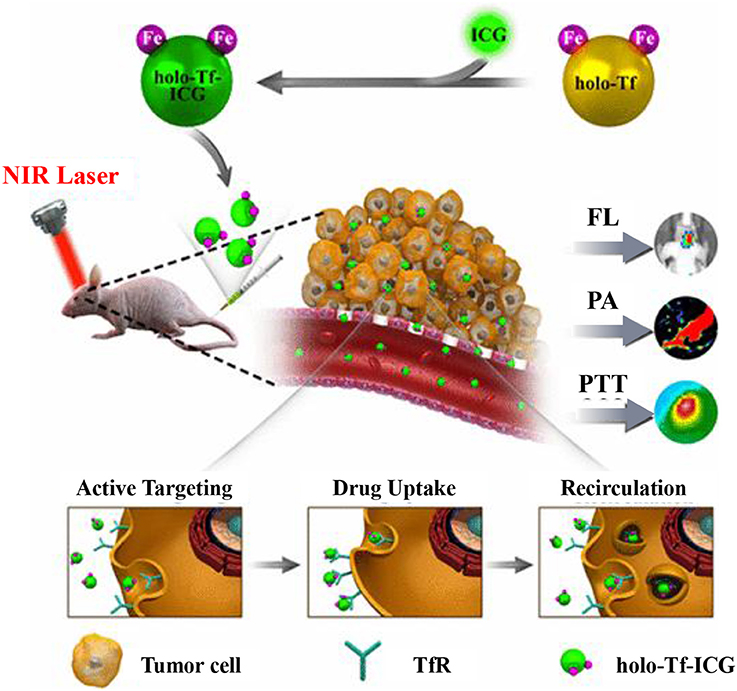
Figure 4. Schematic illustration of holo-Transferrin-ICG complex for dual-modal imaging-guided cancer photothermal treatment in vivo. Reproduced with permission (Zhu et al., 2017). Copyright (2017) American Chemical Society.
Notably, transferrin was employed not only as an delivery vehicle but also as a targeting agent in these works. It has been reported than transferrin-functionalized silica NPs lost targeting abilities completely in serum-rich media, due to shielding effects of the adsorbed proteins (Salvati et al., 2013). The bioavailability of the large transferrin determines the targeting functionality in complex biological media. It is a challenge to maintain the targeting efficiency of transferrin-based NPs in vivo.
Others
With the exception of the foregoing proteins, several other proteins, e.g., silk fibroin (Bian et al., 2016; Khalid et al., 2016; Liu et al., 2016), zein (Wang et al., 2017b), lipoprotein (Mathew et al., 2013; Alberti et al., 2015) and lactoferrin (Kamalapuram et al., 2016; Kanwar et al., 2016), have also been successfully employed for cancer theranostics, as detailed in Table 1. For instance, multimodular zinc-doped Fe3O4-saturated bovine lactoferrin NPs (Zn-Fe-bLf NPs) were fabricated for a targeted theranostics application through oral administration (Kamalapuram et al., 2016).
Although natural proteins were used in most studies, the disadvantages of these protein-based platforms were obvious, such as poor permeability, ease of degradation, and potential immune responses. Moreover, after functionalization or conjugation of other molecules, the protein properties and functions might be altered. In recent years, encapsulation of proteins by in situ polymerization was developed as an alternative strategies for protein modification (Ye et al., 2016b). The protein nanogels were obtained by incorporation of cross-linkers to protein surface and polymerization. Such protein nanogels can maintain the protein properties and functions in complex chemical or biological environment, and avoid the immunogenicity of proteins. The out layer polymers were cleavable and degradable, while the inner proteins could be released by internal and external stimuli. Numerous therapeutic proteins have been developed using this method for cancer therapy (Ye et al., 2016a, 2017).
Conclusion and Prospects
Natural proteins as biocompatible nanocarriers are broadly adopted in delivering therapeutic and diagnostic agents simultaneously. These nanoformulations are fabricated progressively complex and “smart” to employ multi functions in one platform. To increase the accuracy of diagnostic and the efficiency of cancer therapy, “all-in-one” nanoplatforms are designed with more than two imaging strategies and more than two therapeutic methods. Surface modification with targeted molecules and controlled drug release are employed to spatially control the localization of administered NPs. The improved efficiencies in tumor imaging and ablation are verified both in vitro and in vivo. Despite these huge advances, the translation of natural protein nanoplatforms from laboratory to clinical trials remains an enormous challenge. More efforts should be made to improve the NP reproducibility, colloidal stability in biological environments, and drug-loading efficiency. Furthermore, live and spleen accumulation is a big barrier in clinical translation of all types of NPs. Accordingly, more understanding of bio-nano interactions is required, while more data of pharmacokinetics should be collected and analyzed. Long-term toxicity of nanoformulation is not well evaluated. In addition to mouse model, more animal models, e.g., beagle dogs, monkeys and gorillas, should serve for assessing the long-term toxicity and therapeutic effect. Engineering complex particles with multiple functions are currently achieved by chemists. Accurate diagnose of early stage of tumor and precision drug delivery to tumor site should be more considerable in the future developing of protein-based cancer theranostics.
Author Contributions
YG, HZ, and GS: designed this work of review; DM, MZ, and LW: performed the literature search of the databases; YG: wrote the manuscript; HZ and GS: revised the manuscript. All authors approved the paper for publication.
Conflict of Interest Statement
The authors declare that the research was conducted in the absence of any commercial or financial relationships that could be construed as a potential conflict of interest.
Acknowledgments
This work was supported by the National Natural Science Foundation of China (91743107, 21405084), Natural Science Foundation of Nantong City in China (MS12015053).
References
Aisen, P., Leibman, A., and Zweier, J. (1978). Stoichiometric and site characteristics of the binding of iron to human transferrin. J. Biol. Chem. 253, 1930–1937.
Alberti, D., Protti, N., Toppino, A., Deagostino, A., Lanzardo, S., Bortolussi, S., et al. (2015). A theranostic approach based on the use of a dual boron/Gd agent to improve the efficacy of Boron Neutron Capture Therapy in the lung cancer treatment. Nanomedicine 11, 741–750. doi: 10.1016/j.nano.2014.12.004
Alkhateeb, A. A., and Connor, J. R. (2013). The significance of ferritin in cancer: anti-oxidation, inflammation and tumorigenesis. Biochim. Biophys. Acta 1836, 245–254. doi: 10.1016/j.bbcan.2013.07.002
An, F. F., and Zhang, X. H. (2017). Strategies for preparing albumin-based nanoparticles for multifunctional bioimaging and drug delivery. Theranostics 7, 3667–3689. doi: 10.7150/thno.19365
Anand, U., and Mukherjee, S. (2013). Binding, unfolding and refolding dynamics of serum albumins. Biochim. Biophys. Acta 1830, 5394–5404. doi: 10.1016/j.bbagen.2013.05.017
Aoun, F., Kourie, H. R., Artigas, C., and Roumeguère, T. (2015). Next revolution in molecular theranostics: personalized medicine for urologic cancers. Future Oncol. 11, 2205–2219. doi: 10.2217/fon.15.104
Banyard, S. H., Stammers, D. K., and Harrison, P. M. (1978). Electron density map of apoferritin at 2.8-A resolution. Nature 271, 282–284. doi: 10.1038/271282a0
Bardhan, R., Lal, S., Joshi, A., and Halas, N. J. (2011). Theranostic nanoshells: from probe design to imaging and treatment of cancer. Acc. Chem. Res. 44, 936–946. doi: 10.1021/ar200023x
Barreto, J. A., O'Malley, W., Kubeil, M., Graham, B., Stephan, H., and Spiccia, L. (2011). Nanomaterials: applications in cancer imaging and therapy. Adv. Mater. Weinheim. 23, H18–H40. doi: 10.1002/adma.201100140
Battogtokh, G., and Ko, Y. T. (2016). Graphene oxide-incorporated pH-responsive folate-albumin-photosensitizer nanocomplex as image-guided dual therapeutics. J. Control. Release 234, 10–20. doi: 10.1016/j.jconrel.2016.05.007
Bellini, M., Mazzucchelli, S., Galbiati, E., Sommaruga, S., Fiandra, L., Truffi, M., et al. (2014). Protein nanocages for self-triggered nuclear delivery of DNA-targeted chemotherapeutics in Cancer Cells. J. Control. Release 196, 184–196. doi: 10.1016/j.jconrel.2014.10.002
Bhushan, B., Kumar, S. U., Matai, I., Sachdev, A., Dubey, P., and Gopinath, P. (2014). Ferritin nanocages: a novel platform for biomedical applications. J. Biomed. Nanotechnol. 10, 2950–2976. doi: 10.1166/jbn.2014.1980
Bian, X., Wu, P., Sha, H., Qian, H., Wang, Q., Cheng, L., et al. (2016). Anti-EGFR-iRGD recombinant protein conjugated silk fibroin nanoparticles for enhanced tumor targeting and antitumor efficiency. Onco. Targets Ther. 9, 3153–3162. doi: 10.2147/OTT.S100678
Brandsma, M. E., Jevnikar, A. M., and Ma, S. (2011). Recombinant human transferrin: beyond iron binding and transport. Biotechnol. Adv. 29, 230–238. doi: 10.1016/j.biotechadv.2010.11.007
Calzolari, A., Oliviero, I., Deaglio, S., Mariani, G., Biffoni, M., Sposi, N. M., et al. (2007). Transferrin receptor 2 is frequently expressed in human cancer cell lines. Blood Cells Mol. Dis. 39, 82–91. doi: 10.1016/j.bcmd.2007.02.003
Charron, D. M., Chen, J., and Zheng, G. (2015). Theranostic lipid nanoparticles for cancer medicine. Cancer Treat. Res. 166, 103–127. doi: 10.1007/978-3-319-16555-4_5
Chen, L., Zhou, X., Nie, W., Feng, W., Zhang, Q., Wang, W., et al. (2017a). Marriage of albumin-gadolinium complexes and MoS2 nanoflakes as cancer theranostics for dual-modality magnetic resonance/photoacoustic imaging and photothermal therapy. ACS Appl. Mater. Interfaces 9, 17786–17798. doi: 10.1021/acsami.7b04488
Chen, M. L., He, Y. J., Chen, X. W., and Wang, J. H. (2013). Quantum-dot-conjugated graphene as a probe for simultaneous cancer-targeted fluorescent imaging, tracking, and monitoring drug delivery. Bioconjug. Chem. 24, 387–397. doi: 10.1021/bc3004809
Chen, Q., Liang, C., Wang, C., and Liu, Z. (2015a). An imagable and photothermal “Abraxane-like” nanodrug for combination cancer therapy to treat subcutaneous and metastatic breast tumors. Adv. Mater. Weinheim. 27, 903–910. doi: 10.1002/adma.201404308
Chen, Q., Liang, C., Wang, X., He, J., Li, Y., and Liu, Z. (2014a). An albumin-based theranostic nano-agent for dual-modal imaging guided photothermal therapy to inhibit lymphatic metastasis of cancer post surgery. Biomaterials 35, 9355–9362. doi: 10.1016/j.biomaterials.2014.07.062
Chen, Q., and Liu, Z. (2016). Albumin carriers for cancer theranostics: a conventional platform with new promise. Adv. Mater. Weinheim. 28, 10557–10566. doi: 10.1002/adma.201600038
Chen, Q., Wang, C., Cheng, L., He, W., Cheng, Z., and Liu, Z. (2014b). Protein modified upconversion nanoparticles for imaging-guided combined photothermal and photodynamic therapy. Biomaterials 35, 2915–2923. doi: 10.1016/j.biomaterials.2013.12.046
Chen, Q., Wang, C., Zhan, Z., He, W., Cheng, Z., Li, Y., et al. (2014c). Near-infrared dye bound albumin with separated imaging and therapy wavelength channels for imaging-guided photothermal therapy. Biomaterials 35, 8206–8214. doi: 10.1016/j.biomaterials.2014.06.013
Chen, Q., Wang, X., Wang, C., Feng, L., Li, Y., and Liu, Z. (2015b). Drug-induced self-assembly of modified albumins as nano-theranostics for tumor-targeted combination therapy. ACS Nano 9, 5223–5233. doi: 10.1021/acsnano.5b00640
Chen, Y., Wu, Y., Sun, B., Liu, S., and Liu, H. (2017b). Two-dimensional nanomaterials for cancer nanotheranostics. Small 13:1603446. doi: 10.1002/smll.201603446
Cheng, Z., Dai, Y., Kang, X., Li, C., Huang, S., Lian, H., et al. (2014). Gelatin-encapsulated iron oxide nanoparticles for platinum (IV) prodrug delivery, enzyme-stimulated release and MRI. Biomaterials 35, 6359–6368. doi: 10.1016/j.biomaterials.2014.04.029
Conti, L., Lanzardo, S., Ruiu, R., Cadenazzi, M., Cavallo, F., Aime, S., et al. (2016). L-Ferritin targets breast cancer stem cells and delivers therapeutic and imaging agents. Oncotarget 7, 66713–66727. doi: 10.18632/oncotarget.10920
Dai, Z., and Yue, X. (2017). Liposomal nanotechnology for cancer theranostics. Curr. Med. Chem. doi: 10.2174/0929867324666170306105350. [Epub ahead of print].
Dong, C., Liu, Z., Wang, S., Zheng, B., Guo, W., Yang, W., et al. (2016). A protein-polymer bioconjugate-coated upconversion nanosystem for simultaneous tumor cell imaging, photodynamic therapy, and chemotherapy. ACS Appl. Mater. Interfaces 8, 32688–32698. doi: 10.1021/acsami.6b11803
Dufès, C., Al Robaian, M., and Somani, S. (2013). Transferrin and the transferrin receptor for the targeted delivery of therapeutic agents to the brain and cancer cells. Ther. Deliv. 4, 629–640. doi: 10.4155/tde.13.21
Elzoghby, A. O. (2013). Gelatin-based nanoparticles as drug and gene delivery systems: reviewing three decades of research. J. Control. Release 172, 1075–1091. doi: 10.1016/j.jconrel.2013.09.019
Foox, M., and Zilberman, M. (2015). Drug delivery from gelatin-based systems. Expert Opin. Drug Deliv. 12, 1547–1563. doi: 10.1517/17425247.2015.1037272
Fürst, W., and Banerjee, A. (2005). Release of glutaraldehyde from an albumin-glutaraldehyde tissue adhesive causes significant in vitro and in vivo toxicity. Ann. Thorac. Surg. 79, 1522–1528. doi: 10.1016/j.athoracsur.2004.11.054
Gao, F. P., Lin, Y. X., Li, L. L., Liu, Y., Mayerhöffer, U., Spenst, P., et al. (2014). Supramolecular adducts of squaraine and protein for noninvasive tumor imaging and photothermal therapy in vivo. Biomaterials 35, 1004–1014. doi: 10.1016/j.biomaterials.2013.10.039
Ge, Z., and Liu, S. (2013). Functional block copolymer assemblies responsive to tumor and intracellular microenvironments for site-specific drug delivery and enhanced imaging performance. Chem. Soc. Rev. 42, 7289–7325. doi: 10.1039/c3cs60048c
Gelperina, S., Kisich, K., Iseman, M. D., and Heifets, L. (2005). The potential advantages of nanoparticle drug delivery systems in chemotherapy of tuberculosis. Am. J. Respir. Crit. Care Med. 172, 1487–1490. doi: 10.1164/rccm.200504-613PP
Geninatti Crich, S., Cadenazzi, M., Lanzardo, S., Conti, L., Ruiu, R., Alberti, D., et al. (2015). Targeting ferritin receptors for the selective delivery of imaging and therapeutic agents to breast cancer cells. Nanoscale 7, 6527–6533. doi: 10.1039/C5NR00352K
Gomme, P. T., McCann, K. B., and Bertolini, J. (2005). Transferrin: structure, function and potential therapeutic actions. Drug Discov. Today 10, 267–273. doi: 10.1016/S1359-6446(04)03333-1
Guo, J., Rahme, K., He, Y., Li, L. L., Holmes, J. D., and O'Driscoll, C. M. (2017). Gold nanoparticles enlighten the future of cancer theranostics. Int. J. Nanomed. 12, 6131–6152. doi: 10.2147/IJN.S140772
Han, H., Wang, J., Chen, T., Yin, L., Jin, Q., and Ji, J. (2017). Enzyme-sensitive gemcitabine conjugated albumin nanoparticles as a versatile theranostic nanoplatform for pancreatic cancer treatment. J. Colloid Interface Sci. 507, 217–224. doi: 10.1016/j.jcis.2017.07.047
Hanahan, D., and Weinberg, R. A. (2011). Hallmarks of cancer: the next generation. Cell 144, 646–674. doi: 10.1016/j.cell.2011.02.013
Hou, L., Shan, X., Hao, L., Feng, Q., and Zhang, Z. (2017). Copper sulfide nanoparticle-based localized drug delivery system as an effective cancer synergistic treatment and theranostic platform. Acta Biomater. 54, 307–320. doi: 10.1016/j.actbio.2017.03.005
Hu, D., Sheng, Z., Gao, G., Siu, F., Liu, C., Wan, Q., et al. (2016). Activatablet albumin-photosensitizer nanoassemblies for triple-modal imaging and thermal-modulated photodynamic therapy of cancer. Biomaterials 93, 10–19. doi: 10.1016/j.biomaterials.2016.03.037
Hu, G., Chun, X., Wang, Y., He, Q., and Gao, H. (2015). Peptide mediated active targeting and intelligent particle size reduction-mediated enhanced penetrating of fabricated nanoparticles for triple-negative breast cancer treatment. Oncotarget 6, 41258–41274. doi: 10.18632/oncotarget.5692
Huang, H. C., Barua, S., Sharma, G., Dey, S. K., and Rege, K. (2011). Inorganic nanoparticles for cancer imaging and therapy. J. Control. Release 155, 344–357. doi: 10.1016/j.jconrel.2011.06.004
Huang, H., Yang, D. P., Liu, M., Wang, X., Zhang, Z., Zhou, G., et al. (2017). pH-sensitive Au-BSA-DOX-FA nanocomposites for combined CT imaging and targeted drug delivery. Int. J. Nanomed. 12, 2829–2843. doi: 10.2147/IJN.S128270
Huang, P., Rong, P., Jin, A., Yan, X., Zhang, M. G., Lin, J., et al. (2014). Dye-loaded ferritin nanocages for multimodal imaging and photothermal therapy. Adv. Mater. Weinheim. 26, 6401–6408. doi: 10.1002/adma.201400914
Janib, S. M., Moses, A. S., and MacKay, J. A. (2010). Imaging and drug delivery using theranostic nanoparticles. Adv. Drug Deliv. Rev. 62, 1052–1063. doi: 10.1016/j.addr.2010.08.004
Jia, Q., Ge, J., Liu, W., Zheng, X., Wang, M., Zhang, H., et al. (2017). Biocompatible Iron Phthalocyanine-Albumin Assemblies as Photoacoustic and Thermal Theranostics in Living Mice. ACS Appl. Mater. Interf. 9, 21124–21132. doi: 10.1021/acsami.7b04360
Jiao, Y., Sun, Y., Tang, X., Ren, Q., and Yang, W. (2015). Tumor-targeting multifunctional rattle-type theranostic nanoparticles for MRI/NIRF bimodal imaging and delivery of hydrophobic drugs. Small 11, 1962–1974. doi: 10.1002/smll.201402297
Kamalapuram, S. K., Kanwar, R. K., Roy, K., Chaudhary, R., Sehgal, R., and Kanwar, J. R. (2016). Theranostic multimodular potential of zinc-doped ferrite-saturated metal-binding protein-loaded novel nanocapsules in cancers. Int. J. Nanomed. 11, 1349–1366. doi: 10.2147/IJN.S95253
Kamaly, N., Xiao, Z., Valencia, P. M., Radovic-Moreno, A. F., and Farokhzad, O. C. (2012). Targeted polymeric therapeutic nanoparticles: design, development and clinical translation. Chem. Soc. Rev. 41, 2971–3010. doi: 10.1039/c2cs15344k
Kang, C. S., Ren, S., Sun, X., and Chong, H. S. (2016). Theranostic Polyaminocarboxylate-Cyanine-Transferrin Conjugate for Anticancer Therapy and Near-Infrared Optical Imaging. Chem. Med. Chem. 11, 2188–2193. doi: 10.1002/cmdc.201600072
Kanwar, J. R., Kamalapuram, S. K., Krishnakumar, S., and Kanwar, R. K. (2016). Multimodal iron oxide (Fe3O4)-saturated lactoferrin nanocapsules as nanotheranostics for real-time imaging and breast cancer therapy of claudin-low, triple-negative (ER(-)/PR(-)/HER2(-)). Nanomedicine (Lond). 11, 249–268. doi: 10.2217/nnm.15.199
Karimi, M., Bahrami, S., Ravari, S. B., Zangabad, P. S., Mirshekari, H., Bozorgomid, M., et al. (2016). Albumin nanostructures as advanced drug delivery systems. Expert Opin. Drug Deliv. 13, 1609–1623. doi: 10.1080/17425247.2016.1193149
Khalid, A., Mitropoulos, A. N., Marelli, B., Tomljenovic-Hanic, S., and Omenetto, F. G. (2016). Doxorubicin loaded nanodiamond-silk spheres for fluorescence tracking and controlled drug release. Biomed. Opt. Express 7, 132–147. doi: 10.1364/BOE.7.000132
Kouchakzadeh, H., and Abbas Shojaosadati, S. (2016). The prominent role of protein-based delivery systems on the development of cancer treatment. Curr. Pharm. Des. 22, 3455–3465. doi: 10.2174/1381612822666160204120643
Lee, C., Lee, S., Thao, L. Q., Hwang, H. S., Kim, J. O., Lee, E. S., et al. (2016). An albumin nanocomplex-based endosomal pH-activatable on/off probe system. Colloids Surf. B Biointerf. 144, 327–334. doi: 10.1016/j.colsurfb.2016.04.022
Li, F., Liang, Z., and Ling, D. (2017). Smart organic-inorganic nanogels for activatable theranostics. Curr. Med. Chem. doi: 10.2174/0929867324666170920164614. [Epub ahead of print].
Li, W. M., Chen, S. Y., and Liu, D. M. (2013). In situ doxorubicin-CaP shell formation on amphiphilic gelatin-iron oxide core as a multifunctional drug delivery system with improved cytocompatibility, pH-responsive drug release and MR imaging. Acta Biomater. 9, 5360–5368. doi: 10.1016/j.actbio.2012.09.023
Li, X., Mu, J., Liu, F., Tan, E. W., Khezri, B., Webster, R. D., et al. (2015). Human transport protein carrier for controlled photoactivation of antitumor prodrug and real-time intracellular tumor imaging. Bioconjug. Chem. 26, 955–961. doi: 10.1021/acs.bioconjchem.5b00170
Lisitskiy, V. A., Khan, H., Popova, T. V., Chubarov, A. S., Zakharova, O. D., Akulov, A. E., et al. (2017). Multifunctional human serum albumin-therapeutic nucleotide conjugate with redox and pH-sensitive drug release mechanism for cancer theranostics. Bioorg. Med. Chem. Lett. 27, 3925–3930. doi: 10.1016/j.bmcl.2017.05.084
Liu, J., Qi, C., Tao, K., Zhang, J., Zhang, J., Xu, L., et al. (2016). Sericin/dextran injectable hydrogel as an optically trackable drug delivery system for malignant melanoma treatment. ACS Appl. Mater. Interfaces 8, 6411–6422. doi: 10.1021/acsami.6b00959
Liu, Y., Zhang, G., Guo, Q., Ma, L., Jia, Q., Liu, L., et al. (2017). Artificially controlled degradable inorganic nanomaterial for cancer theranostics. Biomaterials 112, 204–217. doi: 10.1016/j.biomaterials.2016.10.028
Maham, A., Tang, Z., Wu, H., Wang, J., and Lin, Y. (2009). Protein-based nanomedicine platforms for drug delivery. Small 5, 1706–1721. doi: 10.1002/smll.200801602
Majorek, K. A., Porebski, P. J., Dayal, A., Zimmerman, M. D., Jablonska, K., Stewart, A. J., et al. (2012). Structural and immunologic characterization of bovine, horse, and rabbit serum albumins. Mol. Immunol. 52, 174–182. doi: 10.1016/j.molimm.2012.05.011
Mathew, S., Murakami, T., Nakatsuji, H., Okamoto, H., Morone, N., Heuser, J. E., et al. (2013). Exclusive photothermal heat generation by a gadolinium bis(naphthalocyanine) complex and inclusion into modified high-density lipoprotein nanocarriers for therapeutic applications. ACS Nano 7, 8908–8916. doi: 10.1021/nn403384k
Mehra, N. K., Jain, A. K., and Nahar, M. (2017). Carbon nanomaterials in oncology: an expanding horizon. Drug Discov. Today. doi: 10.1016/j.drudis.2017.09.013. [Epub ahead of print].
Mo, Y., Barnett, M. E., Takemoto, D., Davidson, H., and Kompella, U. B. (2007). Human serum albumin nanoparticles for efficient delivery of Cu, Zn superoxide dismutase gene. Mol. Vis. 13, 746–757.
Mohammadi, M., Ramezani, M., Abnous, K., and Alibolandi, M. (2017). Biocompatible polymersomes-based cancer theranostics: Towards multifunctional nanomedicine. Int. J. Pharm. 519, 287–303. doi: 10.1016/j.ijpharm.2017.01.037
Moon, H., Kang, J., Sim, C., Kim, J., Lee, H., Chang, J. H., et al. (2015). Multifunctional theranostic contrast agent for photoacoustics- and ultrasound-based tumor diagnosis and ultrasound-stimulated local tumor therapy. J. Control. Release 218, 63–71. doi: 10.1016/j.jconrel.2015.09.060
Muthu, M. S., Kutty, R. V., Luo, Z., Xie, J., and Feng, S. S. (2015). Theranostic vitamin E TPGS micelles of transferrin conjugation for targeted co-delivery of docetaxel and ultra bright gold nanoclusters. Biomaterials 39, 234–248. doi: 10.1016/j.biomaterials.2014.11.008
Nezhadi, S. H., Choong, P. F., Lotfipour, F., and Dass, C. R. (2009). Gelatin-based delivery systems for cancer gene therapy. J. Drug Target. 17, 731–738. doi: 10.3109/10611860903096540
Ng, K. K., Lovell, J. F., and Zheng, G. (2011). Lipoprotein-inspired nanoparticles for cancer theranostics. Acc. Chem. Res. 44, 1105–1113. doi: 10.1021/ar200017e
Ninan, G., Jose, J., and Abubacker, Z. (2011). Preparation and characterization of gelatin extracted from the skins of rohu (labeo rohita) and common carp (cyprinus carpio). J. Food Process. Pres. 35, 143–162. doi: 10.1111/j.1745-4549.2009.00467.x
Opoku-Damoah, Y., Wang, R., Zhou, J., and Ding, Y. (2016). Versatile nanosystem-based cancer theranostics: design inspiration and predetermined routing. Theranostics 6, 986–1003. doi: 10.7150/thno.14860
Paliwal, R., and Palakurthi, S. (2014). Zein in controlled drug delivery and tissue engineering. J. Control. Release 189, 108–122. doi: 10.1016/j.jconrel.2014.06.036
Pan, J., Wang, Y., Pan, H., Zhang, C., Zhang, X., Fu, Y.-Y., et al. (2017). Mimicking drug–substrate interaction: a smart bioinspired technology for the fabrication of theranostic nanoprobes. Adv. Funct. Mater. 27:1603440. doi: 10.1002/adfm.201603440
Panyam, J., and Labhasetwar, V. (2003). Biodegradable nanoparticles for drug and gene delivery to cells and tissue. Adv. Drug Deliv. Rev. 55, 329–347. doi: 10.1016/S0169-409X(02)00228-4
Parkkinen, J., von Bonsdorff, L., Ebeling, F., and Sahlstedt, L. (2002). Function and therapeutic development of apotransferrin. Vox Sang 83 (Suppl. 1), 321–326. doi: 10.1111/j.1423-0410.2002.tb05327.x
Patel, Z. S., Yamamoto, M., Ueda, H., Tabata, Y., and Mikos, A. G. (2008). Biodegradable gelatin microparticles as delivery systems for the controlled release of bone morphogenetic protein-2. Acta Biomater. 4, 1126–1138. doi: 10.1016/j.actbio.2008.04.002
Peer, D., Karp, J. M., Hong, S., Farokhzad, O. C., Margalit, R., and Langer, R. (2007). Nanocarriers as an emerging platform for cancer therapy. Nat. Nanotechnol. 2, 751–760. doi: 10.1038/nnano.2007.387
Pene, F., Courtine, E., Cariou, A., and Mira, J. P. (2009). Toward theragnostics. Crit. Care Med. 37, S50–S58. doi: 10.1097/CCM.0b013e3181921349
Peng, H., Tang, J., Zheng, R., Guo, G., Dong, A., Wang, Y., et al. (2017). Nuclear-targeted multifunctional magnetic nanoparticles for photothermal therapy. Adv. Healthc. Mater. 6:1601289. doi: 10.1002/adhm.201601289
Pérez-Tomás, R. (2006). Multidrug resistance: retrospect and prospects in anti-cancer drug treatment. Curr. Med. Chem. 13, 1859–1876. doi: 10.2174/092986706777585077
Rong, P., Huang, P., Liu, Z., Lin, J., Jin, A., Ma, Y., et al. (2015). Protein-based photothermal theranostics for imaging-guided cancer therapy. Nanoscale 7, 16330–16336. doi: 10.1039/C5NR04428F
Sahoo, N., Sahoo, R. K., Biswas, N., Guha, A., and Kuotsu, K. (2015). Recent advancement of gelatin nanoparticles in drug and vaccine delivery. Int. J. Biol. Macromol. 81, 317–331. doi: 10.1016/j.ijbiomac.2015.08.006
Sahu, A., Lee, J. H., Lee, H. G., Jeong, Y. Y., and Tae, G. (2016). Prussian blue/serum albumin/indocyanine green as a multifunctional nanotheranostic agent for bimodal imaging guided laser mediated combinatorial phototherapy. J. Control. Release 236, 90–99. doi: 10.1016/j.jconrel.2016.06.031
Salvati, A., Pitek, A. S., Monopoli, M. P., Prapainop, K., Bombelli, F. B., Hristov, D. R., et al. (2013). Transferrin-functionalized nanoparticles lose their targeting capabilities when a biomolecule corona adsorbs on the surface. Nat. Nanotechnol. 8, 137–143. doi: 10.1038/nnano.2012.237
Semkina, A., Abakumov, M., Grinenko, N., Abakumov, A., Skorikov, A., Mironova, E., et al. (2015). Core-shell-corona doxorubicin-loaded superparamagnetic Fe3O4 nanoparticles for cancer theranostics. Colloids Surf. B Biointerfaces 136, 1073–1080. doi: 10.1016/j.colsurfb.2015.11.009
Sharma, H., Mishra, P. K., Talegaonkar, S., and Vaidya, B. (2015). Metal nanoparticles: a theranostic nanotool against cancer. Drug Discov. Today 20, 1143–1151. doi: 10.1016/j.drudis.2015.05.009
Sheng, Z., Hu, D., Zheng, M., Zhao, P., Liu, H., Gao, D., et al. (2014). Smart human serum albumin-indocyanine green nanoparticles generated by programmed assembly for dual-modal imaging-guided cancer synergistic phototherapy. ACS Nano 8, 12310–12322. doi: 10.1021/nn5062386
Sheng, Z., Song, L., Zheng, J., Hu, D., He, M., Zheng, M., et al. (2013). Protein-assisted fabrication of nano-reduced graphene oxide for combined in vivo photoacoustic imaging and photothermal therapy. Biomaterials 34, 5236–5243. doi: 10.1016/j.biomaterials.2013.03.090
Sim, C., Kim, H., Moon, H., Lee, H., Chang, J. H., and Kim, H. (2015). Photoacoustic-based nanomedicine for cancer diagnosis and therapy. J. Control. Release 203, 118–125. doi: 10.1016/j.jconrel.2015.02.020
Sinha, V. R., and Trehan, A. (2003). Biodegradable microspheres for protein delivery. J. Control. Release 90, 261–280. doi: 10.1016/S0168-3659(03)00194-9
Sk, U. H., and Kojima, C. (2015). Dendrimers for theranostic applications. Biomol. Concepts 6, 205–217. doi: 10.1515/bmc-2015-0012
Sonali Singh, R. P., Singh, N., Sharma, G., Vijayakumar, M. R., Koch, B., and Muthu, M. S. (2016). Transferrin liposomes of docetaxel for brain-targeted cancer applications: formulation and brain theranostics. Drug Deliv. 23, 1261–1271. doi: 10.3109/10717544.2016.1162878
Suarasan, S., Focsan, M., Potara, M., Soritau, O., Florea, A., Maniu, D., et al. (2016). Doxorubicin-incorporated nanotherapeutic delivery system based on gelatin-coated gold nanoparticles: formulation, drug release, and multimodal imaging of cellular internalization. ACS Appl. Mater. Interfaces 8, 22900–22913. doi: 10.1021/acsami.6b07583
Sugio, S., Kashima, A., Mochizuki, S., Noda, M., and Kobayashi, K. (1999). Crystal structure of human serum albumin at 2.5 A resolution. Protein Eng. 12, 439–446. doi: 10.1093/protein/12.6.439
Szakács, G., Paterson, J. K., Ludwig, J. A., Booth-Genthe, C., and Gottesman, M. M. (2006). Targeting multidrug resistance in cancer. Nat. Rev. Drug Discov. 5, 219–234. doi: 10.1038/nrd1984
Szoke, D., and Panteghini, M. (2012). Diagnostic value of transferrin. Clin. Chim. Acta 413, 1184–1189. doi: 10.1016/j.cca.2012.04.021
Tekade, R. K., and Sun, X. (2017). The Warburg effect and glucose-derived cancer theranostics. Drug Discov. Today 22, 1637-1653. doi: 10.1016/j.drudis.2017.08.003
Torre, L. A., Bray, F., Siegel, R. L., Ferlay, J., Lortet-Tieulent, J., and Jemal, A. (2015). Global cancer statistics, 2012. CA Cancer J. Clin. 65, 87–108. doi: 10.3322/caac.21262
Tortorella, S., and Karagiannis, T. C. (2014). Transferrin receptor-mediated endocytosis: a useful target for cancer therapy. J. Membr. Biol. 247, 291–307. doi: 10.1007/s00232-014-9637-0
Tran, T. T., Tran, P. H., Amin, H. H., and Lee, B. J. (2017a). Biodistribution and in vivo performance of fattigation-platform theranostic nanoparticles. Mater. Sci. Eng. C Mater. Biol. Appl. 79, 671–678. doi: 10.1016/j.msec.2017.05.029
Tran, T. T., Tran, P. H., Yoon, T. J., and Lee, B. J. (2017b). Fattigation-platform theranostic nanoparticles for cancer therapy. Mater. Sci. Eng. C Mater. Biol. Appl. 75, 1161–1167. doi: 10.1016/j.msec.2017.03.012
Tros de Ilarduya, C., and Düzgüneş, N. (2013). Delivery of therapeutic nucleic acids via transferrin and transferrin receptors: lipoplexes and other carriers. Expert Opin. Drug Deliv. 10, 1583–1591. doi: 10.1517/17425247.2013.837447
Truffi, M., Fiandra, L., Sorrentino, L., Monieri, M., Corsi, F., and Mazzucchelli, S. (2016). Ferritin nanocages: a biological platform for drug delivery, imaging and theranostics in cancer. Pharmacol. Res. 107, 57–65. doi: 10.1016/j.phrs.2016.03.002
Tsai, L. C., Hsieh, H. Y., Lu, K. Y., Wang, S. Y., and Mi, F. L. (2016). EGCG/gelatin-doxorubicin gold nanoparticles enhance therapeutic efficacy of doxorubicin for prostate cancer treatment. Nanomedicine 11, 9–30. doi: 10.2217/nnm.15.183
Turino, L. N., Ruggiero, M. R., Stefanìa, R., Cutrin, J. C., Aime, S., and Geninatti Crich, S. (2017). Ferritin decorated PLGA/paclitaxel loaded nanoparticles endowed with an enhanced toxicity toward MCF-7 breast tumor cells. Bioconjug. Chem. 28, 1283–1290. doi: 10.1021/acs.bioconjchem.7b00096
Wang, D., Li, Y., Tian, Z., Cao, R., and Yang, B. (2014a). Transferrin-conjugated nanodiamond as an intracellular transporter of chemotherapeutic drug and targeting therapy for cancer cells. Ther. Deliv. 5, 511–524. doi: 10.4155/tde.14.17
Wang, D., Zhu, L., Pu, Y., Wang, J. X., Chen, J. F., and Dai, L. (2017a). Transferrin-coated magnetic upconversion nanoparticles for efficient photodynamic therapy with near-infrared irradiation and luminescence bioimaging. Nanoscale 9, 11214–11221. doi: 10.1039/C7NR03019C
Wang, F., Chen, L., Zhang, R., Chen, Z., and Zhu, L. (2014b). RGD peptide conjugated liposomal drug delivery system for enhance therapeutic efficacy in treating bone metastasis from prostate cancer. J. Control. Release 196, 222–233. doi: 10.1016/j.jconrel.2014.10.012
Wang, H., Zhu, W., Huang, Y., Li, Z., Jiang, Y., and Xie, Q. (2017b). Facile encapsulation of hydroxycamptothecin nanocrystals into zein-based nanocomplexes for active targeting in drug delivery and cell imaging. Acta Biomater. 61, 88–100. doi: 10.1016/j.actbio.2017.04.017
Wang, K., Zhang, Y., Wang, J., Yuan, A., Sun, M., Wu, J., et al. (2016a). Self-assembled IR780-loaded transferrin nanoparticles as an imaging, targeting and PDT/PTT agent for cancer therapy. Sci. Rep. 6:27421. doi: 10.1038/srep27421
Wang, L., An, Y., Yuan, C., Zhang, H., Liang, C., Ding, F., et al. (2015). GEM-loaded magnetic albumin nanospheres modified with cetuximab for simultaneous targeting, magnetic resonance imaging, and double-targeted thermochemotherapy of pancreatic cancer cells. Int. J. Nanomed. 10, 2507–2519. doi: 10.2147/IJN.S77642
Wang, Z., Huang, P., Jacobson, O., Wang, Z., Liu, Y., Lin, L., et al. (2016b). Biomineralization-inspired synthesis of copper sulfide-ferritin nanocages as cancer theranostics. ACS Nano 10, 3453–3460. doi: 10.1021/acsnano.5b07521
Wei, K. C., Lin, F. W., Huang, C. Y., Ma, C. C., Chen, J. Y., Feng, L. Y., et al. (2016). 1,3-Bis(2-chloroethyl)-1-nitrosourea-loaded bovine serum albumin nanoparticles with dual magnetic resonance-fluorescence imaging for tracking of chemotherapeutic agents. Int. J. Nanomed. 11, 4065–4075. doi: 10.2147/IJN.S113589
Wen, L., Yang, S., Zhong, J., Zhou, Q., and Xing, D. (2017). Thermoacoustic imaging and therapy guidance based on ultra-short pulsed microwave pumped thermoelastic effect induced with superparamagnetic iron oxide nanoparticles. Theranostics 7, 1976–1989. doi: 10.7150/thno.17846
Yang, W., Guo, W., Le, W., Lv, G., Zhang, F., Shi, L., et al. (2016). Albumin-bioinspired Gd:CuS nanotheranostic agent for in vivo photoacoustic/magnetic resonance imaging-guided tumor-targeted photothermal therapy. ACS Nano 10, 10245–10257. doi: 10.1021/acsnano.6b05760
Ye, Y. Q., Yu, J. C., and Gu, Z. (2016b). Versatile protein nanogels prepared by in situ polymerization. Macromol. Chem. Phys. 217, 333–343. doi: 10.1002/macp.201500296
Ye, Y., Wang, C., Zheng, X., Hu, Q., Zhang, Y., Liu, Q., et al. (2017). A melanin-mediated cancer immunotherapy patch. Sci. Immunol. 2:eaan5692. doi: 10.1126/sciimmunol.aan5692
Ye, Y., Wang, J., Hu, Q., Hochu, G. M., Xin, H., Wang, C., et al. (2016a). Synergistic transcutaneous immunotherapy enhances antitumor immune responses through delivery of checkpoint inhibitors. ACS Nano 10, 8956–8963. doi: 10.1021/acsnano.6b04989
Yewale, C., Baradia, D., Vhora, I., and Misra, A. (2013). Proteins: emerging carrier for delivery of cancer therapeutics. Expert Opin. Drug Deliv. 10, 1429–1448. doi: 10.1517/17425247.2013.805200
Yu, J., Ju, Y., Zhao, L., Chu, X., Yang, W., Tian, Y., et al. (2016). Multistimuli-regulated photochemothermal cancer therapy remotely controlled via Fe5C2 nanoparticles. ACS Nano 10, 159–169. doi: 10.1021/acsnano.5b04706
Yue, X., Zhang, Q., and Dai, Z. (2017). Near-infrared light-activatable polymeric nanoformulations for combined therapy and imaging of cancer. Adv. Drug Deliv. Rev. 115, 155–170. doi: 10.1016/j.addr.2017.04.007
Zhen, Z., Tang, W., Chen, H., Lin, X., Todd, T., Wang, G., et al. (2013a). RGD-modified apoferritin nanoparticles for efficient drug delivery to tumors. ACS Nano 7, 4830–4837. doi: 10.1021/nn305791q
Zhen, Z., Tang, W., Guo, C., Chen, H., Lin, X., Liu, G., et al. (2013b). Ferritin nanocages to encapsulate and deliver photosensitizers for efficient photodynamic therapy against cancer. ACS Nano 7, 6988–6996. doi: 10.1021/nn402199g
Zhou, L., Yang, T., Wang, J., Wang, Q., Lv, X., Ke, H., et al. (2017). Size-Tunable Gd2O3@albumin nanoparticles conjugating chlorin e6 for magnetic resonance imaging-guided photo-induced therapy. Theranostics 7, 764–774. doi: 10.7150/thno.15757
Zhu, H., Zhang, S., Ling, Y., Meng, G., Yang, Y., and Zhang, W. (2015). pH-responsive hybrid quantum dots for targeting hypoxic tumor siRNA delivery. J. Control. Release 220, 529–544. doi: 10.1016/j.jconrel.2015.11.017
Keywords: protein nanoparticles, cancer therapeutics, theranostics, drug delivery, cancer diagnostics
Citation: Gou Y, Miao D, Zhou M, Wang L, Zhou H and Su G (2018) Bio-Inspired Protein-Based Nanoformulations for Cancer Theranostics. Front. Pharmacol. 9:421. doi: 10.3389/fphar.2018.00421
Received: 01 March 2018; Accepted: 11 April 2018;
Published: 27 April 2018.
Edited by:
Qingxin Mu, University of Washington, United StatesReviewed by:
Yanqi Ye, University of North Carolina at Chapel Hill, United StatesShumei Zhai, Shandong University, China
Copyright © 2018 Gou, Miao, Zhou, Wang, Zhou and Su. This is an open-access article distributed under the terms of the Creative Commons Attribution License (CC BY). The use, distribution or reproduction in other forums is permitted, provided the original author(s) and the copyright owner are credited and that the original publication in this journal is cited, in accordance with accepted academic practice. No use, distribution or reproduction is permitted which does not comply with these terms.
*Correspondence: Hongyu Zhou, hyzhou001@126.com
Gaoxing Su, sugaoxing@ntu.edu.cn