 Agustín Hernández1,2*
 Rosana Herrera-Palau1
 Juan M. Madroñal1Tomás Albi1
 Guillermo López-Lluch3
 José R. Perez-Castiñeira1
 Plácido Navas3
 Federico Valverde1
 Aurelio Serrano1*
- 1Instituto de Bioquímica Vegetal y Fotosíntesis, Consejo Superior de Investigaciones Científicas, Universidad de Sevilla, Sevilla, Spain
- 2Departamento de Parasitologia, Instituto de Ciências Biomédicas, Universidade de São Paulo, São Paulo, Brazil
- 3Centro Andaluz de Biología del Desarrollo and Centre of Biomedical Research in Rare Diseases, ISCIII, Consejo Superior de Investigaciones Científicas, Universidad Pablo de Olavide, Sevilla, Spain
Amine fungicides are widely used as crop protectants. Their success is believed to be related to their ability to inhibit postlanosterol sterol biosynthesis in fungi, in particular sterol-Δ8,Δ7-isomerases and sterol-Δ14-reductases, with a concomitant accumulation of toxic abnormal sterols. However, their actual cellular effects and mechanisms of death induction are still poorly understood. Paradoxically, plants exhibit a natural resistance to amine fungicides although they have similar enzymes in postcicloartenol sterol biosynthesis that are also susceptible to fungicide inhibition. A major difference in vacuolar ion homeostasis between plants and fungi is the presence of a dual set of primary proton pumps in the former (V-ATPase and H+-pyrophosphatase), but only the V-ATPase in the latter. Abnormal sterols affect the proton-pumping capacity of V-ATPases in fungi and this has been proposed as a major determinant in fungicide action. Using Saccharomyces cerevisiae as a model fungus, we provide evidence that amine fungicide treatment induced cell death by apoptosis. Cell death was concomitant with impaired H+-pumping capacity in vacuole vesicles and dependent on vacuolar proteases. Also, the heterologous expression of the Arabidopsis thaliana main H+-pyrophosphatase (AVP1) at the fungal vacuolar membrane reduced apoptosis levels in yeast and increased resistance to amine fungicides. Consistently, A. thaliana avp1 mutant seedlings showed increased susceptibility to this amine fungicide, particularly at the level of root development. This is in agreement with AVP1 being nearly the sole H+-pyrophosphatase gene expressed at the root elongation zones. All in all, the present data suggest that H+-pyrophosphatases are major determinants of plant tolerance to amine fungicides.
Introduction
Ion-translocating pyrophosphatases are a type of primary transporters that exemplifies a key difference between plants and fungi. In the latter, similar to what is found in animals, PPi is hydrolysed to orthophosphate solely by means of soluble pyrophosphatases and the energy in the phosphoanhydride bond is released as heat. On the other hand, in plants and other organisms, ion-translocating pyrophosphatases couple the hydrolysis of PPi to the transport of ions across membranes. Additionally, other enzymes use pyrophosphate. PPi-dependent phosphofructokinase (PFP, or diphosphate:D-fructose-6-phosphate 1-phosphotransferase) and UDP-glucose pyrophosphorylase have a direct role in PPi and sucrose homeostasis (van der Merwe et al., 2010; Wang et al., 2013). This has obvious benefits for the plant cell in terms of energetics but also in terms of ion homeostasis. In all eukaryotes, V-ATPases are a different type of H+ pumps that drive the generation of a H+-gradient utilizing ATP as a substrate. Notoriously, these pumps are found in plant cells in the same organelles where H+-PPases are present, in brief, the vacuole, Golgi apparatus, and endosomes (Segami et al., 2010, 2014). To date, the implications of the presence of a double set of pumps in plant cells is still an unresolved issue, although a role in stress resistance has been proposed (Atwell et al., 2015).
Fungal pests are the greatest biological challenge in crop protection since they comprise the largest number of plant pathogens. Treatment of fungal plant diseases often relies on fungicides as one of its most effective defenses and, in this respect, ergosterol biosynthesis inhibitors (EBIs) are among the most important of fungicides in terms of market share. Two main kinds of EBI are usually considered: azoles, which target the product of the ERG11 gene, and amines (formerly morpholines) that exert their inhibition on the products of the genes ERG24 and ERG2. It has been shown that fungicides inducing the accumulation of Δ14-sterols and 14α-methylated sterols, through Erg24p and Erg11p inhibition respectively, exert part of their effects via inhibition of the vacuolar H+-ATPase in Saccharomyces cerevisiae and Candida albicans (Zhang and Rao, 2010; Zhang et al., 2010). Similarly, yeast mutants bearing mutations in the ERG2 gene has been shown to display faulty V-ATPase-dependent proton pumping and intraorganellar acidification leading to a plethora of cellular defects (Hernandez et al., 2015). The S. cerevisiae ERG2 gene encodes the yeast sterol-Δ8,Δ7-isomerase, an ortholog of Arabidopsis thaliana HYD1 (Souter et al., 2002) and it is a target for amine fungicides like tridemorph. Noticeably, this fungicide is specific for Erg2p in yeast (Baloch and Mercer, 1987), while fenpropimorph and other amine fungicides also target Erg24p. Effects of a gene mutation and its corresponding fungicide do not always concur. Thus, Ustilago maydis strain P51, an erg2- mutant, showed a reduced change in fatty acid unsaturation and a lesser activation of the plasma membrane H+-ATPase than an amine fungicide-treated wild type as a result of the accumulation of 8-dehydrosterols (Hernandez et al., 1997). It is therefore necessary to evaluate the effects of fungicides in appropriate systems. To date, the mechanism of proliferation control by amine fungicides is still unclear. In addition, the prevalent hypothesis on the mechanism of fungal cell death induction by EBIs posits that abnormal sterols induce permeabilisation and dysfunction of the plasma membrane (Van den Bossche et al., 1983). However, this hypothesis was formulated before the introduction of modern concepts in programmed cell death processes (PCDs) like apoptosis, necrosis or mitotic catastrophe and, since then, PCD has been described in some instances of EBI use on parasitic fungi (Ramsdale, 2008). Paradoxically, plants and fungi have been known for a long time to display similar in vitro inhibitor sensitivity in their sterol biosynthetic enzymes (Rahier et al., 1986), but the latter organisms are far more sensitive to inhibition of this pathway and no cell death-associated mechanism has been described in plants. On the other hand, EBIs have been described to exert a toll on plant growth and development (Buchenauer and Röhner, 1981; Köller, 1987; Cerdon et al., 1996).
In the present report, we provide evidence showing the stimulation of apoptotic cell death by the amine fungicide tridemorph in yeast and that its mechanism is, in part, related to inhibition of vacuole acidification. Also, we present data showing that H+-PPases in plants may play as fail-safe devices in tridemorph tolerance and help counteracting fungicide harmful effects such as inhibition of root development. We also provide evidence on the distribution of the expression of the three different H+-PPase gene isoforms in the model plant A. thaliana which could help to understand plant tolerance to amine fungicides.
Materials and Methods
Yeast Strains, Plasmids, and Growth Conditions
All strains are derivatives of S. cerevisiae strain W303-1a (MATa leu2-3,112 trp1-1 can1-100 ura3-1 ade2-1 his3-11,15). Relevant genotypes are described in Table 1. Yeast soluble PPase IPP1 or TcGFP-AVP1, a chimera consisting of the main H+-PPase vacuolar isoform AVP1 from A. thaliana fused to the N-terminal sequence of the H+-PPase TcVP from the protist Tripanosoma cruzi and the sequence of the GFP from Aequorea victoria, were expressed from a pRS699 multicopy plasmid as described (Drake et al., 2010). Introduction of plasmids into yeast cells was done by the lithium-acetate method (Gietz and Woods, 2002). Cells were routinely grown on standard YP or synthetic media supplemented with appropriate carbon sources (Sherman, 1991). Unless otherwise stated, all determinations were done on exponentially growing cells (0.5 < A600 < 0.8). For drop tests, cells were grown to early stationary phase; A600 of cultures was adjusted to 0.4 (ca 4 × 10⁶ cells/ml) with water and three 10-fold serial dilutions in water prepared from them. Two-point-five microliter aliquots from each dilution were placed onto appropriate agar plates, resulting in ca 104, 103, 102, or 10 cells per spot, accordingly. Liquid cultures were routinely treated with 3 μM tridemorph for 3 h (annexin V experiments) or 5 h (cell cycle analysis) prior analysis.
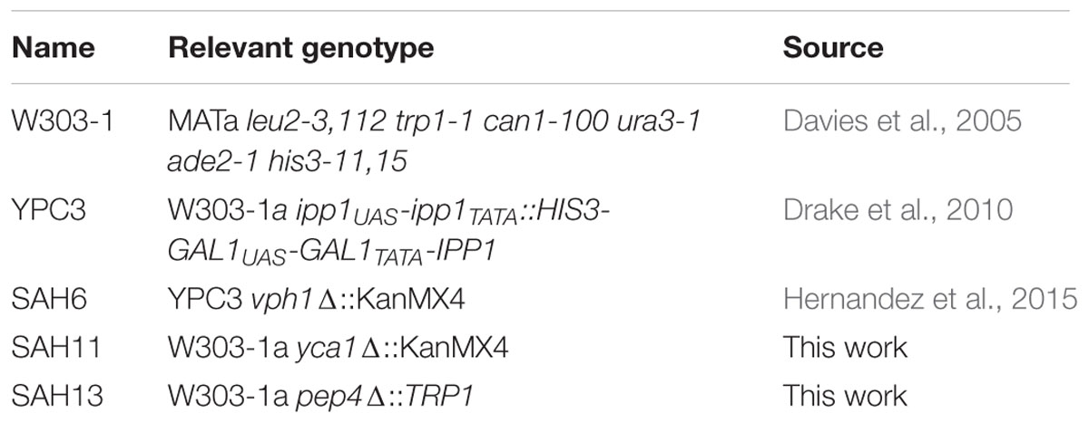
TABLE 1. Yeast strains.
Vacuole Vesicles and ATPase Assays
Intact vacuoles were isolated essentially as described (Hernandez et al., 2009) with the only modification of using a single 8% (w/w) Ficoll gradient. After isolation, vacuoles were vesiculated by resuspension in TKE buffer [10 mM Tris/HCl pH 7.5, 2 mM KCl, 1 mM EDTA and protease inhibitor cocktail (Sigma-Aldrich)]; vesicles were recovered after centrifugation at 100,000 × g for 30 min and resuspended in TKE. The formation of a ΔpH was evaluated as the ACMA fluorescence quenching produced by the activity of the tonoplast H+-ATPase using the following reaction mixture: 1 μM ACMA, 20 mM MOPS-Tris pH 7.2, 25 mM KCl, 2 mM MgCl2, and vacuolar membrane preparation (up to 50 μg of protein). The assay was initiated with the addition of 1.5 mM ATP. Recovery of fluorescence, as a proof of gradient formation, was induced by adding 3 μM gramicidin D. ATPase hydrolytic activities were assayed as described (Hernandez et al., 1994).
Flow Cytometry
Annexin-V-FITC and PI staining test were done essentially as described in Madeo et al. (1997). Briefly, after treatment, cells were collected, washed once in distilled water, once in SPMB (1.2 M sorbitol, 50 mM phosphate buffer pH 7.5, 1 mM MgCl2) and resuspended in SPMB with 30 units of lyticase (Sigma, St. Louis, MO, USA) per 107 cells. After protoplasting was complete (0.5–1 h), protoplasts were collected by centrifugation for 5 min at 500 × g and washed once with fresh SPMB and a second time with annexin-V labeling buffer (1.2 M sorbitol, 10 mM HEPES-NaOH pH 7.4, 140 mM NaCl, 2.5 mM CaCl2). Annexin-V-FITC and PI (final concentrations 1 μg/ml and 2.5 μg/ml, respectively) were incubated with protoplasts in labeling buffer for 20 min prior to flow cytometry. For DNA content analysis, cells were stained with PI essentially as described by Sazer and Sherwood (1990), and analyzed on a Coulter Epics XL apparatus as previously described (Hernandez et al., 2008). Although the extent of death induction by tridemorph was somewhat batch-dependent, the phenomena were consistent and reproducible within each batch.
Plant Materials and Growth Conditions
Plant mutant genotypes are described in Table 2. All A. thaliana wild-type and mutants were from Columbia ecotype. Seeds were sterilized using vapor phase-chlorine gas for 5 h (Clough and Bent, 1998) and sown individually using sterile forceps on 1X Murashige and Skoog (MS) plates containing 1% sucrose and 3.5 g l-1 Phytagel (Sigma-Aldrich).
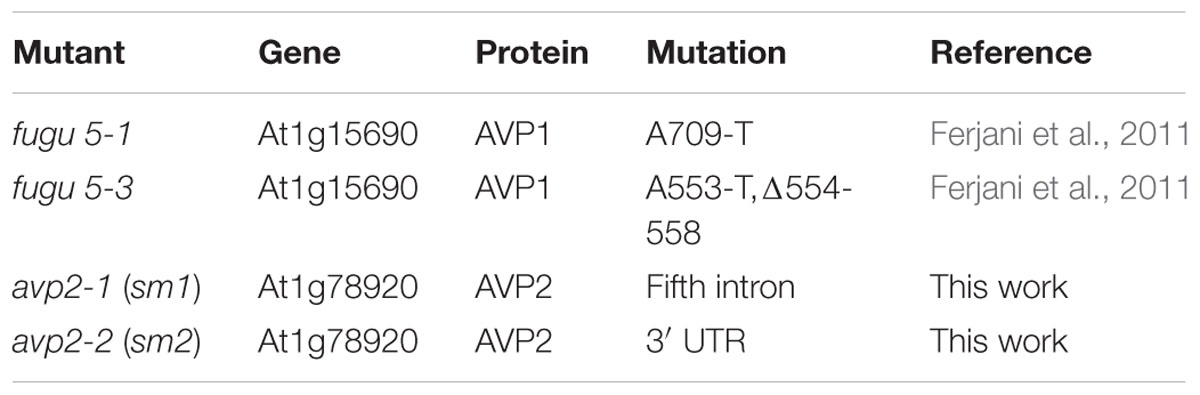
TABLE 2. Plant mutants.
Homozygous mutant lines for gene At1g15690 encoding the vacuole-localized H+-PPase AVP1 isoform (fugu 5-1 and fugu 5-3) were a kind gift of Dr Ferjani (Tokyo Gakugei University, Tokyo, Japan). Mutants for gene At1g78920 encoding the AVP2 isoform were obtained from the collection held at the Salk Institute Genomic Analysis Laboratory (USA) and named for this work as sm1 and sm2. Homozygosis of the T-DNA insertion was confirmed by PCR. Details on mutants are listed on Table 2.
Histological Determination of Gene Expression
Promotor sequences from genes AVP1 (At1g15690, 1,930 bp upstream of ATG), AVP2 (At1g78920, 1,100 bp), and AVP3 (At1g16780, 900 bp) were amplified by PCR from genomic DNA and cloned into vector pMDC162 (Curtis and Grossniklaus, 2003). Columbia wild-type A. thaliana plants were transformed by the floral dipping method (Clough and Bent, 1998). Plants carrying gene insertions were selected on kanamycin-supplemented plates. All experiments were performed with F3 seeds. The transgene insertions in all lines were sequenced to check for integrity of the inserted constructs. Staining was done by immersing plants or seedlings in GUS buffer [2 mM 5-bromo-4-chloro-3-indol-β-D-glucuronic acid, 50 mM potassium phosphate buffer (pH 7.2), 2 mM potassium ferrocyanide, 2 mM potassium ferrycyanide, 0.2% (w/v) triton X-100] for 1–3 days. Chlorophyll was washed-off using several 96% (v/v) ethanol washes at 37°C. Images were collected using a Nikon Eclipse 80i microscope coupled to a Nikon Digital Sight DS L1 camera or with a Leica MZFLIII binocular coupled to a Leica DC camera.
Semi-quantitative Reverse Transcription-PCR
Total RNA (1 μg) was subjected to cDNA synthesis using a “QuantiTect® Reverse Transcription kit” (Qiagen) according to manufacturer’s instructions. Final cDNA (1 μl) was subjected to PCR using primers amplifying 300–400 bp fragments at the 3′ UTR regions of the cDNA corresponding to genes At1g15690 (AVP1), At1g78920 (AVP2), At1g16780 (AVP3), and At4g05320 (UBIQUITIN10, UBQ10). PCR was run for a total of 20 cycles. Primers annealing on the UBQ10 cDNA were used to normalize equal cDNA loading onto PCR mixes. Saturation of the reaction and specificity of primers were checked using a five-fold excess of cDNA.
Plant Extracts
Seedlings were frozen in liquid nitrogen and kept at -80°C until use. Seedlings were ground with pestle and mortar in liquid nitrogen. The powder was resuspended in 1 ml of extraction buffer containing 50 mM MOPS adjusted to pH 7.5 with NaOH, 330 mM sorbitol, 2 mM MgCl2, 1 mM DTT, 1 mM PMSF and Plant Protease Inhibitor Cocktail (Sigma-Aldrich). The mixture was centrifuged for 15 min at 16,000 rpm at 4°C, the supernatant transferred to a new tube and reserved for Western blot analysis.
Hypocotyl and Root Length Measurements
To measure hypocotyl and root elongation in the darkness, seeds were stratified for 5 days at 4°C in the dark before being placed vertically in a growth chamber equipped with fluorescence light (light intensity 100 μE m-2 s-1) at 22°C for 4 h. To ensure dark conditions after that period, plates were wrapped in four layers of aluminum foil and plates incubated vertically at 22°C for 6 days. When appropriate, plates were supplemented with 10 μM tridemorph. Fungicide treatments and mock controls were carried out simultaneously in three replicate experiments. Hypocotyl and root length were quantified using Image J software (Schneider et al., 2012).
Protein Determination and Western Blotting
Protein determination was done using a dye-binding based assay from Bio-Rad (Hercules, CA, USA), according to manufacturer instructions or a modified Lowry assay (Thermo Scientific, Rockford, IL, USA), using ovoalbumin as a standard. Proteins were separated in SDS-PAGE gels using standard procedures (Sambrook et al., 1989). Proteins were then transferred to nitrocellulose filters and probed with polyclonal antibodies raised in rabbit against VHA-A (Cosmobio, Tokyo) and AVP1 (Agrisera, Vånnås) at a 1:1000 dilution. Blots were visualized on X-ray films using horseradish peroxidase-coupled secondary antibodies and a chemiluminescence kit (Millipore, Madrid). Quantification of blot band intensity was done using a Bio-Rad GS-800 densitometer.
Results
The Amine Fungicide Tridemorph Induces Metacaspase-Independent Apoptosis
We aimed at determining if inhibition of ERG2 gene product was fungistatic or if it induced cell death, and in the latter case, which type of cell death. Therefore, we treated W303-1a cells with 3 μM tridemorph for 5 h prior to annexin V-FITC/PI flow cytometric tests (Figure 1A). Both annexin-V-FITC (AV) and PI are membrane impermeable molecules; AV is a protein that binds phosphatidyl serine, while PI dyes nucleic acids. Briefly, cells dying from apoptosis expose phosphatidyl serine on the outer leaflet of the plasma membrane early in the process and only become permeable to PI at late stages, while necrotic cells are characterized by early breakage of the plasma membrane permeability barrier and little or no translocation of phosphatidyl serine (Madeo et al., 1997). Yeast cells treated with a growth inhibitory dose of tridemorph for 3 h displayed a differential staining with AV but little staining with PI in non-permeabilized protoplasts, compared with an untreated control (Figure 1A and Table 3). In comparison, cells treated with 160 mM acetic acid, a condition known to induce both apoptosis and necrosis in yeast (Ludovico et al., 2001) yielded a significant fraction of the protoplasts permeable to PI (Figure 1A). Accumulation of abnormal sterols has been associated to cell cycle perturbations (Marcireau et al., 1990; Fernández et al., 2002); hence, we also investigated if cell cycle arrest occurred concomitantly to apoptosis. Experiments using PI staining of permeabilised cells previously treated with tridemorph revealed that genomic DNA fragmentation takes place upon fungicide treatment with an increase in cells at the G1 phase of the cell cycle (Figure 1B). On their turn, erg2Δ (JRY7773) cells showed no differences with respect to a wild-type strain in subG1 extent (below 4%) and only a slight accumulation of cells in G2/M phases of the cell cycle, compared with W303-1a. Deletion of YCA1, the only yeast metacaspase, on a W303-1a background yielded cells with no differential sensitivity toward tridemorph as assessed by drop test assays (Figure 1C) or PI-flow cytometry of permeabilised cells (data not shown).
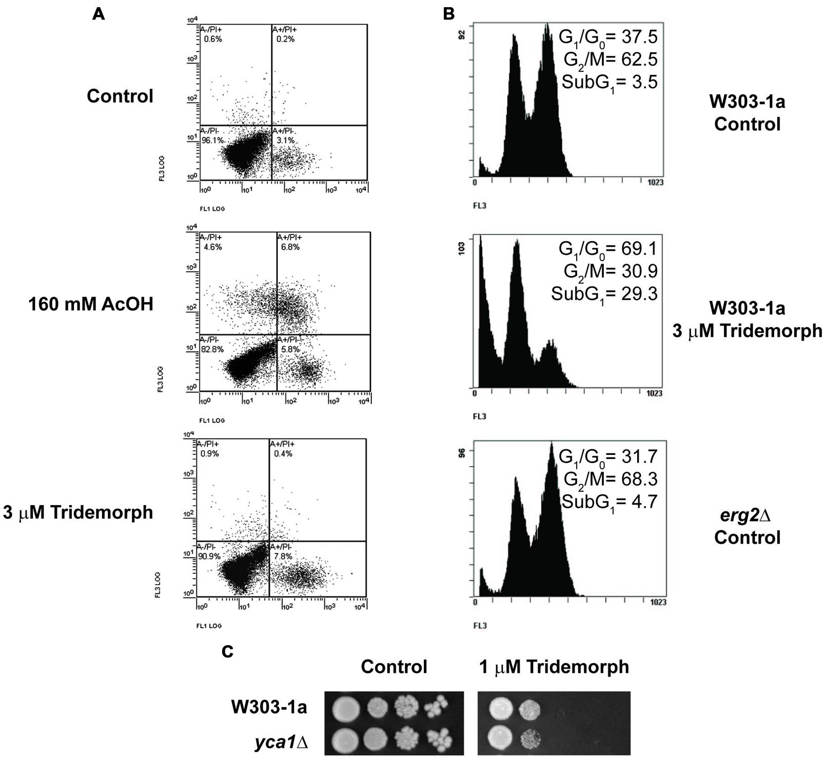
FIGURE 1. Cell death and cell cycle arrest induction in tridemorph treated yeast cells. (A) Determination of early apoptosis and necrosis in tridemorph-treated yeast. Externalization of phosphatydilserine and plasma membrane permeability to propidium iodide (Annexin V-FITC/PI assays) assessed by flow cytometry in non-permeabilised yeast protoplasts obtained from cells treated with 3 μM tridemorph for 3 h. Acetic acid treatment is included as a control inducing both Annexin V-FITC and PI fluorescence. (B) Cell cycle profiles in tridemorph treated yeast cells; quantitation of cell death. Flow cytometry determination of DNA contents by propidium iodide staining in ethanol-permeabilised yeast cells. Percentages of cells in every stage are shown in each panel. Total cells analyzed per case: 10,000. (C) Tridemorph-induced apoptosis dependence from metacaspases. Early stationary yeast cultures were diluted and spotted onto YPD agar plates (control, left) and YPD plus 1 μm tridemorph (right) at densities distributing ca 104, 103, 102, and 10 cells per spot, from left to right, respectively.
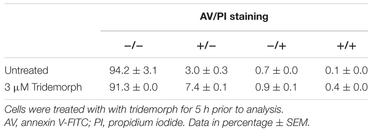
TABLE 3. Annexin V-FITC and propidium iodide staining of W303-1a yeast cells.
Tridemorph Causes V-ATPase Dysfunction
Tridemorph is a fungicide that phenocopies a defect on ERG2, which causes V-ATPase dysfunction (Hernandez et al., 2015). Bearing in mind the differences found in terms of cell death induction between tridemorph treatment and an erg2Δ mutation, we were interested in ascertaining if inhibition of V-ATPase proton pumping was reproduced by tridemorph (Figure 2). Vacuole vesicles extracted from cells treated with 3 μM tridemorph for 5 h were not capable to form proton gradients (Figure 2A). This impairment was similar using erg2Δ-derived vesicles (Hernandez et al., 2015). Also in agreement with previous results, hydrolytic activity assays showed that V-ATPase activity was inhibited to a minor extent (435 ± 97 vs. 341 ± 8 μmol Pi min-1 mg-1 protein ± SEM for vesicles obtained from control and tridemorph-treated yeast cells respectively).
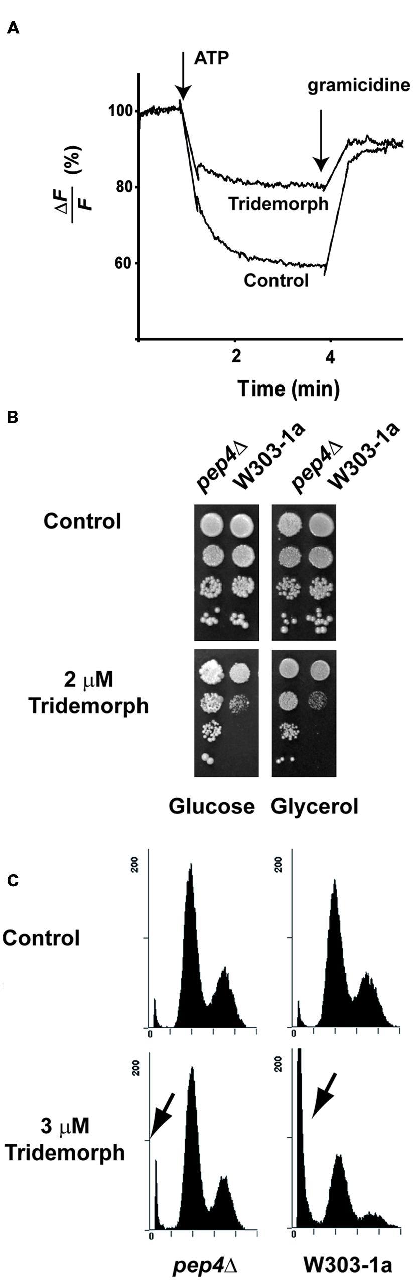
FIGURE 2. Vacuolar V-ATPase inhibition by tridemorph treatment. (A) Proton pumping assays using vacuolar vesicles. Proton transport in vacuole vesicles isolated from control yeasts (lower trace) and treated with 3 μM tridemorph for 5 h (top trace) followed by quenching of ACMA fluorescence in the presence of Mg⋅ATP (B) Enhancement of tridemorph tolerance in yeast by deletion of the gene corresponding to Proteinase A (PEP4). Early stationary yeast cultures were diluted and spotted onto control (upper panels) or 2 μm tridemorph-supplemented (lower panels) YPD (Glucose) or YPGlycerol agar plates at densities distributing ca 104, 103, 102, and 10 cells per spot, up to down, respectively. (C) Quantitation of cell death in pep4Δ mutant cells by flow cytometry. Flow cytometry determination of DNA contents by propidium iodide staining in ethanol-permeabilised pep4Δ mutant (left) and W303-1a (right) yeast cells in control (upper panels) and 3 μm tridemorph-treated cultures (lower panels). Arrows point to dead cell populations (Sub-G1). A total of 10,000 cells were analyzed per case per experiment.
In a previous report (Hernandez et al., 2015), we described that the presence of 8-dehydrosterols in yeast membranes induced a change in subunit a of the V-ATPase that made it more susceptible to proteolytic attack. We hypothesized that limiting the proteolytic turnover of vacuolar proteins might help yeast to withstand tridemorph treatment. To evaluate this hypothesis, we deleted PEP4, the gene encoding vacuolar protease A, since this deletion is known to affect a wide range of proteolytic activities in this organelle (Ammerer et al., 1986; Woolford et al., 1986). In addition, defects on vacuole acidification are associated to deficiencies to grow on non-fermentable carbon sources (Kane, 2006). Spot tests using W303-1a and its pep4Δ derivative showed that deletion of this protease provided a proliferative advantage when cells were challenged with tridemorph on agar plates, both on glucose and on glycerol carbon sources (Figure 2B). We also tested if cell death induction or cell cycle were affected in these cells. Analysis of PI-stained permeabilised yeast cells by flow cytometry showed again a modest accumulation of cells in G1/G0 phases, irrespectively of the genotype (Figure 2C, upper panels). However, while proportions of cells in subG1 raised from 4.5 ± 0.4% to 47.6 ± 0.5% in the case of W303-1a cells treated with vehicle or tridemorph, respectively, pep4Δ cells showed only 4.9 ± 0.3% and 7.3 ± 0.4% for vehicle and fungicide-treatments, respectively (Figure 2C, lower panels).
A Heterologously Expressed Plant H+-PPase Gene Alleviates Tridemorph Growth Inhibition and Cell Death in Yeast
Since tridemorph induced cell death in yeast cells but deletion of ERG2 did not reproduce this effect, we were interested to know if an abnormal vacuolar acidification could be involved in the stimulation of cell death by tridemorph. To this end, YPC3 cells were transformed with pTcGFP-AVP1 or pIPP1 plasmids and kept on glucose medium to repress the expression of the genomic IPP1 gene encoding a soluble pyrophosphatase (Figure 3). Overexpression of the A. thaliana H+-PPase chimera TcGFP-AVP1 conferred resistance to concentrations of this amine fungicide above those able to restrict growth to wild-type cells (Figure 3A). This resistance could also be observed and was slightly greater in strains lacking a vacuole-localized V-ATPase (Figure 3A, rightmost panel). Under the present conditions, tridemorph treatment did not affect substantially the H+-pumping ability of TcGFP-AVP1 (Supplementary Figure S1).
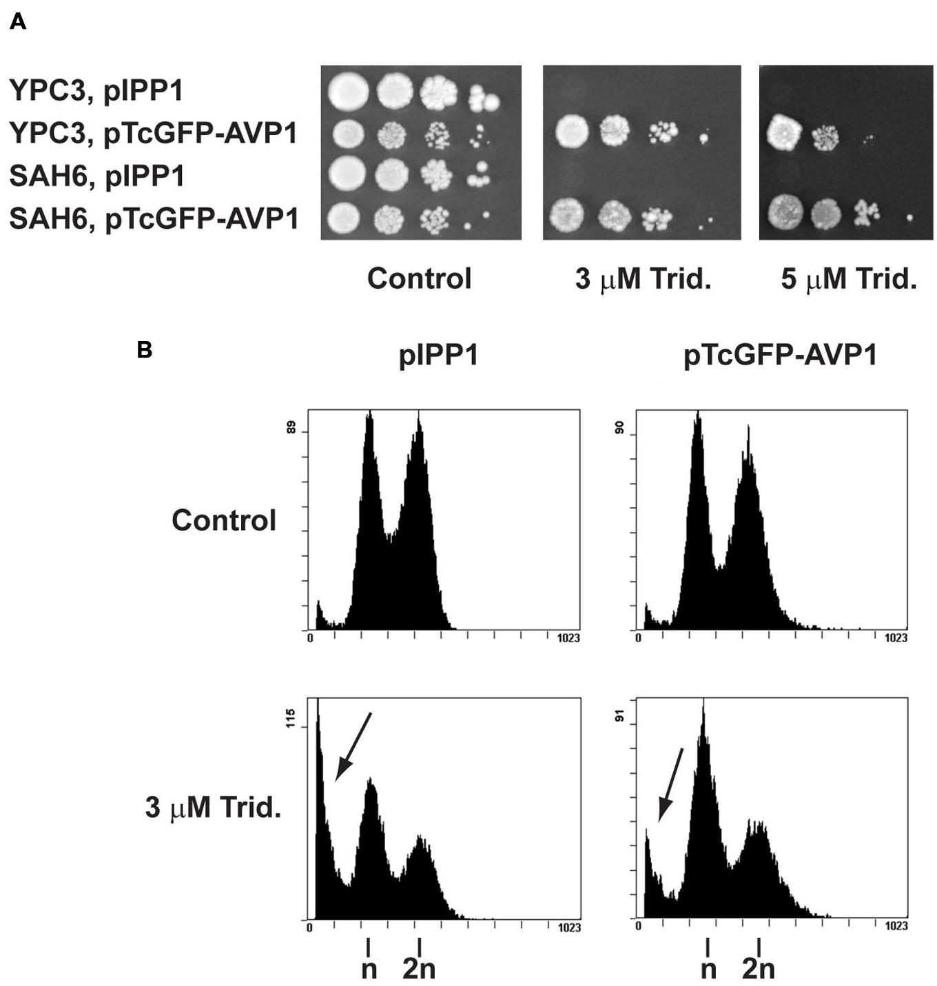
FIGURE 3. Influence of the heterologous expression of a H+-PPase gene on tridemorph tolerance in yeast cells. (A) Evaluation of tolerance to tridemorph in wild-type type (YPC3) and V-ATPase (SAH6) mutants. Early stationary yeast cultures were diluted and spotted onto control (left), 3 μm tridemorph-supplemented (middle) and 5 μm tridemorph-supplemented (right) YPD plates at densities distributing ca 104, 103, 102, and 10 cells per spot, respectively. (B) Quantitation of cell death in yeasts expressing a functional plant H+-PPase gene by flow cytometry. Flow cytometry determination of DNA contents by propidium iodide staining in ethanol-permeabilised yeast cells expressing episomally IPP1 (left panels) and plant AVP1 (right panels). Arrows point to dead cell populations (Sub-G1). A total of 10,000 cells were analyzed per case per experiment.
In order to obtain a more direct and quantifiable measurement of apoptosis, YPC3 cells transformed with pTcGFP-AVP1 or pIPP1 plasmids were treated with tridemorph in liquid culture for 5 h. Cell cycle was then analyzed by PI-flow citometry (Figure 3B). Tridemorph treatment induced changes in cell cycle profiles leading to an accumulation of cells in the G1/G0 phase of the cell cycle, irrespectively of the plasmid borne. This was akin to what was observed in W303-1a cells (Figure 1B), although reduced in extent. In particular, the proportion of cells in G1/G0 was 42.8 ± 1.8% and 55.3 ± 0.8% for IPP1-expressing cells untreated or treated with 3 μM tridemorph, respectively. Cells expressing TcGFP-AVP1 showed similar proportions: 45.7 ± 2.2% vs. 52.4 ± 0.1% in G1/G0 for untreated and tridemorph-treated cells, respectively. As previously shown, tridemorph induced a dramatic increase in cell death as assessed by accumulation of cells with subG1 complements of genomic DNA. Thus, pIPP1-transformed YPC3 strain showed 3.7 ± 0.2% of cells in subG1 when untreated but this proportion raised up to 29.0 ± 2.8% upon tridemorph treatment. However, presence of an H+-PPase as an alternative proton pump in YPC3 alleviated this cell death induction (4.2 ± 0.2% vs. 14.3 ± 2.8% in untreated and tridemorph-treated pTcGFP-AVP1 YPC3 cells, respectively).
Expression of H+-PPases Gene Isoforms in Arabidopsis thaliana
Arabidopsis thaliana genome includes three different genes encoding membrane-bound H+-translocating pyrophosphatases (AVPs), with gene products named AVP1 (type I or K+-stimulated isoform), and AVP2, AVP3 (type II, K+-independent isoforms). Bearing in mind the observations made in yeast, we hypothesized that these proteins could play a major role in fungicide tolerance in this model plant. However, in order to make meaningful functional assignments, a previous comparative study on the expression of these genes was necessary. Although some studies have already been published (Mitsuda et al., 2001a,b; Segami et al., 2010; Pizzio et al., 2015), none of them made a comparative study, nor included AVP3 expression. We met this task by making transgenic lines with AVP-promoter-β-glucuronidase (GUS) reporter system, where the different AVPs promoters were genomic fragments encompassing 1930, 1100, and 930 bp upstream the ATG codon of AVP1, AVP2, and AVP3 genes, respectively. Qualitative results are summarized on Table 4.
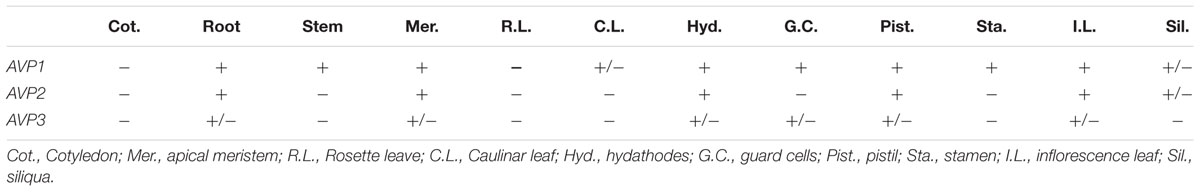
TABLE 4. Location of H+-PPase mRNA expression.
In adult plants (Figure 4), AVP1 was expressed mostly at the vascular tissues but at low levels in leaf mesophyll. In inflorescences, its expression was evident in pistils, stamen pedicels and the vascular system of sepals, while its expression in siliqua was only significantly observed at its base (Figures 4A–D). GUS staining corresponding to AVP2 (Figures 4E–H) was also present in the vascular system, albeit to a smaller degree than AVP1 (Figure 4E). Leaf expression was again very low in mesophyll and, in addition with vascular bundles, only clearly observed in guard cells (Figure 4F). In reproductive tissues, its signal was perceptible at the top and bottom parts of the pistil, in stamen pedicels and vascular tissue of sepals (Figure 4G), while in siliqua, the signal was nearly undetectable (Figure 4H). GUS signal corresponding to AVP3 expression was very weak throughout the plant and down to undetectable levels in many tissues. However, some signal was apparent in reproductive tissues, particularly a weak expression was observed in pistils, stamen pedicels and sepals (Figure 4K).
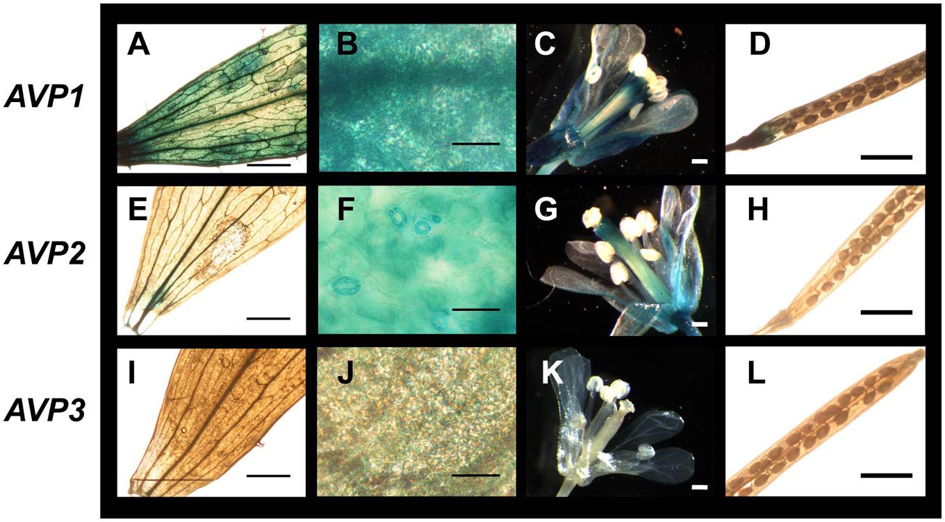
FIGURE 4. Expression of H+-PPase genes in adult organs and tissues of Arabidopsis thaliana. (A,E,I) Cauline leaves; (B,F,J) detail of cauline leaves (mesophyll); (C,G,K) inflorescences; (D,H,L) silique. Transgenic plants expressing β-glucuronidase as a reporter of the expression driven from a 1.9, 1.1, or 0.9 kb fragment from the AVP1, AVP2, or AVP3 promotor sequence, respectively. Bars correspond to 0.5 mm (A,C–E,G–I,K,L) or 50 μm (B,F,J).
In general, expression was more evident in developing seedlings for all three isoforms (Figure 5). AVP1 was robustly expressed in roots and stems and, especially, at the root columella and apical meristem (Figures 5A–D). In roots, the signal was easily detectable up to the transition zone with the shoot (Figure 5D). On the other hand, developing leaves and meristems showed high levels of GUS staining, particularly at the hydathodes (Figure 5B). AVP2 signal (Figures 5E–H) was found in leaf and root apices, although at a lower intensity to AVP1 and often showing intermissions in non-dividing tissue (Figure 5E). Also, it was detected in developing apical meristems (Figure 5G). Leaf expression was again observed in developing organs, remarkably at the hydathodes and in comparable levels to that of AVP1 (compare Figures 5B–F). In the case of AVP3 expression (Figures 5I–L), a clear signal was only observed in the apical meristem (Figure 5K) and in the root vascular system (Figure 5L); in leaves, no clear signal was detected (Figure 5J).
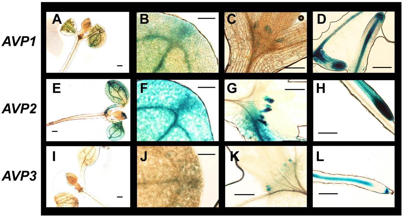
FIGURE 5. Expression of H+-PPase genes in seedling organs and tissues of A. thaliana. (A,E,I) Whole seedling; (B,F,J) detail of rosette leaves showing hydathodes; (C,G,H) shoot apical meristems; (D,H,L) root. Transgenic plants expressing β-glucuronidase as a reporter of the expression driven from a 1.9, 1.1, or 0.9 kb fragment from AVP1 (upper panel), AVP2 (middle panel), or AVP3 (lower panel) promotor sequences, respectively. Plants were incubated with X-Gluc to develop the signal prior to being photographed. Bars correspond to 0.1 mm (A,D,E,H,I,L) or 50 μm (B,C,F,G,J,K).
Since GUS staining indicated some clear differences in the expression of the three AVPs, and in order to confirm the data, semi-quantitative RT-PCR experiments were used (Figure 6). First, we confirmed that there were large differences in the expression of the isoforms. We chose tissues of adult plants grown on soil where the most diverse differences in GUS staining had been observed and, in particular, two tissues where the AVPs expression mostly differed: roots and stems (Table 4). The expression of the gene UBQ10 encoding Ubiquitin-10 was used as a loading control. AVP1 mRNA was more abundant than any of the other two isoforms in roots, but AVP1 and AVP2 transcripts showed comparable levels in stems. On the other hand, AVP3 mRNA was nearly undetectable in both tissues (Figure 6A).
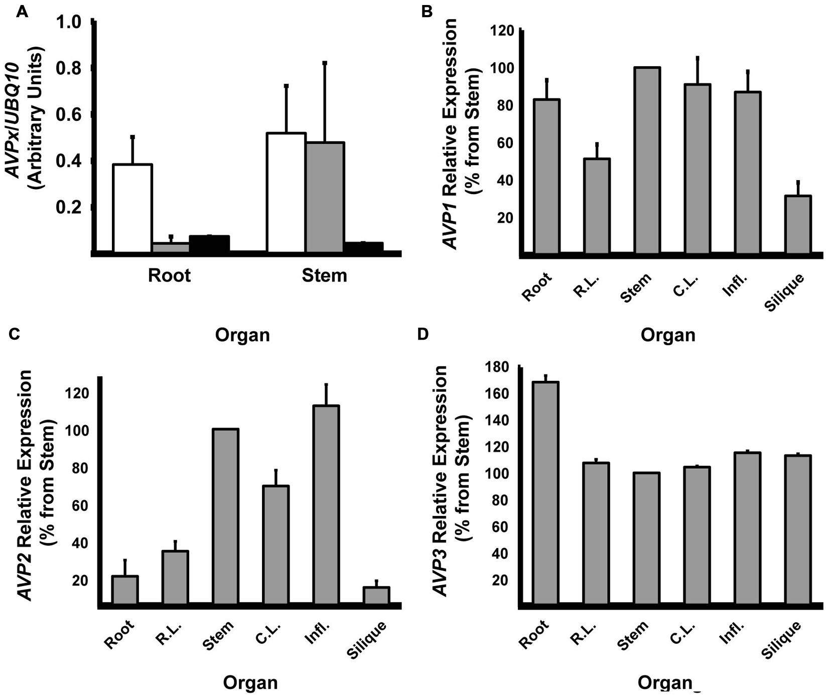
FIGURE 6. Relative expression of the different H+-PPase isoforms. (A) Expression of AVP1-3 in adult root and stem tissues. Open bars: AVP1; Shaded bars: AVP2; Black bars: AVP3. (B) Relative expression of AVP1 in different plant organs. (C) Relative expression of AVP2 in different plant organs. (D) Relative expression of AVP3 in different plant organs. Semi-quantitation of mRNA abundance estimated by RT-PCR. Data were normalized against the expression of UBQ10 gene. (B–D) Average expression in stems was taken as 100%. Expression (AVPx/UBQ10) in stems were AVP1: 0.52 ± 0.21; AVP2: 0.48 ± 0.36; AVP3: 0.04 ± 0.00. Data are mean ± SE of three independent experiments.
We next wanted to confirm that the expression of each AVP gene varied widely between organs. On the whole, AVP1 mRNA relative amount estimates agreed with GUS data. Thus, it showed a high level of expression in roots, stems and influorescences and minimal in siliques and rosette leaves. Curiously, cauline leaves showed a strong AVP1 expression, in contrast to GUS data. All in all, little differences in expression were found among organs and tissues with values near those found in stems (Figure 6B). AVP2 mRNA showed a relatively robust expression in stems and inflorescences and again in cauline leaves. Minimal levels of expression were observed in roots, rosette leaves and siliques. In this case, the differences between high- and low-expression tissues were greater than in the case of AVP1 (Figure 6C). Regarding AVP3 expression, it was only found unambiguously in roots, while levels in other tissues were nearly negligible (Figure 6D).
Role of H+-PPases in Plant Tridemorph Tolerance
Once the expression of the three AVP isoforms was settled, we met the question of whether H+-PPases were involved in A. thaliana fungicide tolerance (Figure 7). We found that tridemorph produced a dose-dependent growth retardation effect in developing seedlings grown in the dark. This effect was noticeable in both hypocotyl and root lengths as reductions in both parameters nearing ca 40% at a concentration of 10 μM tridemorph (Figure 7A). To answer which of the H+-PPases was involved in fungicide tolerance, we treated wild-type (Columbia ecotype) and two null alleles of either avp1 (fugu 5-1 and fugu 5-3) or avp2 (sm1 and sm2) mutants with 10 μM tridemorph. Plants mutant for AVP3 were cast-off from this study since expression levels of this gene, even in developing tissues, was nearly undetectable. As shown on Figure 7B, all seedlings displayed a remarkable reduction in both root and hypocotyl length upon tridemorph treatment. However, fugu 5-1 and 5-3 mutants showed a significantly greater reduction in root length compared with either wild-type or avp2 mutants, while these latter ones showed no differences between them (Figure 7B). In the case of hypocotyl length, none of the mutants showed any differences compared with a wild-type.
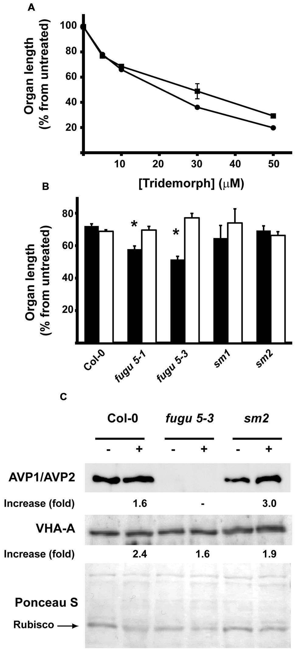
FIGURE 7. Effect of tridemorph addition on seedling root and hypocotyl elongation. (A) Length of roots and hypocotyls on wild-type A. thaliana as a function of fungicide concentration. Circles, root length; squares, hypocotyl length. (B) Length of roots and hypocotyls on wild-type and H+-PPase mutants of A. thaliana. Solid bars, roots; empty bars, hypocotyls. (C) Polypeptide levels of AVP1 and V-ATPase in tridemorph treated wild-type and pyrophosphatase mutants. Seeds were germinated and grown in the dark in phytagel-MS medium supplemented with 1% sucrose for 14 days prior analysis. Length measurements done by image analysis using NIH-ImageJ. Asterisks denote significant differences at p ≤ 0.05 using unpaired t-tests. Equal amounts of whole plant extracts were separated by SDS-PAGE, electroblotted onto nitrocellulose and decorated with antibodies recognizing the subunit A from V-ATPase (VHA-A) or AVP1. The protein band corresponding to RUBISCO large subunit (arrow) and the whole lanes stained with Ponceau S were used as loading controls with similar results. Those obtained with whole lanes are shown.
Next, we wanted to find out if H+ PPase or V-ATPase polypeptides levels varied as a response to tridemorph treatment. For this purpose, we measured the levels of polypeptides in whole seedling extracts by Western blot compared to levels of the small subunit of the ribulose 1,5 biphosphate carboxilase-oxygenase (RuBisCO; Figure 7C). Unfortunately, we were unable to perceive detectable levels of AVP2 even in avp1 null backgrounds, despite the use of two different polyclonal rabbit antibodies recognizing H+-PPases (Figure 7C, upper panel and data not shown). Effects on V-ATPases, as measured using the signal from the polypeptide corresponding to their catalytic subunit A as a marker of the holoenzyme, were apparent but modest in all mutants (Figure 7C, lower panel). Similarly, H+-PPase abundance increased discreetly in the case of wild-type plants treated with tridemorph; however, the accumulation of AVP1 augmented distinctly in the case of avp2 null mutants (Figure 7C, upper panel).
Discussion
In this report, we have shown that tridemorph, an amine-type fungicide, can induce a cell death process in yeast that shows typical markers of apoptosis. In addition, tridemorph was able to induce cell cycle arrest to a certain extent. Apoptotic demise in fungi has been controversial for some time but it is now widely accepted, among other reasons, because ortholog genes of the basic machinery have been found and proved to be functional in yeast (Carmona-Gutierrez et al., 2010). Remarkably, death induction by tridemorph was independent of YCA1, the only metacaspase identified in the yeast genome. PCD in yeast has been shown to depend on YCA1 in some cases (e.g., de Castro et al., 2011), but it is dispensable for others (Guaragnella et al., 2010; Hoeberichts et al., 2010), much in the same way as it happens in mammalian cells (Guaragnella et al., 2010; Hoeberichts et al., 2010). At any rate, little is known about the mechanisms of this type of cell death in yeast. Thus, tridemorph can be instrumental in the delineation of the mechanisms behind Yca1p-independent apoptosis in yeast.
For many years, it was considered that the main effect of the accumulation of 8-dehydrosterols, like any other abnormal sterols, was an increase in the permeability of the plasma membrane and that this latter effect was the actual cause for their death induction capability (Bard et al., 1978; Abe and Hiraki, 2009). There is no doubt that plasma membrane physical characteristics are affected upon extensive replacement of naturally occurring ergosterol by abnormal lipid species. However, little has been done along these years to determine if plasma membrane-associated effects are the actual cause of cell death and which mechanism it encompasses. Some years ago, it was shown that changes in physicochemical characteristics of the plasma membrane by abnormal sterols in U. maydis did not provide an explanation for the fungicidal action of these lipids (Hernandez et al., 1997). Following this, the present report showed that tridemorph-treated cells did not accumulate PI initially, which ruled out necrosis as the plausible mechanism of cell death under these conditions, but also questioned the long-held view of an increase in permeability at the plasma membrane level. Furthermore, the ability to reduce fungicide-induced cell death dramatically by expression of an alternative H+ pump that localizes preferentially in internal membranes, such as the TcGFP-AVP1 construct (Drake et al., 2010), proved that at least part of the actual cellular mode of action of 8-dehydrosterols is located at the vacuole, though probably not exclusively. It must be mentioned here that, although erg2Δ cells showed many phenotypes in common with tridemorph treatment, the former did not show any detectable PCD. The reasons for this discrepancy are unknown, but may be related to either secondary mutations that help these cells to cope with the presence of abnormal sterols or because tridemorph has other targets independently of the Erg2p protein.
It has been proposed that 14α-methylated sterols exert part of their effects through inhibition of the vacuolar H+-ATPase in budding yeast and C. albicans (Zhang and Rao, 2010; Zhang et al., 2010). However, this kind of data are not necessarily applicable to all types of abnormal sterols; for example, the proton transport across plasma membrane vesicles was found to be uncoupled by 8-dehydrosterols but showed no differences by the presence of biologically active levels of 14α-methylated sterols in U. maydis (Hernandez et al., 1998). Here we showed that tridemorph, a fungicide inducing the accumulation of 8-dehydrosterols in yeast (Baloch and Mercer, 1987), causes the same effects on V-ATPase as a mutation in ERG2 gene (Hernandez et al., 2015), i.e., a reduction in the proton transport capacity due to uncoupling from ATPase hydrolysis, but without the concomitant inhibition in ATP hydrolytic activity observed with 14α-methylated sterols. This uncoupling is also concomitant to protease sensitivity (Hernandez et al., 2015). Not surprisingly then, a mutation in PEP4, the gene encoding the master protease at the yeast vacuole, resulted in increased tolerance to tridemorph. Fungicide resistance is a growing concern in agriculture and, thus, information on possible mutational targets can be of great importance in order to understand field results on pest resistance.
In plant vacuoles, V-ATPases and H+-PPases share a common location. Recently, we have shown that the latter type of pumps is sufficient for acidifying internal compartments (Perez-Castiñeira et al., 2011). When grown on glucose, YPC3 yeast cells expressing TcGFP-AVP1 reflects closely the situation found in a plant cell in terms of PPi homeostasis and proton transport across the tonoplast. We previously reported that this chimera is able to pump protons and, thus, functionally substitute vacuolar V-ATPase (Perez-Castiñeira et al., 2011); moreover, it could provide a means to test the influence of abnormal sterols in endomembrane traffic (Hernandez et al., 2015). We therefore hypothesized that this transporter may be an important factor in plant EBI fungicide tolerance. Indeed, the greater tolerance to an amine fungicide found in these metabolically engineered fungal cells concurs with what is usually observed in plants. Conversely, A. thaliana mutants lacking AVP1 polypeptides were more sensitive to tridemorph, specifically at the roots. This is in agreement with earlier reports showing that A. thaliana hydra1 mutants (null mutants for the gene encoding sterol-Δ8, Δ7-isomerase, one of the two identified targets of tridemorph, the other being the sterol-Δ14-reductase) were affected in seedling root development (Souter et al., 2002). Also, it is consistent with AVP1 gene being the most expressed gene isoform throughout the plant and especially in root tissues. This underlines the importance of the vacuole and, hence, their luminal acidification mechanisms, in root elongation (Barrada et al., 2015). On the other hand, AVP2 is distinctly expressed in stems and, therefore, an influence on hypocotyl length on tridemorph treated avp2 seedlings could be foreseen. However, since expression of AVP2 is in the same order as that of AVP1 in this organ, lack of any effect on hypocotyl elongation can be ascribed to compensation by AVP1 activity, in agreement with greater polypeptide amounts observed in avp2 mutants upon tridemorph treatment. A contribution by AVP3 cannot be discarded; however, the levels of AVP3 mRNA compared with those of AVP1 where far too dissimilar to credit AVP3 with any major influence. All in all, the presence of a double set of H+ pumps can be a factor that helps to explain why plants can tolerate this kind of compounds, even though they affect the sterol composition of their cell membranes (He et al., 2003).
Author Contributions
AH contributed with the design of the study, experimental work, and manuscript writing; RH-P, JM, TA, GL-L, and JP-C contributed experimental work; AH, PN, FV and AS coordinated and financed the present study.
Funding
This work was funded by the Andalusian Regional Government through their support to PAIDI groups BIO-261 and BIO-177, and grant P07-CVI-03082; likewise, the Spanish Ministry of Science and Innovation provided funds through grants BFU2004-00843, BFU2007-61887, and BFU2010-15622. All the above were partially supported by the EU FEDER program. Also thanks to Fundação de Ajuda a Pesquisa do Estado de São Paulo (FAPESP) for funding through grant 2014/10443-0. FV and TA were funded by MINECO project BIO2014-52425-P. PAIDI group BIO-261 belongs to the CeiA3 and AndaluciaTECH University Campuses of International Excellence.
Conflict of Interest Statement
The authors declare that the research was conducted in the absence of any commercial or financial relationships that could be construed as a potential conflict of interest.
Acknowledgments
Authors deeply thank Dr. Rosa Laura Lopez Marques (Department of Plant and Environmental Sciences, University of Copenhagen, Denmark) for her help and advice with in situ gene expression experiments, Dr. MT Ruiz-Pérez for critical reading of the manuscript and Ms. I. Jiménez for her invaluable technical assistance.
Supplementary Material
The Supplementary Material for this article can be found online at: http://journal.frontiersin.org/article/10.3389/fpls.2016.00085
Abbreviations
A600, absorbance at 600 nm; GFP, green fluorescent protein; H+-PPase, H+- translocating pyrophosphatase; IP, immunoprecipitation; PI, propidium iodide; PPi, inorganic pyrophosphate; UPR, unfolded protein response.
References
Abe, F., and Hiraki, T. (2009). Mechanistic role of ergosterol in membrane rigidity and cycloheximide resistance in Saccharomyces cerevisiae. Biochim. Biophys. Acta 1788, 743–752. doi: 10.1016/j.bbamem.2008.12.002
Ammerer, G., Hunter, C. P., Rothman, J. H., Saari, G. C., Valls, L. A., and Stevens, T. H. (1986). PEP4 gene of Saccharomyces cerevisiae encodes proteinase A, a vacuolar enzyme required for processing of vacuolar precursors. Mol. Cell. Biol. 6, 2490–2490. doi: 10.1128/MCB.6.7.2490
Atwell, B. J., Greenway, H., and Colmer, T. D. (2015). Efficient use of energy in anoxia-tolerant plants with focus on germinating rice seedlings. New Phytol. 206, 36–56. doi: 10.1111/nph.13173
Baloch, R. I., and Mercer, E. I. (1987). Inhibition of sterol delta8, delta7-isomerase and delta14-reductase by fenpropimorph, tridemorph and fenpropidin in cell-free enzyme systems from Saccharomyces cerevisiae. Phytochemistry 26, 663–668. doi: 10.1016/S0031-9422(00)84762-7
Bard, M., Lees, N. D., Burrows, L. S., and Kleinhans, F. W. (1978). Differences in crystal violet uptake and cation-induced death among yeast sterol mutants. J. Bacteriol. 135, 1146–1148.
Barrada, A., Montane, M. H., Robaglia, C., and Menand, B. (2015). Spatial regulation of root growth: placing the plant TOR pathway in a developmental perspective. Int. J. Mol. Sci. 16, 19671–19697. doi: 10.3390/ijms160819671
Buchenauer, H., and Röhner, E. (1981). Effect of triadimefon and triadimenol on growth of various plant species as well as on gibberellin content and sterol metabolism in shoots of barley seedlings. Pestic. Biochem. Physiol. 15, 58–70. doi: 10.1016/0048-3575(81)90034-1
Carmona-Gutierrez, D., Eisenberg, T., Buttner, S., Meisinger, C., Kroemer, G., and Madeo, F. (2010). Apoptosis in yeast: triggers, pathways, subroutines. Cell Death Differ. 17, 763–773. doi: 10.1038/cdd.2009.219
Cerdon, C., Rahier, A., Taton, M., and Sauvaire, Y. (1996). Effect of tridemorph and fenpropimorph on sterol composition in fenugreek. Phytochemistry 41, 423–431. doi: 10.1016/0031-9422(95)00550-1
Clough, S. J., and Bent, A. F. (1998). Floral dip: a simplified method for Agrobacterium-mediated transformation of Arabidopsis thaliana. Plant J. 16, 735–743. doi: 10.1046/j.1365-313x.1998.00343.x
Curtis, M. D., and Grossniklaus, U. (2003). A gateway cloning vector set for high-throughput functional analysis of genes in planta. Plant Physiol. 133, 462–469. doi: 10.1104/pp.103.027979
Davies, B. S., Wang, H. S., and Rine, J. (2005). Dual activators of the sterol biosynthetic pathway of Saccharomyces cerevisiae: similar activation/regulatory domains but different response mechanisms. Mol. Cell. Biol. 25, 7375–7385. doi: 10.1128/MCB.25.16.7375-7385.2005
de Castro, P. A., Savoldi, M., Bonatto, D., Barros, M. H., Goldman, M. H. S., Berretta, A. A., et al. (2011). Molecular characterization of propolis-induced cell death in Saccharomyces cerevisiae. Eukaryot. Cell 10, 398–411. doi: 10.1128/EC.00256-10
Drake, R., Serrano, A., and Perez-Castiñeira, J. R. (2010). N-terminal chimaeras with signal sequences enhance the functional expression and alter the subcellular localization of heterologous membrane-bound inorganic pyrophosphatases in yeast. Biochem. J. 426, 147–157. doi: 10.1042/BJ20091491
Ferjani, A., Segami, S., Horiguchi, G., Muto, Y., Maeshima, M., and Tsukaya, H. (2011). Keep an eye on PPi: the vacuolar-type H+-pyrophosphatase regulates postgerminative development in Arabidopsis. Plant Cell 23, 2895–2908. doi: 10.1105/tpc.111.085415
Fernández, C., Suárez, Y., Ferruelo, A. J., Gómez-Coronado, D., and Lasunción, M. A. (2002). Inhibition of cholesterol biosynthesis by Delta22-unsaturated phytosterols via competitive inhibition of sterol Delta24-reductase in mammalian cells. Biochem. J. 366, 109–119. doi: 10.1042/bj20011777
Gietz, R. D., and Woods, R. A. (2002). Transformation of yeast by lithium acetate/single-stranded carrier DNA/polyethylene glycol method. Methods Enzymol. 350, 87–96. doi: 10.1016/S0076-6879(02)50957-5
Guaragnella, N., Bobba, A., Passarella, S., Marra, E., and Giannattasio, S. (2010). Yeast acetic acid-induced programmed cell death can occur without cytochrome c release which requires metacaspase YCA1. FEBS Lett. 584, 224–228. doi: 10.1016/j.febslet.2009.11.072
He, J.-X., Fujioka, S., Li, T.-C., Kang, S. G., Seto, H., Takatsuto, S., et al. (2003). Sterols regulate development and gene expression in Arabidopsis. Plant Physiol. 131, 1258–1269. doi: 10.1104/pp.014605
Hernandez, A., Cooke, D. T., and Clarkson, D. T. (1994). Lipid composition and proton transport in Penicillium cyclopium and Ustilago maydis plasma membrane vesicles isolated by two-phase partitioning. Biochim. Biophys. Acta 1195, 103–109. doi: 10.1016/0005-2736(94)90015-9
Hernandez, A., Cooke, D. T., and Clarkson, D. T. (1998). Effects of abnormal-sterol accumulation on Ustilago maydis plasma membrane H+ -ATPase stoichiometry and polypeptide pattern. J. Bacteriol. 180, 412–415.
Hernandez, A., Cooke, D. T., Lewis, M., and Clarkson, D. T. (1997). Fungicides and sterol-deficient mutants of Ustilago maydis: plasma membrane physico-chemical characteristics do not explain growth inhibition. Microbiology 143, 3165–3174. doi: 10.1099/00221287-143-10-3165
Hernandez, A., Jiang, X., Cubero, B., Nieto, P. M., Bressan, R. A., Hasegawa, P. M., et al. (2009). Mutants of the Arabidopsis thaliana cation/H+ antiporter AtNHX1 conferring increased salt tolerance in yeast: the endosome/prevacuolar compartment is a target for salt toxicity. J. Biol. Chem. 284, 14276–14285. doi: 10.1074/jbc.M806203200
Hernandez, A., Lopez-Lluch, G., Bernal, J. A., Navas, P., and Pintor-Toro, J. A. (2008). Dicoumarol down-regulates human PTTG1/Securin mRNA expression through inhibition of Hsp90. Mol. Cancer Ther. 7, 474–482. doi: 10.1158/1535-7163.MCT-07-0457
Hernandez, A., Serrano-Bueno, G., Perez-Castineira, J. R., and Serrano, A. (2015). 8-Dehydrosterols induce membrane traffic and autophagy defects through V-ATPase dysfunction in Saccharomyces cerevisae. Biochim. Biophys. Acta 1853, 2945–2956. doi: 10.1016/j.bbamcr.2015.09.001
Hoeberichts, F. A., Perez-Valle, J., Montesinos, C., Mulet, J. M., Planes, M. D., Hueso, G., et al. (2010). The role of K(+) and H(+) transport systems during glucose- and H(2)O(2)-induced cell death in Saccharomyces cerevisiae. Yeast 27, 713–725. doi: 10.1002/yea.1767
Kane, P. M. (2006). The where, when, and how of organelle acidification by the yeast vacuolar H+-ATPase. Microbiol. Mol. Biol. Rev. 70, 177–191. doi: 10.1128/MMBR.70.1.177-191.2006
Köller, W. (1987). Isomers of sterol synthesis inhibitors: fungicidal effects and plant growth regulator activities. Pestic. Sci. 18, 129–147. doi: 10.1002/ps.2780180206
Ludovico, P., Sousa, M. J., Silva, M. T., Leão, C., and Côrte-Real, M. (2001). Saccharomyces cerevisiae commits to a programmed cell death process in response to acetic acid. Microbiology 147, 2409–2415. doi: 10.1099/00221287-147-9-2409
Madeo, F., Frohlich, E., and Frohlich, K. U. (1997). A yeast mutant showing diagnostic markers of early and late apoptosis. J. Cell Biol. 139, 729–734. doi: 10.1083/jcb.139.3.729
Marcireau, C., Guilloton, M., and Karst, F. (1990). In vivo effects of fenpropimorph on the yeast Saccharomyces cerevisiae and determination of the molecular basis of the antifungal property. Antimicrob. Agents and Chemother. 34, 989–993. doi: 10.1128/AAC.34.6.989
Mitsuda, N., Enami, K., Nakata, M., Takeyasu, K., and Sato, M. H. (2001a). Novel type Arabidopsis thaliana H(+)-PPase is localized to the Golgi apparatus. FEBS Lett. 488, 29–33. doi: 10.1016/S0014-5793(00)02400-5
Mitsuda, N., Takeyasu, K., and Sato, M. H. (2001b). Pollen-specific regulation of vacuolar H+-PPase expression by multiple cis-acting elements. Plant Mol. Biol. 46, 185–192. doi: 10.1023/A:1010681129557
Perez-Castiñeira, J. R., Hernandez, A., Drake, R., and Serrano, A. (2011). A plant proton-pumping inorganic pyrophosphatase functionally complements the vacuolar ATPase transport activity and confers bafilomycin resistance in yeast. Biochem. J. 437, 269–278. doi: 10.1042/BJ20110447
Pizzio, G. A., Paez-Valencia, J., Khadilkar, A. S., Regmi, K., Patron-Soberano, A., Zhang, S., et al. (2015). Arabidopsis type I proton-pumping pyrophosphatase expresses strongly in phloem, where it is required for pyrophosphate metabolism and photosynthate partitioning. Plant Physiol. 167, 1541–1553. doi: 10.1104/pp.114.254342
Rahier, A., Schmitt, P., Huss, B., Benveniste, P., and Pommer, E. H. (1986). Chemical structure-activity relationships of the inhibition of sterol biosynthesis by N-substituted morpholines in higher plants. Pestic. Biochem. Physiol. 25, 112–124. doi: 10.1016/0048-3575(86)90038-6
Ramsdale, M. (2008). Programmed cell death in pathogenic fungi. Biochim. Biophys. Acta 1783, 1369–1380. doi: 10.1016/j.bbamcr.2008.01.021
Sambrook, J., Fritsch, E. F., and Maniatis, T. (1989). Molecular Cloning: A Laboratory Manual. New York, NY: Cold Spring Harbor Laboratory Press.
Sazer, S., and Sherwood, S. W. (1990). Mitochondrial growth and DNA synthesis occur in the absence of nuclear DNA replication in fission yeast. J. Cell Sci. 97, 509–516.
Schneider, C. A., Rasband, W. S., and Eliceiri, K. W. (2012). NIH image to imageJ: 25 years of image analysis. Nat. Methods 9, 671–675. doi: 10.1038/nmeth.2089
Segami, S., Makino, S., Miyake, A., Asaoka, M., and Maeshima, M. (2014). Dynamics of vacuoles and H+-pyrophosphatase visualized by monomeric green fluorescent protein in Arabidopsis: artifactual bulbs and native intravacuolar spherical structures. Plant Cell 26, 3416–3434. doi: 10.1105/tpc.114.127571
Segami, S., Nakanishi, Y., Sato, M. H., and Maeshima, M. (2010). Quantification, organ-specific accumulation and intracellular localization of type II H(+)-pyrophosphatase in Arabidopsis thaliana. Plant Cell Physiol. 51, 1350–1360. doi: 10.1093/pcp/pcq096
Sherman, F. (1991). Getting started with yeast. Methods Enzymol. 194, 3–21. doi: 10.1016/0076-6879(91)94004-V
Souter, M., Topping, J., Pullen, M., Friml, J., Palme, K., Hackett, R., et al. (2002). hydra Mutants of Arabidopsis are defective in sterol profiles and auxin and ethylene signaling. Plant Cell 14, 1017–1031. doi: 10.1105/tpc.001248
Van den Bossche, H., Willemsens, G., Cools, W., Marichal, P., and Lauwers, W. (1983). Hypothesis on the molecular basis of the antifungal activity of N-substituted imidazoles and triazoles. Biochem. Soc. Trans. 11, 665–667. doi: 10.1042/bst0110665
van der Merwe, M. J., Groenewald, J.-H., Stitt, M., Kossmann, J., and Botha, F. C. (2010). Downregulation of pyrophosphate: D-fructose-6-phosphate 1-phosphotransferase activity in sugarcane culms enhances sucrose accumulation due to elevated hexose-phosphate levels. Planta 231, 595–608. doi: 10.1007/s00425-009-1069-1
Wang, J., Nayak, S., Koch, K., and Ming, R. (2013). Carbon partitioning in sugarcane (Saccharum species). Front. Plant Sci. 4:201. doi: 10.3389/fpls.2013.00201
Woolford, C. A., Daniels, L. B., Park, F. J., Jones, E. W., Van Arsdell, J. N., and Innis, M. A. (1986). The PEP4 gene encodes an aspartyl protease implicated in the posttranslational regulation of Saccharomyces cerevisiae vacuolar hydrolases. Mol. Cell. Biol. 6, 2500–2500. doi: 10.1128/MCB.6.7.2500
Zhang, Y.-Q., Gamarra, S., Garcia-Effron, G., Park, S., Perlin, D. S., and Rao, R. (2010). Requirement for ergosterol in V-ATPase function underlies antifungal activity of azole drugs. PLoS Pathog. 6:e1000939. doi: 10.1371/journal.ppat.1000939
Keywords: pyrophosphate, cell death, abnormal sterols, vacuole, H+-pyrophosphatase, V-ATPase, fungicide, crop protection
Citation: Hernández A, Herrera-Palau R, Madroñal JM, Albi T, López-Lluch G, Perez-Castiñeira JR, Navas P, Valverde F and Serrano A (2016) Vacuolar H+-Pyrophosphatase AVP1 is Involved in Amine Fungicide Tolerance in Arabidopsis thaliana and Provides Tridemorph Resistance in Yeast. Front. Plant Sci. 7:85. doi: 10.3389/fpls.2016.00085
Received: 14 October 2015; Accepted: 17 January 2016;
Published: 09 February 2016.
Edited by:
Ali Ferjani, Tokyo Gakugei University, JapanReviewed by:
Omar Pantoja, Universidad Nacional Autónoma de México, MexicoMarco Zancani, University of Udine, Italy
Copyright © 2016 Hernández, Herrera-Palau, Madroñal, Albi, López-Lluch, Perez-Castiñeira, Navas, Valverde and Serrano. This is an open-access article distributed under the terms of the Creative Commons Attribution License (CC BY). The use, distribution or reproduction in other forums is permitted, provided the original author(s) or licensor are credited and that the original publication in this journal is cited, in accordance with accepted academic practice. No use, distribution or reproduction is permitted which does not comply with these terms.
*Correspondence: Agustín Hernández, agustin.hernandez@usp.br; Aurelio Serrano, aurelio@ibvf.csic.es